UGT activity assay - generic protocol
advertisement
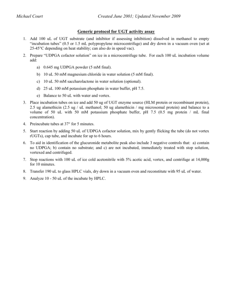
Michael Court Created June 2001; Updated November 2009 Generic protocol for UGT activity assay 1. Add 100 uL of UGT substrate (and inhibitor if assessing inhibition) dissolved in methanol to empty “incubation tubes” (0.5 or 1.5 mL polypropylene microcentrifuge) and dry down in a vacuum oven (set at 25-45°C depending on heat stability; can also do in speed vac). 2. Prepare “UDPGA cofactor solution” on ice in a microcentrifuge tube. For each 100 uL incubation volume add: a) 0.645 mg UDPGA powder (5 mM final). b) 10 uL 50 mM magnesium chloride in water solution (5 mM final). c) 10 uL 50 mM saccharolactone in water solution (optional). d) 25 uL 100 mM potassium phosphate in water buffer, pH 7.5. e) Balance to 50 uL with water and vortex. 3. Place incubation tubes on ice and add 50 ug of UGT enzyme source (HLM protein or recombinant protein), 2.5 ug alamethicin (2.5 ug / uL methanol; 50 ug alamethicin / mg microsomal protein) and balance to a volume of 50 uL with 50 mM potassium phosphate buffer, pH 7.5 (0.5 mg protein / mL final concentration). 4. Preincubate tubes at 37° for 5 minutes. 5. Start reaction by adding 50 uL of UDPGA cofactor solution, mix by gently flicking the tube (do not vortex rUGTs), cap tube, and incubate for up to 6 hours. 6. To aid in identification of the glucuronide metabolite peak also include 3 negative controls that: a) contain no UDPGA; b) contain no substrate; and c) are not incubated, immediately treated with stop solution, vortexed and centrifuged. 7. Stop reactions with 100 uL of ice cold acetonitrile with 5% acetic acid, vortex, and centrifuge at 14,000g for 10 minutes. 8. Transfer 190 uL to glass HPLC vials, dry down in a vacuum oven and reconstitute with 95 uL of water. 9. Analyze 10 - 50 uL of the incubate by HPLC.
