Preparing a Wet Mount
advertisement
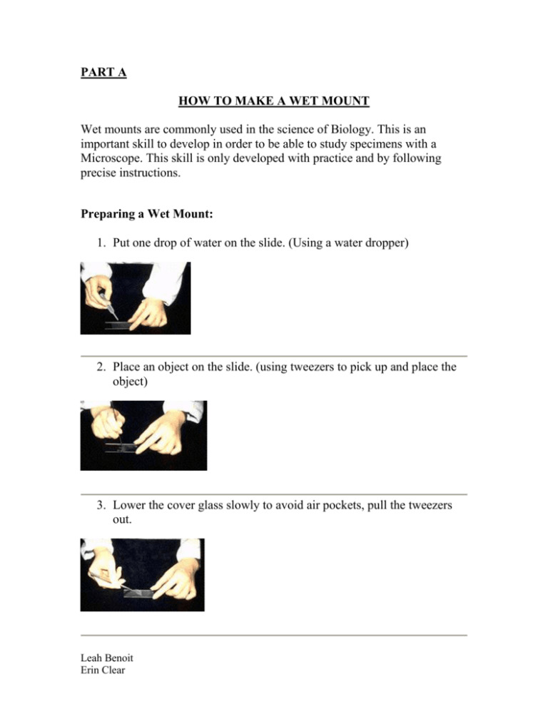
PART A HOW TO MAKE A WET MOUNT Wet mounts are commonly used in the science of Biology. This is an important skill to develop in order to be able to study specimens with a Microscope. This skill is only developed with practice and by following precise instructions. Preparing a Wet Mount: 1. Put one drop of water on the slide. (Using a water dropper) 2. Place an object on the slide. (using tweezers to pick up and place the object) 3. Lower the cover glass slowly to avoid air pockets, pull the tweezers out. Leah Benoit Erin Clear 4. After placing the cover glass, the excess water should be absorbed with paper. http://markun.cs.shinshu-u.ac.jp/learn/kenbikyou/purepa/purepa-e.html 5. Place your prepared wet mount on the “stage” of the microscope Proceed to student directed activities sheet. Leah Benoit Erin Clear PART B CSI: CRIME SCENE INVESTIGATION (student directed activity sheet) Introduction: In everyday life we are surrounded by particles that are naked to the human eye. The way we view these tiny particles is with a microscope. Imagine, you are a forensic scientist and have to use science to help you solve a murder case. You have collected DNA from the crime scene in the form of epithelial cells (skin cells). In order to catch the criminal, you must study the epithelial cells under a high powered microscope. For this method to be successful you must use a “wet mount”, on the microscope to properly identify the DNA you have found. Safety Concerns: If the object that you are using to prepare a wet mount is too thick is has to be sliced. Caution must be taken while using a scalpel to prepare your specimen. If your specimen is too thick, then the cover-slip will wobble on top of the sample like a see-saw (http://shs.westport.k12.ct.us/mjvl/biology/microscope/microscope.htm). Curriculum Objectives: GLO’s: C1, C2, D1 SLO’s: 8-1-06: Demonstrate proper use and care of a microscope to observe the general structure of plant and animal cells. Includes: preparing wet mounts beginning with the least powerful lenses; focusing; drawing specimens; indicating magnification. Leah Benoit Erin Clear Applying your new science skills: A) 1. Take a tooth pick and gently scratch it on the inside of your cheek. This is removing some of your epithelial cells (skin cells) from your mouth. 2. Prepare a wet mount with those cells 3. Remove the skin of the provided onion and carefully slice a small specimen to observe. 4. Prepare it in a wet mount. B) 1. Observe and draw your skin cells (wet mount) at the frequency: 10X 50X 100X Leah Benoit Erin Clear 2. Observe and draw the prepared wet mount containing the onion cells at the frequency: 10X 50X 100X 3. List the similarities and differences that you observed at each frequency. 10X 50X Leah Benoit Erin Clear 100X 4. Imagine now, that you are a forensic scientist. With your new understanding of wet mounts, explain how you could use this skill to analyze DNA from a crime scene? Leah Benoit Erin Clear
