RFLP-2 - Locating Genes in Large Genomes Using RFLPs
advertisement
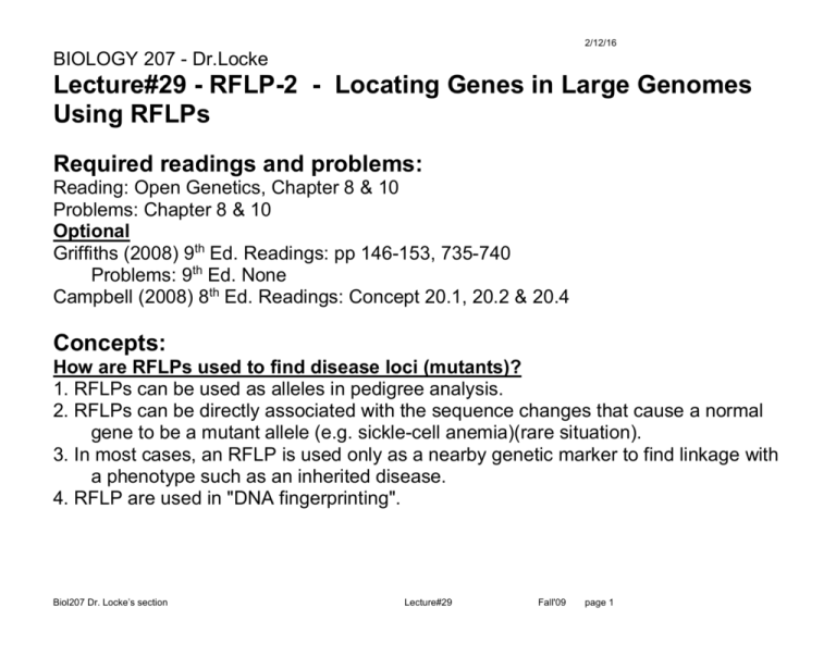
2/12/16 BIOLOGY 207 - Dr.Locke Lecture#29 - RFLP-2 - Locating Genes in Large Genomes Using RFLPs Required readings and problems: Reading: Open Genetics, Chapter 8 & 10 Problems: Chapter 8 & 10 Optional Griffiths (2008) 9th Ed. Readings: pp 146-153, 735-740 Problems: 9th Ed. None Campbell (2008) 8th Ed. Readings: Concept 20.1, 20.2 & 20.4 Concepts: How are RFLPs used to find disease loci (mutants)? 1. RFLPs can be used as alleles in pedigree analysis. 2. RFLPs can be directly associated with the sequence changes that cause a normal gene to be a mutant allele (e.g. sickle-cell anemia)(rare situation). 3. In most cases, an RFLP is used only as a nearby genetic marker to find linkage with a phenotype such as an inherited disease. 4. RFLP are used in "DNA fingerprinting". Biol207 Dr. Locke’s section Lecture#29 Fall'09 page 1 2/12/16 RFLPs in Pedigree analysis Advantages of RFLP analysis in human applications 1. An RFLP can be found at almost every location in a genome. - not dependent on a gene with a phenotype. - probe has to be unique (not repeated DNA sequences) - try many different restriction-enzyme/probe combinations - any randomly chosen unique DNA probe can usually serve as an RFLP marker. 2. RFLP analysis requires a small amount of DNA. - a blood sample is usually enough to do many tests - can culture and grow more white blood cells if more DNA is needed. 3. RFLP analysis can be done before any disease symptoms appear. - or before expected to appear -> predictive - may even be done before birth (pre-natal). 4. Same advantages are present in non-human applications. Why map disease genes? - find linkage to known diseases and can then predict inheritance of those diseases - find and clone gene for more study and better treatments/cure Biol207 Dr. Locke’s section Lecture#29 Fall'09 page 2 2/12/16 RFLPs can be directly associated with the base pair mutation causing a disease: http://www.flickr.com/photos/euthman/5610746554/sizes/z/in/photostream/ Rare situation, but demonstrates a concept. Sickle-Cell Anemia – Fig Sickle-cell anemia is an inherited genetic disease that causes round red blood cells to become sickled in shape. The mutation is a DNA sequence change that alters the normal glutamine to a valine amino acid in the globin polypeptide chain (GAG to GTG codon). This change results in the loss of a Mst II . The sickle-cell hemoglobin gene lacks the Mst II site. Biol207 Dr. Locke’s section Lecture#29 Fall'09 page 3 2/12/16 This provides Absolute linkage between the RFLP and a mutant gene (the mutation itself). Not the usual situation. Biol207 Dr. Locke’s section Lecture#29 Fall'09 page 4 2/12/16 RFLP linkage with an inherited disease (phenotype) – Fig RFLPs can be used as marker loci. Test crosses can then find linkage between an RFLP and a disease gene locus. Example: Probe “P” detects two different alleles: Morph#1 (small band) or Morph#2 (large band). Examine a pedigree of an affected individual -> disease “D” is due to a dominant mutation. D/d - affected has morph 1, 2 (heterozygote mother) d/d - unaffected has morph 2 only homozygote (father) Biol207 Dr. Locke’s section ->(classic test cross) Lecture#29 Fall'09 page 5 2/12/16 Progeny from Cross: Dd x dd gives 8 progeny -> each child has one allele as morph 2 (normal Father – red, d/d ) and either morph 1 or 2 (from affected Mother – black D/d ). Note: This example is a small sample size. Real human genetics research uses many families and combines the data. If the disease locus and the RFLP are: - unlinked, then D would have 50% morph 1 50% morph 2. - absolutely linked, then D would have 100% morph 1 Morph 1 2 Allele D d 4 0 1 3 observed Unlinked D d 2 2 2 2 expected Absolutely linked D d 5 0 0 3 expected Conclusion: The single discordance can be explained as a cross-over between the D/d locus and the RFLP locus in the one child. So: D/d disease locus appears linked, but not absolutely linked -> Partial linkage 1 recombinant / 8 total = 12.5% RF Biol207 Dr. Locke’s section Lecture#29 Fall'09 page 6 2/12/16 Linkage Phase (parental, original combinations) Mother’s heritage is (or could be) different than Father’s. Each parent in each pedigree must be treated independently regarding allele combinations. Re-examine assumption of “D” with morph#1 in the mother: Note: We canNOT assume that because the Father is d/d and is 2/2 that the morph 2 in Mother is also d , and D then must be morph 1. It could be: ---morph#1----D---o------morph#2----d---o---- or ---morph#1----d---o------morph#2----D---o---- Mother’s heritage is (or could be) different than Father’s. Thus, the "linkage phase" could be either one way or the other. Progeny will reveal the phase combination. Similar disease mutations could arise on chromosomes with different morphs (RFLP patterns) -> different phase. Linkage Phase in this example: D phase morph 1 and d phase morph 2 Note: If unlinked, then the linkage phase is not possible to determine. Class exercise: 1- What would the Southern blot look like if the phase was reversed but the affected progeny were the same? 2- What would the pedigree look like (affected vs unaffected) if the phase was reversed and the Southern blot was the same? Biol207 Dr. Locke’s section Lecture#29 Fall'09 page 7 2/12/16 Real mapping of human disease genes: 1- A large scale search uses many RFLP and many progeny. - it has been possible to find linkage with most human disease genes. - currently this is the method of choice for mapping a human genetic disease. 2- One can test 100-1000 of RFLP sites on various chromosomes for linkage. - first find weak linkage with a RFLP probe - then refine the location with other RFLP sites nearby - "Zero-in" on genetic disease gene. 3- Then use RFLP locations to identify candidate genes in a region. - test candidate genes to see if they are the one that is mutant (e.g. sequence mutant alleles) 4- There are "statistical aberrations" > mislead investigators to look in the wrong location. Biol207 Dr. Locke’s section Lecture#29 Fall'09 page 8 2/12/16 RFLP, VNTRs, and DNA fingerprinting RFLP can arise due to VNTR's ( Variable Number Tandem Repeat) First VNTR example found in the human myoglobin gene. Short sequence of 33 base pairs, repeated 4 times in the normal myoglobin gene Other examples of VNTRs vary from 15-100 bp and are repeated a variable number of times. Direct repeats - highly polymorphic in the population - many allele morphs - allelic series Use this repeat sequence as a DNA probe to genomic southern blot. - there are several loci that hybridize to this probe -> multiple loci in genome - each locus may have many different morphs (repeat numbers). - each individual has a unique combination of alleles at each of the multiple loci, -> band pattern is unique for each individual -> a DNA fingerprint (Identical twins have identical alleles & patterns). Biol207 Dr. Locke’s section Lecture#29 Fall'09 page 9 2/12/16 DNA fingerprint – Fig - Bloodstain matches a suspect. Note: The technique is much more powerful at excluding people, than including. If the banding pattern doesn't "match" then the samples must be different (e.g. from different individuals). Twins?? – expect a matching set of bands (same genotype) Clone one locus out and use it to identify a single specific locus probe where there is only one VNTR present at the locus. Biol207 Dr. Locke’s section Lecture#29 Fall'09 page 10 2/12/16 VNTR probing to find linkage to a disease locus 1- Use a VNTR repeat to identify polymorphic alleles at multiple loci in a single cross. 2- Identify affected and unaffected individuals. 3- See if the inheritance of any bands (alleles at a locus) are linked to the disease inheritance Observations: 1- 200 & 400 always inherited together -> Linked? 2- 900 & 800 always either -> allelic? 3- 900 & 600 4 combinations > IA (unlinked) 4- Disease linked to 100 & 700 (red arrow) Biol207 Dr. Locke’s section Lecture#29 Fall'09 page 11 2/12/16 Add PCR to a VNTR locus 1- Have parents with all 4 alleles identified with VNTR morphs 2- Can follow each allele through to each child 3- Can find linkage with Disease (D) allele 4- This type of analysis is easier and faster than is possible with classical Southern based RFLP analysis. Biol207 Dr. Locke’s section Lecture#29 Fall'09 page 12 2/12/16 Summary of ways to identify variations in DNA sequences: Biol207 Dr. Locke’s section Lecture#29 Fall'09 page 13
