paper
advertisement
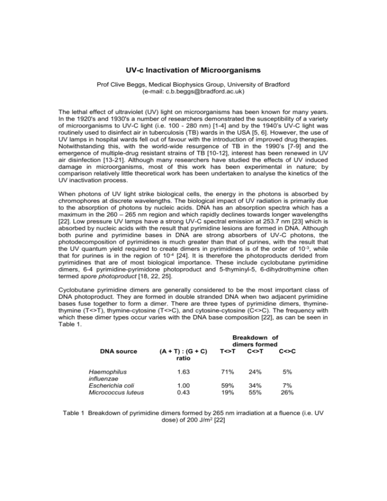
UV-c Inactivation of Microorganisms Prof Clive Beggs, Medical Biophysics Group, University of Bradford (e-mail: c.b.beggs@bradford.ac.uk) The lethal effect of ultraviolet (UV) light on microorganisms has been known for many years. In the 1920's and 1930's a number of researchers demonstrated the susceptibility of a variety of microorganisms to UV-C light (i.e. 100 - 280 nm) [1-4] and by the 1940’s UV-C light was routinely used to disinfect air in tuberculosis (TB) wards in the USA [5, 6]. However, the use of UV lamps in hospital wards fell out of favour with the introduction of improved drug therapies. Notwithstanding this, with the world-wide resurgence of TB in the 1990’s [7-9] and the emergence of multiple-drug resistant strains of TB [10-12], interest has been renewed in UV air disinfection [13-21]. Although many researchers have studied the effects of UV induced damage in microorganisms, most of this work has been experimental in nature; by comparison relatively little theoretical work has been undertaken to analyse the kinetics of the UV inactivation process. When photons of UV light strike biological cells, the energy in the photons is absorbed by chromophores at discrete wavelengths. The biological impact of UV radiation is primarily due to the absorption of photons by nucleic acids. DNA has an absorption spectra which has a maximum in the 260 – 265 nm region and which rapidly declines towards longer wavelengths [22]. Low pressure UV lamps have a strong UV-C spectral emission at 253.7 nm [23] which is absorbed by nucleic acids with the result that pyrimidine lesions are formed in DNA. Although both purine and pyrimidine bases in DNA are strong absorbers of UV-C photons, the photodecomposition of pyrimidines is much greater than that of purines, with the result that the UV quantum yield required to create dimers in pyrimidines is of the order of 10 -3, while that for purines is in the region of 10-4 [24]. It is therefore the photoproducts derided from pyrimidines that are of most biological importance. These include cyclobutane pyrimidine dimers, 6-4 pyrimidine-pyrimidone photoproduct and 5-thyminyl-5, 6-dihydrothymine often termed spore photoproduct [18, 22, 25]. Cyclobutane pyrimidine dimers are generally considered to be the most important class of DNA photoproduct. They are formed in double stranded DNA when two adjacent pyrimidine bases fuse together to form a dimer. There are three types of pyrimidine dimers, thyminethymine (T<>T), thymine-cytosine (T<>C), and cytosine-cytosine (C<>C). The frequency with which these dimer types occur varies with the DNA base composition [22], as can be seen in Table 1. DNA source Haemophilus influenzae Escherichia coli Micrococcus luteus (A + T) : (G + C) ratio Breakdown of dimers formed T<>T C<>T C<>C 1.63 71% 24% 5% 1.00 0.43 59% 19% 34% 55% 7% 26% Table 1 Breakdown of pyrimidine dimers formed by 265 nm irradiation at a fluence (i.e. UV dose) of 200 J/m2 [22] Like cyclobutane dimers, 6-4 pyrimidine-pyrimidone adducts are formed between adjacent pyrimidine bases in DNA. The frequency with which 6-4 pyrimidine-pyrimidone photoproducts are formed depends on the base composition of the DNA. In the Escherichia coli (E. coli) lacI and lacZ genes cyclobutane pyrimidine dimers and 6-4 pyrimidine-pyrimidone photoproducts form in a 2:1 ratio during UV irradiation [25]. Spore photoproduct is a thymine lesion which is the predominant photoproduct formed when bacterial spores are irradiated with UV light [22, 26]. Spore photoproduct has also been reported in isolated vegetative cell DNA at low relative humidity (RH) levels [27, 28] and in aerosolised bacteria at humidities below 65% RH [18]. When investigating the kinetics of UV inactivation it is helpful to consider a UV field as a series of photon ‘bullets’, each containing a specific package of energy, which are fired at a randomly moving series of target microorganisms (or stationary targets if the microorganisms are on solid media). The energy contained in each photon of light can be determined using equation 1. Q Where; Q h c = = = = hc (1) Energy of a single photon (J) Wavelength of the light (m) Planck’s constant (i.e. 6.623 10-34 Js) Speed of light (i.e. 2.998 108 m/s) In many instances a log-linear plot of the number of microorganisms surviving against UV fluence (i.e. UV dose in J/m 2) reveals a straight line which passes through unity on the y axis. This pattern of inactivation is often referred to as a one-hit model and is characterised by the simple exponential equation: Nt kt e No Where: Nt No k t = = = = (2) Number of living microorganisms at time t seconds (cfu) Number of living microorganisms at time 0 seconds (cfu) Inactivation rate constant (s-1) Duration of exposure to UV irradiation (s) Equation 2 can be modified to incorporate the UV fluence rate. Nt ZEt e No where: E Z = = (3) UV fluence rate (W/m 2) UV susceptibility constant of particular microorganism (m 2/J) In the one-hit model it is assumed that; (i) a single hit is sufficient to inactivate a cell, (ii) the number of hits achieved is directly proportional to the UV fluence applied, and that (iii) the microorganism population is uniformly susceptible to the UV irradiation [29]. Although the equations 2 and 3 can be successfully applied to many UV inactivation systems, the one-hit model does not fully explain the inactivation process. In reality, a great many photon hits are required before a microorganism finally becomes inactivated. In addition, the various DNA repair mechanisms play an important role in repairing damage sustained by UV photons. The number of photons absorbed by a target microorganism can be determined using equation 4 and the quantum yield using equation 5. P Where; Pabs abs = = Et Number of photons absorbed by the target microorganism Inactivation cross-sectional area (m 2) Where; M (4) hc = = M Pa bs (5) Quantum yield Number of molecules altered Jagger [24] estimates that at a UV fluence of 0.1 J/m 2 at 260 nm one thymine dimer is produced per 200 microns of E. coli DNA. This equates to approximately 5 dimers per E.coli chromosome. Similarly, Harm [30] states that for E. coli a fluence of 1.0 J/m 2, at 254 nm, will produce 65 pyrimidine dimers in a chromosome. By applying equations 4 and 5 to a 2.5 m 1 m E. coli bacterium it is possible to analyse and compare Jagger and Harm’s assertions (see Table 2). Researcher Jagger (24) Harm (30) UV fluence Wavelength (J/m2) (nm) 0.1 1.0 260 254 Number of photons absorbed by a single E.coli target Average number of photon hits required to create a dimer Quantum Yield 327361 3198065 65472 49201 1.527 10-5 2.032 10-5 Table 2 Analysis of Jagger and Harm’s data From Table 2 it can be seen that the quantum yields derived from Jagger and Harm’s data are of a similar order of magnitude and that they are both well within the expected range of 10-3 to 10-6 [31]. This implies that many thousands of photon hits occur per bacterium before a cyclobutane pyrimidine dimer is formed. Given that the quantum yield of a pyrimidine dimer is about 1 [32], it can be concluded that most hits cause no photochemical reaction at all. This is not at all surprising given that the target area presented by a bacterium is much greater than that of a single dimer. Given the discussion above it is clear that the one-hit model does not satisfactorily describe the UV inactivation process and that a superior model is required. This is especially the case for UV inactivation curves which exhibit a shoulder, such as the survival curve shown in Figure 1. It is important to note that in many experiments, microorganisms do not exhibit a shoulder simply because the UV fluence rates used are too high and the microorganisms are very quickly overwhelmed by the photons. If lower fluence rates are used then in many instances a shoulder will be observed. Shoulders are often observed in microorganisms which have efficient DNA repair mechanisms. 83.8 79.8 75.8 71.8 67.8 63.8 59.9 55.9 51.9 47.9 43.9 39.9 35.9 31.9 27.9 20 16 12 7.98 3.99 0 23.9 tc 1 0.1 Survival fraction 0.01 0.001 0.0001 0.00001 0.000001 0.0000001 UV fluence (J/m2) Figure 1 Typical shouldered UV Survival Curve From Figure 1 it can be seen that at low UV doses there is a shoulder in the inactivation curve and that after a certain threshold dose the curve becomes a straight line which is similar to that for the one-hit model [29]. This type of curve is often referred to as a multi-target model [29]. The shoulder in the curve represents the period in which the microorganisms receive a sub-lethal dose and little or no inactivation occurs. The UV field can be considered as a series of photon ‘bullets’ which are fired at a randomly moving series of target microorganisms. Each target microorganism will receive many photon hits before one final hit eventually inactivates it. So if the nth photon to hit a target microorganism is lethal, it follows that all the photon hits up to and including n – 1, will produce a sub-lethal effect. If it is assumed that all the target microorganisms are equally susceptible to UV damage and that the photons strike all the targets at roughly the same rate, then they will all accumulate photon hits until each has received n – 1 hits. Thereafter, the first microorganism to be struck by the nth photon will become inactivated. Similarly, the second microorganism to receive a nth hit will be inactivated, and so on, until all the microorganisms present are inactivated. However, as more and more microorganisms are removed, it becomes increasingly difficult for the photons to hit targets. The first tranche of photons will easily inactivate microorganisms, but as the process continues successive tranches of photons will be aiming at fewer and fewer targets. Consequently, more and more photons must be fired in order to maintain the same kill rate. So although only one photon eventually kills any particular microorganism, there is a cumulative component to the inactivation process, since many non-lethal photon hits are required before the first lethal hits occur. Equations 6 and 7 are expressions derived by Kowalski et al. [33] to describe the multi-target inactivation curve. These equations utilise a quasi-threshold term, tc, (see Figure 1) which represents the time at the point where the extended straight line decay curve intersects the unity line. t 2tc If; Then; Nt ( zE / 4tc )t 2 e No where: tc And if; t 2tc = (6) Quasi-threshold time (s) Then; Nt zE (t tc ) e No (7) REFERENCES 1. F.L. Gates, A study of the bacterial action of ultraviolet light, J. Gen. Physiol. 13 (1929) 231 - 260 2. D.G. Sharp, A quantitative method of determining the lethal effect of ultraviolet light on bacteria suspended in air, (1937) 589 – 599 3. D.G. Sharp, The lethal action of short ultraviolet rays on several common pathogenic bacteria, (1938) 447 – 460 4. H.C. Rentschler, R. Nagy, Bacterial action of ultraviolet radiation on air-borne organisms, (1941) 85 - 91 5. L.J. Buttolph, H. Haynes, Ultraviolet air sanitation. General Electric Lamp Division, Cleveland, Ohio, USA, 1953 6. W.W. Stead, et al. Probable role of ultraviolet irradiation in preventing transmission of tuberculosis: A case study. Infection Control & Hospital Epidemiology, 17 (1996) 11 – 13 7. J.M. Grange, A. Zumia, Paradox of the global emergency of tuberculosis. Lancet, 353 (1999) 996 8. WHO. TB - a global emergency: WHO report on the TB epidemic. WHO, Geneva, 1994 9. WHO. Global Tuberculosis Programme. Global tuberculosis control. WHO report, WHO, Geneva. WHO/TB/98 – 237, 1998 10. V. Gleissberg, The threat of multidrug resistance: is tuberculosis ever untreatable or uncontrollable? Lancet, 353 (1999) 998 – 999 11. T.R. Freiden, et al. A multi-institutional outbreak of highly drug resistant tuberculosis: Epidemiology and Clinical Outcomes, Journal of the American Medical Association. 276 (1996) 1229 - 1235. 12. A.S. Breathnach, A. de Ruiter, G.M. Holdsworth, N.T. Bateman, D.G. O’Sullivan, P.J. Rees, D. Snashall, H.J. Milburn, B.S. Peters, J. Watson, F.A. Drobniewski, G.L. French, An outbreak of multi-drug-resistant tuberculosis in a London teaching hospital. Journal of Hospital Infection. 39 (1998) 111 - 117. 13. J.M. Macher, L.E. Alvantis, Y.L. Chang, K.S. Liu, Effect of ultraviolet germicidal lamps on airborne microorganisms in an outpatient waiting room, Applied. Occupational and Environmental Hygiene. 7 (1992) 505 – 513 14. S.L. Miller, J.M. Macher, Evaluation of a methodology for quantifying the effect of room air ultraviolet germicidal irradiation on airborne bacteria, Aerosol Science and Technology. 33 (2000) 274 – 295 15. C.B. Beggs, K.G. Kerr, JK. Donnelly, P.A. Sleigh, D.D. Mara, G. Cairns, An engineering approach to the control of Mycobacterium tuberculosis and other airborne pathogens: a UK hospital based pilot study, Transactions of the Royal Society of Tropical Medicine and Hygiene. 94 (2000) 141 – 146 16. G. Ko, MW First, HA Burge, Influence of relative humidity on particle size and UV sensitivity of Serratia marcescens and Mycobacterium bovis BCG aerosols. Tubercle and Lung Disease. 80 (2000) 217 – 228 17. J.L. Peccia, H.M. Werth, M.T. Hernandez, Effects of relative humidity on the UVinduced inactivation of bacterial bioaerosols. Journal of Aerosol Science. 31 (2000) Suppl. 1, S959 – S960 18. J.L. Peccia, H.M. Werth, S.M. Miller, M.T. Hernandez, Effects of relative humidity on the ultraviolet induced inactivation of airborne bacteria. Aerosol Science and Technology. 35 (2001) 728 – 740 19. C.B. Beggs, P.A. Sleigh, A quantitative method for evaluating the germicidal effect of upper room UV fields. Journal of Aerosol Science. 23 (2002) 1681-1699 20. C.B. Beggs, C.J. Noakes, P.A. Sleigh, L.A. Fletcher, K.G. Kerr, Methodology for determining the susceptibility of airborne microorganisms to irradiation by an upperroom UVGI system. Journal of Aerosol Science. 37 (2006) 885-902 21. C.J. Noakes, P.A. Sleigh, A.R. Escombe, C.B. Beggs, Use of CFD analysis in modifying a TB Ward in Lima, Peru. Indoor and Built Environment. 15 (2006) 41-47 22. W. Harm, Biological effects of ultraviolet radiation. Cambridge University Press, Cambridge, Chapter 3, 1980 23. Philips Lighting. Disinfection by UV-radiation: Germicidal lamps, 1993 24. J. Jagger, Introduction to research in ultraviolet photobiology. Prentice-Hall, New Jersey, Chapter 4, 1967 25. D. Chandrasekhar, B. Van Houten. In vivo formation and repair of cyclobutane pyrimidine dimers and 6-4 photoproducts measured at the gene and nucleotide level in Escherichia coli. Mutation Research, 450 (2000) 19 – 40. 26. W.L Nicholson, T.A. Slieman, DNA in dormant Bacillus subtilis spores exposed to solar radiation accumulates strand breaks and cyclobutane pyrimidine dimers in addition to spore photoproduct. 27. M.H. Patrick, D.M. Grey, Independence of photoproduct formation on DNA conformation. Photochem. Photobiol. 24 (1976) 507 – 513. 28. R.O. Rahn, L.C. Landry, Pyrimidine dimer formation in poly (d-dT) and apurinic acid. Biochim. Biophys. Acta. 247 (1971) 197 – 206. 29. D.L. David, W.D.J. Jones, C.M. Newman, Ultraviolet light inactivation and photoreactivation in the mycobacteria. Infect. Immun. 4 (1971) 319 – 329 30. W. Harm W, Biological effects of ultraviolet radiation. Cambridge University Press, Cambridge, Chapter 7, 1980 31. W. Harm, Biological effects of ultraviolet radiation. Cambridge University Press, Cambridge, Chapter 6, 1980 32. J. Jagger, Introduction to research in ultraviolet photobiology. Prentice-Hall, New Jersey, Chapter 3, 1967 33. W.J. Kowalski, Design and optimisation of UVGI air disinfection systems. PhD Thesis, Pennsylvania State University, (2001) CB Beggs 24th October 2006
