Cardiology Fellows Nuclear Rotation
advertisement
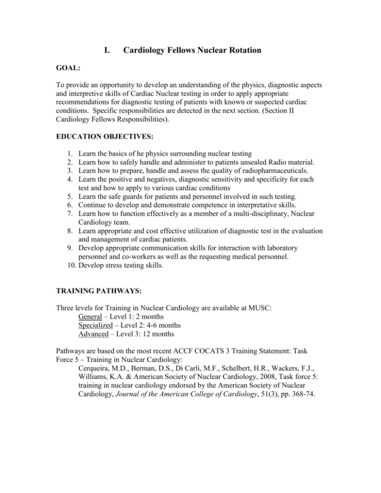
I. Cardiology Fellows Nuclear Rotation GOAL: To provide an opportunity to develop an understanding of the physics, diagnostic aspects and interpretive skills of Cardiac Nuclear testing in order to apply appropriate recommendations for diagnostic testing of patients with known or suspected cardiac conditions. Specific responsibilities are detected in the next section. (Section II Cardiology Fellows Responsibilities). EDUCATION OBJECTIVES: 1. 2. 3. 4. Learn the basics of he physics surrounding nuclear testing Learn how to safely handle and administer to patients unsealed Radio material. Learn how to prepare, handle and assess the quality of radiopharmaceuticals. Learn the positive and negatives, diagnostic sensitivity and specificity for each test and how to apply to various cardiac conditions 5. Learn the safe guards for patients and personnel involved in such testing. 6. Continue to develop and demonstrate competence in interpretative skills. 7. Learn how to function effectively as a member of a multi-disciplinary, Nuclear Cardiology team. 8. Learn appropriate and cost effective utilization of diagnostic test in the evaluation and management of cardiac patients. 9. Develop appropriate communication skills for interaction with laboratory personnel and co-workers as well as the requesting medical personnel. 10. Develop stress testing skills. TRAINING PATHWAYS: Three levels for Training in Nuclear Cardiology are available at MUSC: General – Level 1: 2 months Specialized – Level 2: 4-6 months Advanced – Level 3: 12 months Pathways are based on the most recent ACCF COCATS 3 Training Statement: Task Force 5 – Training in Nuclear Cardiology: Cerqueira, M.D., Berman, D.S., Di Carli, M.F., Schelbert, H.R., Wackers, F.J., Williams, K.A. & American Society of Nuclear Cardiology, 2008, Task force 5: training in nuclear cardiology endorsed by the American Society of Nuclear Cardiology, Journal of the American College of Cardiology, 51(3), pp. 368-74. Table 1. Classification of Nuclear Cardiology Procedures 1. Standard nuclear cardiology procedures a. Myocardial perfusion imaging i. SPECT with technetium-99m agents and/or thallium-201, with or without attenuation correction ii. PET with rubidium-82 and/or nitrogen-13 ammonia iii. Planar with technetium-99m agents and/or thallium-201 iv. ECG gating of perfusion images for assessment of global and regional ventricular function v. Imaging protocols vi. Stress protocols 1. Exercise stress 2. Pharmacologic stress vii. Viability assessment including reinjection and delayed imaging of thallium-201 and/or metabolic imaging where available b. Equilibrium radionuclide angiocardiography and/or “first-pass” radionuclide angiography at rest c. Qualitative and quantitative methods of image display and analysis 2. Less commonly used nuclear cardiology procedures a. Combined myocardial perfusion imaging with cardiac CT for attenuation correction or anatomic localization b. Equilibrium radionuclide angiocardiography and/or “first-pass” radionuclide angiography during exercise or pharmacologic stress c. Metabolic imaging using single-photon and/or positron-emitting radionuclides d. Myocardial infarct imaging e. Cardiac shunt studies Table 2. Nuclear Cardiology Training Components 1. Didactic program a. Lectures and self-study b. Radiation safety 2. Interpretation of clinical cases 3. Hands-on experience a. Clinical cases b. Radiation safety Table 3. Summary of Training Requirements for Nuclear Cardiology Level Minimum Months Total Examinations 1 2 100* 2 4-6 300* 3 12 600* *A minimum of 35 cases with hands-on experience must be performed and interpreted under supervision. General Training—Level 1 (Minimum of 2 Months) The trainee is exposed to the fundamentals of nuclear cardiology for a minimum period of 2 months during training. This 2-month experience provides familiarity with nuclear cardiology technology and its clinical applications in the general clinical practice of adult cardiology, but it is not sufficient for the specific practice of nuclear cardiology. The 3 components of training include a didactic program that includes lectures, self-study, radiation safety and regulations, interpretation of nuclear cardiology studies, and handson experience. Didactic Program Lectures and self-study. This component consists of lectures on the basic aspects of nuclear cardiology and parallel self-study material consisting of reading and viewing case files. The material presented should integrate the role of nuclear cardiology into total patient management. Such information can be included within a weekly noninvasive or invasive cardiology conference, with presentation and discussion of nuclear cardiology image data as part of diagnostic and therapeutic management. Knowledge and appreciation of radiation safety. The didactic program should include reading and practical experience with the effects of radiation and provide the fellow with an understanding of radiation safety as it relates to patient selection and administration of radiopharmaceuticals and utilization of CT systems. Interpretation of Nuclear Cardiology Studies During the 2-month rotation, fellows should actively participate in daily nuclear cardiology study interpretation (minimum of 100 cases). Experience in all the areas listed in Table 1 is recommended. If some procedures are not available or are performed in low volume, an adequate background for general fellowship training can be satisfied by appropriate reading or review of case files. The teaching file should consist of perfusion and ventricular function studies with angiographic/cardiac catheterization documentation of disease. Hands-On Experience Fellows should perform complete nuclear cardiology studies alongside a qualified technologist or other qualified laboratory personnel. They should, under supervision, observe and participate in a large number of the standard procedures and as many of the less commonly performed procedures as possible. Fellows should have experience in the practical aspects of radiation safety associated with performing clinical patient studies. Specialized Training—Level 2 (Minimum of 4-6 Months) **Training which meets the Certification Board of Nuclear Cardiology (CBNC) requirements (appended below) and NRC requirements for “Authorized User” status. Fellows who wish to practice the specialty of nuclear cardiology are required to have at least 4 months of training. Level 2 training includes a minimum of 700 h of radiation safety experience in nuclear cardiology. There needs to be didactic, clinical study interpretation, and hands-on involvement in clinical cases. In training programs with a high volume of procedures, clinical experience may be acquired in as short a period as 4 months. In programs with a lower volume of procedures, a total of 6 months of clinical experience will be necessary to achieve Level 2 competency. The additional training required of Level 2 trainees is to enhance their clinical skills, knowledge, and hands-on experience in radiation safety and qualify them to become authorized users of radioactive materials in accordance with the regulations of the NRC and/or the Agreement States. Didactic Program Lectures and self-study. The didactic training should include in-depth details of all aspects of the procedures listed in Table 1. This program may be scheduled over a 12-to 24-month period concurrent and integrated with other fellowship assignments. Radiation safety. Classroom and laboratory training needs to include extensive review of radiation physics and instrumentation, radiation protection, mathematics pertaining to the use and measurement of radioactivity, chemistry of byproduct material for medical use, radiation biology, the effects of ionizing radiation, and radiopharmaceuticals. There should be a thorough review of regulations dealing with radiation safety for the use of radiopharmaceuticals and ionizing radiation. This experience should total a minimum of 80 h and be separately documented. Interpretation of Clinical Cases Fellows should participate in the interpretation of all nuclear cardiology imaging data for the 4- to 6-month training period. It is imperative that the fellows have experience in correlating catheterization or CT angiographic data with radionuclide-derived data for a minimum of 30 patients. A teaching conference in which the fellow presents the clinical material and nuclear cardiology results is an appropriate forum for such experience. A total of 300 cases should be interpreted under preceptor supervision, from direct patient studies (Table 3). Hands-On Experience Clinical cases. Fellows acquiring Level 2 training should have hands-on supervised experience with a minimum of 35 patients: 25 patients with myocardial perfusion imaging and 10 patients with radionuclide angiography. Such experience should include pre-test patient evaluation; radiopharmaceutical preparation (including experience with relevant radio-nuclide generators and CT systems); performance of studies with and without attenuation correction; administration of the dosage, calibration, and setup of the gamma camera and CT system; setup of the imaging computer; processing the data for display; interpretation of the studies; and generating clinical reports. Radiation safety work experience. This experience should total 620 h and be acquired during training in the clinical environment where radioactive materials are being used and under the supervision of an authorized user who meets the NRC requirements of Part 35.290 or Part 35.290(c)(ii)(G) and Part 35.390 or the equivalent Agreement State requirements, and must include: a. Ordering, receiving, and unpacking radioactive materials safely and performing the related radiation surveys; b. Performing quality control procedures on instruments used to determine the activity of dosages and per-forming checks for proper operation of survey meters; c. Calculating, measuring, and safely preparing patient or human research subject dosages; d. Using administrative controls to prevent a medical event involving the use of unsealed byproduct material; e. Using procedures to safely contain spilled radioactive material and using proper decontamination procedures; f. Administering dosages of radioactive material to patients or human research subjects; and g. Eluting generator systems appropriate for preparation of radioactive drugs for imaging and localization studies, measuring and testing the eluate for radionuclide purity, and processing the eluate with reagent kits to prepare labeled radioactive drugs. Additional Experience The training program for Level 2 must also provide experience in computer methods for analysis. This should include perfusion and functional data derived from thallium or technetium agents and ejection fraction and regional wall motion measurements from radionuclide angiographic studies. This pathway will require completion of all physics/radiation safety/radiation biology training modules available on the MUSC Moodle site, 4-6 months Nuclear Cardiology clinical rotations (MUSC plus VAMC), and completion of a clinical Nuclear Cardiology research project. Please review “II. Cardiology Fellows Responsibilities” while on Nuclear Cardiology. Advanced Training—Level 3 (Minimum of 12 Months) For fellows planning an academic career in nuclear cardiology or a career directing a clinical nuclear cardiology laboratory, an extended program is required. This may be part of the standard 3-year cardiology fellowship. In addition to the recommended program for Level 2, the Level 3 program should include advanced quality control of nuclear cardiology studies and active participation and responsibility in ongoing laboratory or clinical research. In parallel with participation in a research program, the trainee should participate in clinical imaging activities for the total training period of 12 months, to include supervised interpretative experience in a minimum of 600 cases. Hands-on experience should be similar to, or greater than, that required for Level 2 training. The fellow should be trained in most of the following areas: Qualitative interpretation of standard nuclear cardiology studies, including SPECT and/or PET myocardial perfusion imaging, ECG-gated perfusion studies, attenuation-corrected studies, gated-equilibrium studies, “first-pass,” and any of the less commonly performed procedures available at the institution Quantitative analysis of SPECT and/or PET myocardial perfusion and/or metabolic studies Quantitative radionuclide angiographic and gated-myocardial perfusion analyses, including measurement of global and regional ventricular function SPECT and/or PET perfusion acquisition, reconstruction, and display ECG-gated SPECT and/or perfusion acquisition, analysis, and display of functional data Imaging of positron-emitting tracers using dedicated PET systems or hybrid PET/CT systems The requirements for Level 1 to 3 training in nuclear cardiology are summarized in Table 3. Specific Training in Cardiac Imaging of Positron-Emitting Radionuclides Cardiac PET and PET/CT imaging of positron-emitting radionuclides are part of nuclear cardiology. An increasing number of nuclear laboratories have access to both conventional SPECT and PET imaging. For institutions that have positron-imaging devices, training guidelines are appropriate. Training in this particular imaging technology should go hand-in-hand and may be concurrent with training in conventional nuclear cardiology. Such training should include those aspects that are unique or specific to the imaging of positron-emitting radionuclides. Depending on the desired level of expertise, training in cardiac PET and imaging with positron-emitting radionuclides should include knowledge of substrate metabolism in the normal and diseased heart; knowledge of positron-emitting tracers for blood flow, metabolism and neuronal activity, medical cyclotrons, radioisotope production, and radiotracer synthesis; and principles of tracer kinetics and their in vivo application for the noninvasive measurements of regional, metabolic, and functional processes. The training should also include the physics of positron decay, aspects of imaging instrumentation specific to the imaging of positron emitters and the use of CT, production of radiopharmaceutical agents, quality control, handling of ultra-short life radioisotopes, appropriate radiation protection and safety, and regulatory aspects. Consistent with the training guidelines for general nuclear cardiology, training should be divided into 3 classes. General Training (2 Months) This level is for cardiology fellows who are associated with an institution where PET and/or PET/CT devices are available and who wish to become conversant with cardiac positron imaging. Training should therefore be the same as for Level 1 training in nuclear cardiology but should include aspects specific to cardiac positron imaging. The additional proficiency to be acquired by physician trainees includes background in substrate metabolism, patient standardization and problems related to diabetes mellitus and lipid disorders, positron-emitting tracers of flow and metabolism, and technical aspects of positron and CT imaging. A didactic program should include the interpretation of cardiac PET studies of myocardial blood flow and substrate metabolism, the interpretation of studies combining SPECT for evaluation of blood flow with PET for evaluation of metabolism, the evaluation of diagnostic accuracy and cost-effectiveness of viability assessment of coronary artery disease detection, and the understanding of radiation safety as specifically related to positron emitters. Hands-on experience should include supervised observation and interpretation of cardiac studies performed with positronemitting radionuclides and PET and PET/CT imaging devices. Specialized Training (Minimum of 4 Months) This level of training is for fellows who wish to perform and interpret cardiac PET or positron imaging studies in addition to nuclear cardiology. This training should include all Level 1 and Level 2 training in nuclear cardiology (4 to 6 months) as well as general training for cardiac PET and PET/CT. Specific aspects of training for PET and for using positron-emitting radionuclides should include radiation dosimetry, radiation protection and safety, dose calibration, physical decay rates of radioisotopes, handling of large doses of high-energy radioactive materials of short physical half-lives, quality assurance procedures, and NRC safety and record-keeping requirements. This level of training requires direct patient experience with a minimum of 40 patient studies of myocardial perfusion, metabolism, or both. Advanced Training (Minimum 1 Year) This level of training is intended for fellows planning an academic career in cardiac PET or who wish to direct a clinical cardiac PET laboratory. Similar to Level 3 training in nuclear cardiology, this training should include active participation in laboratory and clinical research in parallel with clinical activities. In addition to the requirements for general and specialized cardiac PET training (including standard nuclear cardiology training, as previously described), advanced training should include the following: 1. Basic principles of cyclotrons, isotope production, radiosynthesis, tracer kinetic principles and models, cardiac innervation and receptors, and methods for quantifying regional myocardial blood flow and substrate metabolism. 2. Imaging instrumentation including dedicated PET systems, hybrid PET/CT systems and SPECT-like positron imaging devices with high-energy photon collimators or coincidence detection. Image acquisition and processing to include review of sinograms, errors in image reconstruction, correction routines for photon attenuation, and patient misalignment. 3. Tissue kinetics of positron-emitting tracers; in vivo application of tracer kinetic principles; tracer kinetic models, generation of tissue time-activity curves, and computer-assisted calculation of region of functional processes of the myocardium. 4. Computer-assisted data manipulation, quantitative image analysis, and image display. PROCEDURES: Fellows will be appropriately supervised for all interpretations by a Cardiology Attending and a Nuclear Medicine attending. METHODOLOGY: Duration: Start Time: End Time Attendings: 8 weeks 7:00 am 6:00 pm Cardiology Division and Nuclear Medicine faculty. PRINCIPAL TEACHING METHODS: Teaching Rounds: These are held daily with discussions of interpretation between 2 pm and 5:30 pm Reading List: A collection of important nuclear cardiology references has been attached Articles: Supplied by faculty and other personnel assigned to the specific procedure. Outlined in WIKI – Obtain Cardiology Attending recommendations. Conferences: Daily Cardiology Division conferences are mandatory. Monthly Heart and Vascular Center Mortality and Morbidity conference is mandatory, Fellows are encouraged to attend Department of Medicine Grand Rounds. Supervision: Fellows are directly supervised by the Cardiology Attending and Nuclear Medicine Attending. FELLOW EVALUATION: Fellows are evaluated by the Nuclear Medicine staff and Attendings each rotation using the eValue, which incorporates the standard Division criteria for each competency. PatientCare is assessed based on direct observation, application of evidence based medicine and chart review. Knoweledge is assessed through direct questioning during interpretation sessions. Fellows must demonstrate competent EKG interpretation and successful stress testing capabilities. Professionalism is assessed based on observation of the resident’s demeanor and behavior on the rotation. Interpersonal and Communication Skills are assess by observing the residents interactions with the patients, families, staff and colleagues. System Based Practice is evaluated based on the residents ability to perform in the team setting, including interactions with the Nuclear Medicine Technologist, Nurses, Practice Based Learning is evaluated based on the residents ability to learn and improve his or her skills based on feedback, study, literature review and appropriate application of evidence based medicine. READING LIST: 1. Diamond GA, Forrester JS. Analysis of probability as an aid in the clinical diagnosis of coronary-artery disease. N Engl J Med. 1979;300:1350-8. 2. Mark DB, Hlatky MA, Harrell FE, Lee KL, Califf RM, Pryor DB. Exercise treadmill score for predicting prognosis in coronary artery disease. Ann Intern Med. 1987;106:793800. 3. Mark DB, Shaw L, Harrell FE, Hlatky MA, Lee KL, Bengtson JR, McCants CB, Califf RM, Pryor DB. Prognostic value of a treadmill exercise score in outpatients with suspected coronary artery disease. N Engl J Med. 1991;325:849-53. 4. DePuey EG, Rozanski A. Using gated technetium-99m-sestamibi SPECT to characterize fixed myocardial defects as infarct or artifact. J Nucl Med. 1995;36:952-5. 5. Hachamovitch R, Berman DS, Kiat H, Cohen I, Cabico JA, Friedman J, Diamond GA. Exercise myocardial perfusion SPECT in patients without known coronary artery disease: incremental prognostic value and use in risk stratification. Circulation. 1996;93:905-14. 6. Johnson LL, Verdesca SA, Aude WY, Xavier RC, Nott LT, Campanella MW, Germano G. Postischemic stunning can affect left ventricular ejection fraction and regional wall motion on post-stress gated sestamibi tomograms. J Am Coll Cardiol. 1997;30:1641-8. 7. Smanio PE, Watson DD, Segalla DL, Vinson EL, Smith WH, Beller GA. Value of gating of technetium-99m sestamibi single-photon emission computed tomographic imaging. J Am Coll Cardiol. 1997;30:1687-92. 8. Rywik TM, Zink RC, Gittings NS, Khan AA, Wright JG, O'Connor FC, Fleg JL. Independent prognostic significance of ischemic ST-segment response limited to recovery from treadmill exercise in asymptomatic subjects. Circulation. 1998;97:211722. 9. Shaw LJ, Peterson ED, Shaw LK, Kesler KL, DeLong ER, Harrell FE, Muhlbaier LH, Mark DB. Use of a prognostic treadmill score in identifying diagnostic coronary disease subgroups. Circulation. 1998;98:1622-30. 10. Farkouh ME, Smars PA, Reeder GS, Zinsmeister AR, Evans RW, Meloy TD, Kopecky SL, Allen M, Allison TG, Gibbons RJ, Gabriel SE. A clinical trial of a chestpain observation unit for patients with unstable angina. Chest Pain Evaluation in the Emergency Room (CHEER) Investigators. N Engl J Med. 1998;339:1882-8. 11. Campbell L, Marwick TH, Pashkow FJ, Snader CE, Lauer MS. Usefulness of an exaggerated systolic blood pressure response to exercise in predicting myocardial perfusion defects in known or suspected coronary artery disease. Am J Cardiol. 1999;84:1304-10. 12. Gibbons RJ, Hodge DO, Berman DS, Akinboboye OO, Heo J, Hachamovitch R, Bailey KR, Iskandrian AE. Long-term outcome of patients with intermediate-risk exercise electrocardiograms who do not have myocardial perfusion defects on radionuclide imaging. Circulation. 1999;100:2140-5. 13. McHam SA, Marwick TH, Pashkow FJ, Lauer MS. Delayed systolic blood pressure recovery after graded exercise: an independent correlate of angiographic coronary disease. J Am Coll Cardiol. 1999;34:754-9. 14. Michaelides AP, Psomadaki ZD, Dilaveris PE, Richter DJ, Andrikopoulos GK, Aggeli KD, Stefanadis CI, Toutouzas PK. Improved detection of coronary artery disease by exercise electrocardiography with the use of right precordial leads. N Engl J Med. 1999;340:340-5. 15. Cole CR, Blackstone EH, Pashkow FJ, Snader CE, Lauer MS. Heart-rate recovery immediately after exercise as a predictor of mortality. N Engl J Med. 1999;341:1351-7. 16. Lewin HC, Hachamovitch R, Harris AG, Williams C, Schmidt J, Harris M, Van Train K, Siligan G, Berman DS. Sustained reduction of exercise perfusion defect extent and severity with isosorbide mononitrate (Imdur) as demonstrated by means of technetium 99m sestamibi. J Nucl Cardiol. 2000;7:342-53. 17. Nishime EO, Cole CR, Blackstone EH, Pashkow FJ, Lauer MS. Heart rate recovery and treadmill exercise score as predictors of mortality in patients referred for exercise ECG. JAMA. 2000;284:1392-8. 18. Borges-Neto S, Javaid A, Shaw LK, Kong DF, Hanson MW, Pagnanelli RA, Ravizzini G, Coleman RE. Poststress measurements of left ventricular function with gated perfusion SPECT: comparison with resting measurements by using a same-day perfusion-function protocol. Radiology. 2000;215:529-33. 19. Tavel ME. Stress testing in cardiac evaluation : current concepts with emphasis on the ECG. Chest. 2001;119:907-25. 20. Prakash M, Myers J, Froelicher VF, Marcus R, Do D, Kalisetti D, Atwood JE. Clinical and exercise test predictors of all-cause mortality: results from > 6,000 consecutive referred male patients. Chest. 2001;120:1003-13. 21. Tanaka H, Monahan KD, Seals DR. Age-predicted maximal heart rate revisited. J Am Coll Cardiol. 2001;37:153-6. 22. Germano G. Technical aspects of myocardial SPECT imaging. J Nucl Med. 2001;42:1499-507. 23. Cerqueira MD, Weissman NJ, Dilsizian V, Jacobs AK, Kaul S, Laskey WK, Pennell DJ, Rumberger JA, Ryan T, Verani MS, American Heart Association Writing Group on Myocardial Segmentation and Registration for Cardiac Imaging. Standardized myocardial segmentation and nomenclature for tomographic imaging of the heart: a statement for healthcare professionals from the Cardiac Imaging Committee of the Council on Clinical Cardiology of the American Heart Association. Circulation. 2002;105:539-42. 24. Rywik TM, O'Connor FC, Gittings NS, Wright JG, Khan AA, Fleg JL. Role of nondiagnostic exercise-induced ST-segment abnormalities in predicting future coronary events in asymptomatic volunteers. Circulation. 2002;106:2787-92. 25. Allman KC, Shaw LJ, Hachamovitch R, Udelson JE. Myocardial viability testing and impact of revascularization on prognosis in patients with coronary artery disease and left ventricular dysfunction: a meta-analysis. J Am Coll Cardiol. 2002;39:1151-8. 26. Bonow RO. Myocardial viability and prognosis in patients with ischemic left ventricular dysfunction. J Am Coll Cardiol. 2002;39:1159-62. 27. Hendel RC, Corbett JR, Cullom SJ, DePuey EG, Garcia EV, Bateman TM. The value and practice of attenuation correction for myocardial perfusion SPECT imaging: a joint position statement from the American Society of Nuclear Cardiology and the Society of Nuclear Medicine. J Nucl Cardiol. 2002;9:135-43. 28. McLaughlin MG, Danias PG. Transient ischemic dilation: a powerful diagnostic and prognostic finding of stress myocardial perfusion imaging. J Nucl Cardiol. 2002;9:663-7. 29. Myers J, Prakash M, Froelicher V, Do D, Partington S, Atwood JE. Exercise capacity and mortality among men referred for exercise testing. N Engl J Med. 2002;346:793-801. 30. Schinkel AF, Elhendy A, van Domburg RT, Bax JJ, Vourvouri EC, Bountioukos M, Rizzello V, Agricola E, Valkema R, Roelandt JR, Poldermans D. Incremental value of exercise technetium-99m tetrofosmin myocardial perfusion single-photon emission computed tomography for the prediction of cardiac events. Am J Cardiol. 2003;91:40811. 31. Elhendy A, Schinkel AF, van Domburg RT, Bax JJ, Poldermans D. Prognostic significance of fixed perfusion abnormalities on stress technetium-99m sestamibi singlephoton emission computed tomography in patients without known coronary artery disease. Am J Cardiol. 2003;92:1165-70. 32. Abidov A, Hachamovitch R, Hayes SW, Ng CK, Cohen I, Friedman JD, Germano G, Berman DS. Prognostic impact of hemodynamic response to adenosine in patients older than age 55 years undergoing vasodilator stress myocardial perfusion study. Circulation. 2003;107:2894-9. 33. Hachamovitch R, Hayes SW, Friedman JD, Cohen I, Berman DS. Comparison of the short-term survival benefit associated with revascularization compared with medical therapy in patients with no prior coronary artery disease undergoing stress myocardial perfusion single photon emission computed tomography. Circulation. 2003;107:2900-7. 34. Berman DS, Kang X, Hayes SW, Friedman JD, Cohen I, Abidov A, Shaw LJ, Amanullah AM, Germano G, Hachamovitch R. Adenosine myocardial perfusion singlephoton emission computed tomography in women compared with men. Impact of diabetes mellitus on incremental prognostic value and effect on patient management. J Am Coll Cardiol. 2003;41:1125-33. 35. Hachamovitch R, Hayes S, Friedman JD, Cohen I, Shaw LJ, Germano G, Berman DS. Determinants of risk and its temporal variation in patients with normal stress myocardial perfusion scans: what is the warranty period of a normal scan? J Am Coll Cardiol. 2003;41:1329-40. 36. Lima RS, Watson DD, Goode AR, Siadaty MS, Ragosta M, Beller GA, Samady H. Incremental value of combined perfusion and function over perfusion alone by gated SPECT myocardial perfusion imaging for detection of severe three-vessel coronary artery disease. J Am Coll Cardiol. 2003;42:64-70. 37. Taillefer R, Ahlberg AW, Masood Y, White CM, Lamargese I, Mather JF, McGill CC, Heller GV. Acute beta-blockade reduces the extent and severity of myocardial perfusion defects with dipyridamole Tc-99m sestamibi SPECT imaging. J Am Coll Cardiol. 2003;42:1475-83. 38. Abidov A, Bax JJ, Hayes SW, Hachamovitch R, Cohen I, Gerlach J, Kang X, Friedman JD, Germano G, Berman DS. Transient ischemic dilation ratio of the left ventricle is a significant predictor of future cardiac events in patients with otherwise normal myocardial perfusion SPECT. J Am Coll Cardiol. 2003;42:1818-25. 39. Shaw LJ, Hendel R, Borges-Neto S, Lauer MS, Alazraki N, Burnette J, Krawczynska E, Cerqueira M, Maddahi J, Myoview Multicenter Registry. Prognostic value of normal exercise and adenosine (99m)Tc-tetrofosmin SPECT imaging: results from the multicenter registry of 4,728 patients. J Nucl Med. 2003;44:134-9. 40. Frolkis JP, Pothier CE, Blackstone EH, Lauer MS. Frequent ventricular ectopy after exercise as a predictor of death. N Engl J Med. 2003;348:781-90. 41. Heller GV, Bateman TM, Johnson LL, Cullom SJ, Case JA, Galt JR, Garcia EV, Haddock K, Moutray KL, Poston C, Botvinick EH, Fish MB, Follansbee WP, Hayes S, Iskandrian AE, Mahmarian JJ, Vandecker W. Clinical value of attenuation correction in stress-only Tc-99m sestamibi SPECT imaging. J Nucl Cardiol. 2004;11:273-81. 42. Gulati M, Black HR, Shaw LJ, Arnsdorf MF, Merz CN, Lauer MS, Marwick TH, Pandey DK, Wicklund RH, Thisted RA. The prognostic value of a nomogram for exercise capacity in women. N Engl J Med. 2005;353:468-75. 43. Bateman TM, Heller GV, McGhie AI, Friedman JD, Case JA, Bryngelson JR, Hertenstein GK, Moutray KL, Reid K, Cullom SJ. Diagnostic accuracy of rest/stress ECG-gated Rb-82 myocardial perfusion PET: comparison with ECG-gated Tc-99m sestamibi SPECT. J Nucl Cardiol. 2006;13:24-33. 44. Hachamovitch R, Rozanski A, Hayes SW, Thomson LE, Germano G, Friedman JD, Cohen I, Berman DS. Predicting therapeutic benefit from myocardial revascularization procedures: are measurements of both resting left ventricular ejection fraction and stressinduced myocardial ischemia necessary? J Nucl Cardiol. 2006;13:768-78. 45. Corbett JR, Akinboboye OO, Bacharach SL, Borer JS, Botvinick EH, DePuey EG, Ficaro EP, Hansen CL, Henzlova MJ, Van Kriekinge S, Quality Assurance Committee of the American Society of Nuclear Cardiology. Equilibrium radionuclide angiocardiography. J Nucl Cardiol. 2006;13:e56-79. 46. Henzlova MJ, Cerqueira MD, Mahmarian JJ, Yao SS, Quality Assurance Committee of the American Society of Nuclear Cardiology. Stress protocols and tracers. J Nucl Cardiol. 2006;13:e80-90. 47. Tilkemeier PL, Wackers FJ, Quality Assurance Committee of the American Society of Nuclear Cardiology. Myocardial perfusion planar imaging. J Nucl Cardiol. 2006;13:e91-6. 48. Tilkemeier PL, Cooke CD, Ficaro EP, Glover DK, Hansen CL, McCallister BD, American Society of Nuclear Cardiology. American Society of Nuclear Cardiology information statement: Standardized reporting matrix for radionuclide myocardial perfusion imaging. J Nucl Cardiol. 2006;13:e157-71. 49. Sampson UK, Dorbala S, Limaye A, Kwong R, Di Carli MF. Diagnostic accuracy of rubidium-82 myocardial perfusion imaging with hybrid positron emission tomography/computed tomography in the detection of coronary artery disease. J Am Coll Cardiol. 2007;49:1052-8. 50. Iskandrian AE, Bateman TM, Belardinelli L, Blackburn B, Cerqueira MD, Hendel RC, Lieu H, Mahmarian JJ, Olmsted A, Underwood SR, Vitola J, Wang W, ADVANCE MPI Investigators. Adenosine versus regadenoson comparative evaluation in myocardial perfusion imaging: results of the ADVANCE phase 3 multicenter international trial. J Nucl Cardiol. 2007;14:645-58. 51. Shaw LJ, Berman DS, Maron DJ, Mancini GB, Hayes SW, Hartigan PM, Weintraub WS, O'Rourke RA, Dada M, Spertus JA, Chaitman BR, Friedman J, Slomka P, Heller GV, Germano G, Gosselin G, Berger P, Kostuk WJ, Schwartz RG, Knudtson M, Veledar E, Bates ER, McCallister B, Teo KK, Boden WE, COURAGE Investigators. Optimal medical therapy with or without percutaneous coronary intervention to reduce ischemic burden: results from the Clinical Outcomes Utilizing Revascularization and Aggressive Drug Evaluation (COURAGE) trial nuclear substudy. Circulation. 2008;117:1283-91. 52. Hachamovitch R, Di Carli MF. Methods and limitations of assessing new noninvasive tests: part I: Anatomy-based validation of noninvasive testing. Circulation. 2008;117:2684-90. 53. Hachamovitch R, Di Carli MF. Methods and limitations of assessing new noninvasive tests: Part II: Outcomes-based validation and reliability assessment of noninvasive testing. Circulation. 2008;117:2793-801. 54. Patton JA, Turkington TG. SPECT/CT physical principles and attenuation correction. J Nucl Med Technol. 2008;36:1-10. 55. Bengel FM, Higuchi T, Javadi MS, Lautamäki R. Cardiac positron emission tomography. J Am Coll Cardiol. 2009;54:1-15. 56. Al Jaroudi W, Iskandrian AE. Regadenoson: a new myocardial stress agent. J Am Coll Cardiol. 2009;54:1123-30. 57. Langah R, Spicer K, Gebregziabher M, Gordon L. Effectiveness of prolonged fasting 18f-FDG PET-CT in the detection of cardiac sarcoidosis. J Nucl Cardiol. 2009;16:80110. 58. Heller GV, Calnon D, Dorbala S. Recent advances in cardiac PET and PET/CT myocardial perfusion imaging. J Nucl Cardiol. 2009;16:962-9. 59. Young LH, Wackers FJ, Chyun DA, Davey JA, Barrett EJ, Taillefer R, Heller GV, Iskandrian AE, Wittlin SD, Filipchuk N, Ratner RE, Inzucchi SE, DIAD Investigators. Cardiac outcomes after screening for asymptomatic coronary artery disease in patients with type 2 diabetes: the DIAD study: a randomized controlled trial. JAMA. 2009;301:1547-55. 60. Holly TA, Abbott BG, Al-Mallah M, Calnon DA, Cohen MC, DiFilippo FP, Ficaro EP, Freeman MR, Hendel RC, Jain D, Leonard SM, Nichols KJ, Polk DM, Soman P, American Society of Nuclear Cardiology. Single photon-emission computed tomography. J Nucl Cardiol. 2010;17:941-73. 61. Hachamovitch R, Rozanski A, Shaw LJ, Stone GW, Thomson LE, Friedman JD, Hayes SW, Cohen I, Germano G, Berman DS. Impact of ischaemia and scar on the therapeutic benefit derived from myocardial revascularization vs. medical therapy among patients undergoing stress-rest myocardial perfusion scintigraphy. Eur Heart J. 2011;32:1012-24. 62. Zoghbi GJ, Iskandrian AE. Selective adenosine agonists and myocardial perfusion imaging. J Nucl Cardiol. 2011. 2011 CERTIFICATION ELIGIBILITY REQUIREMENTS FOR CANDIDATES TRAINED IN THE UNITED STATES (all criteria must be met to receive a U.S. certificate) Requirement 1: Training/Experience in the provision of Nuclear Cardiology Services (Level 2 nuclear cardiology training, including 80 hours of Classroom and Laboratory Training (CLT) must be completed prior to submission of application) Formal Training Pathway (effective as of 2009) • Candidates must document Level 2 training in nuclear cardiology in accordance with the ACCF/ASNC COCATS Guidelines in Nuclear Cardiology, Revised 2008 by providing a preceptor attestation letter. Training in nuclear cardiology must occur at a center that has an ACGME or AOA accredited training program in Cardiovascular Disease, Nuclear Medicine, or Radiology. • All applicants must document Authorized User status or a minimum of 80 hours of Classroom and Laboratory Training (CLT) in radiation safety that meets the NRC topic requirements. Documentation may be a copy of the facility RAM license listing the applicant's name or a copy of a certificate of completion of an 80 hour CLT course that meet NRC topic requirements. ◦ The CLT hours must have been taken no more than seven (7) years prior to the date of the exam for which an individual is applying. CLT must be repeated if more than 7 years have elapsed since initial CLT and the applicant is not an Authorized user. Classroom and Laboratory Training (CLT) may be taken externally from the fellowship program. ◦ If CLT hours were taken directly within the fellowship program, the preceptor must state this in the preceptor attestation letter by including the sentence "Dr.__________ completed a minimum of 80 hours of Radioisotope Handling Classroom and Laboratory Training which meets the requirements of the Nuclear Regulatory Commission within his/her fellowship program". If using the online preceptor letter template the box that states that this part of training was completed as an "integral" part of the fellowship should be checked. • A preceptor attestation letter must be provided from an individual who can verify the candidate's total training in nuclear cardiology. All components of Level 2 training in nuclear cardiology, including the 80 hours of Classroom and Laboratory Training (CLT), must be completed prior to applying to sit for the exam. The preceptor letter must document the dates of the applicant's training, be written on organizational letterhead and dated no earlier than 36 months prior to application. Important Notice: Beginning in 2011 all preceptors must have a Program Verification on file with CBNC before a preceptor attestation letter can be accepted. Check with your preceptor prior to sending your documents as your application cannot be approved without this document on file. Preceptor Information and Guidance A preceptor for this pathway must be one of the following: • Program Director of an accredited residency or fellowship in Cardiovascular Disease, Nuclear Medicine, or Radiology • Director of Nuclear Cardiology laboratory at an institution with an accredited residency or fellowship in Cardiovascular Disease, Nuclear Medicine, or Radiology. • If the preceptor is not an authorized user, an authorized user at the training institution must cosign the letter to verify that the candidate has had appropriate training in radiation safety. Recentness of Training If nuclear cardiology training was completed seven (7) or more years prior to the date of the CBNC exam for which you are applying, you must provide: • documentation of at least 300 cases within the last 24 months at time of application. • 25 CME Category I hours in nuclear cardiology taken within the last 36 months at time of application. See Guidance on CME Credit "Experience" Pathway Candidates [no new candidates accepted] Candidates who did not receive nuclear cardiology training within the context of an accredited fellowship or residency program and who have sat previously for the CBNC exam must provide: • Documentation of Authorized User status (e.g., by copy of current facility radioactive materials license listing the applicant's name) or Authorized User eligibility (e.g., by certificate of completion of a course with a minimum of 80 hours which included all topic areas required by the Nuclear Regulatory Commission, taken no more than 7 years prior to the date of the exam for which you are applying). • A preceptor attestation letter. The preceptor must be certified by one of the following Boards: CBNC, ABNM, ABR, AOBNM or AOBR. ABIM certification alone does not qualify. If the preceptor is not an Authorized User, an Authorized User must co-sign to verify that the candidate has had appropriate training in radiation safety. The preceptor letter must be dated no earlier than 36 months prior to application and must document the training dates of the applicant. The preceptor verifying training/experience for this pathway must include in the preceptor letter his or her NRC or Agreement State License Number. • Ongoing experience as evidenced by interpretation of a minimum of 300 cases (current Nuclear Cardiology COCATS requirement for Level 2) in the preceding 24 months of application; See Letter Templates and • At least 25 hours of CME specific to nuclear cardiology within the preceding 36 months of application. Special NOTE: All U.S. applicants must submit evidence of either Authorized User status, (e.g., a copy of the facility's radioactive materials license listing the candidate's name), OR of Authorized User eligibility, (e.g., a certificate of completion of a radioisotope handling and radiation safety course with a minimum of 80 hours which included all topic areas required by the Nuclear Regulatory Commission and dated no more than 7 years prior to the date of the exam for which you are applying.) If the Classroom and Laboratory Training hours were an integral part of the fellowship program, the candidate's preceptor should include the following text in his/her preceptor attestation: Dr. __________ completed a minimum of 80 hours of Radioisotope Handling Classroom and Laboratory Training which meets the requirements of the Nuclear Regulatory Commission within his/her fellowship program. Training and experience requirements for licensure by the Nuclear Regulatory Commission (NRC) or Agreement States vary from state to state; therefore, candidates seeking licensure should check with their regional NRC office or the office responsible for licensure in the Agreement State in which they practice. Information is also available on the NRC website: nrcstp.ornl.gov/. Requirement 2: Licensure To be certified, applicants must hold a current, unconditional, unrestricted license to practice medicine in the U.S. and must provide a copy of the current license. • Individuals with limited or training medical licenses may apply to sit for the examination. Certification will only be granted, however, when all requirements are met within 6 years of the examination, including holding a current unrestricted medical license. Such candidates who pass the examination will be listed as "testamurs" until all requirements for certification are fulfilled. Individuals in this situation should call for direction on documentation to be submitted. Requirement 3: Board Certification To be certified, applicants must be physicians who are Board Certified in Cardiology, Nuclear Medicine or Radiology by a board which holds membership in either the American Board of Medical Specialties or the Bureau of Osteopathic Specialists of the American Osteopathic Association and must provide a copy of the current board certification. • Individuals enrolled in an ACGME or AOA fellowship or residency program in Cardiology, Nuclear Medicine, or Radiology may apply to sit for the examination. Certification will only be granted, however, when all requirements are met within 6 years of the examination, including board certification in Cardiology, Nuclear Medicine, or Radiology. Such candidates who pass the examination will be listed as "testamurs" until all requirements for certification are fulfilled. Special Note Regarding Testamur Status: As noted above, individuals who pass the CBNC exam under Testamur status have 6 years from passing the CBNC to document full licensure and successful certification in Cardiology, Nuclear Medicine or Radiology in order to have their Testamur status changed to Diplomate. This certification will expire 10 years from the date of passing the CBNC examination. Special Note: • Beginning in 2011 all preceptors who write attestation letters for individuals who completed Level 2 in a training program must have a Program Verification form on file with the CBNC office before their Preceptor Attestation Letter can be accepted. Guidance and information for preceptors can be found on this page of the CBNC website. • CBNC is not accepting NEW applicants whose Level 2 equivalent was completed by experience. • Preceptor attestation letters and other supporting documentation must accompany application. Preceptor templates may be found and completed on this link of the CBNC website. Preceptors are STRONGLY encouraged to use these to ensure that the language in their letters complies with CBNC requirements. • Preceptor letters must be dated no earlier than 36 months prior to application. • Preceptor letters must be on organizational letterhead and the author's relationship to the applicant provided (e.g., Program Director). • The letter must include the applicant's nuclear cardiology training dates. • If the preceptor is not an Authorized User, an Authorized User at the training institution must co-sign the letter to verify that the candidate has had appropriate training in radiation safety.
![[Medical Director] [Name of Contractor] [Address] [Address] [Date](http://s3.studylib.net/store/data/007181140_1-9fc41a11423fb81baaffea8254c27e1e-300x300.png)
