Cardio Study Guide
advertisement
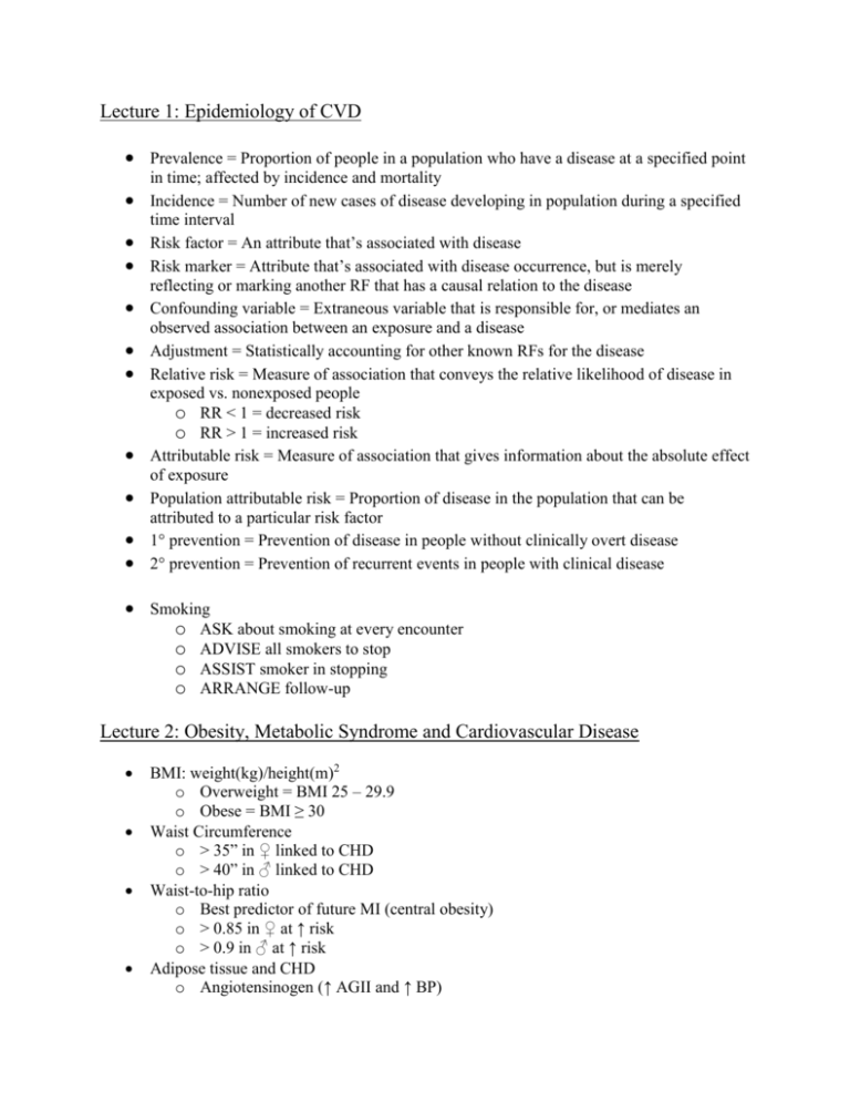
Lecture 1: Epidemiology of CVD Prevalence = Proportion of people in a population who have a disease at a specified point in time; affected by incidence and mortality Incidence = Number of new cases of disease developing in population during a specified time interval Risk factor = An attribute that’s associated with disease Risk marker = Attribute that’s associated with disease occurrence, but is merely reflecting or marking another RF that has a causal relation to the disease Confounding variable = Extraneous variable that is responsible for, or mediates an observed association between an exposure and a disease Adjustment = Statistically accounting for other known RFs for the disease Relative risk = Measure of association that conveys the relative likelihood of disease in exposed vs. nonexposed people o RR < 1 = decreased risk o RR > 1 = increased risk Attributable risk = Measure of association that gives information about the absolute effect of exposure Population attributable risk = Proportion of disease in the population that can be attributed to a particular risk factor 1° prevention = Prevention of disease in people without clinically overt disease 2° prevention = Prevention of recurrent events in people with clinical disease Smoking o ASK about smoking at every encounter o ADVISE all smokers to stop o ASSIST smoker in stopping o ARRANGE follow-up Lecture 2: Obesity, Metabolic Syndrome and Cardiovascular Disease BMI: weight(kg)/height(m)2 o Overweight = BMI 25 – 29.9 o Obese = BMI ≥ 30 Waist Circumference o > 35” in ♀ linked to CHD o > 40” in ♂ linked to CHD Waist-to-hip ratio o Best predictor of future MI (central obesity) o > 0.85 in ♀ at ↑ risk o > 0.9 in ♂ at ↑ risk Adipose tissue and CHD o Angiotensinogen (↑ AGII and ↑ BP) o FFA (Toxic to blood vessels, ↑ insulin resistance) o CRP (↑ inflammation, ↑ CV risk) o Plasminogen activator inhibitor-1 (↑ thrombosis) o IL-1, TNF-α (↑ inflammation) o Resistin (↑ insulin resistance) Metabolic Syndrome (THeHOG) o ≥ 3 of the following 5: Abdominal obesity ↑ Triglycerides; ≥ 150 mg/dL (or being treated for) ↓ HDL (or being treated for) < 50 mg/dL in ♀ < 40 mg/dL in ♂ HTN (or being treated for) ≥ 130 mmHg systolic ≥ 80 mmHg diastolic ↑ fasting BG (or being treated for) ≥ 100 mg/dL o Most important parts are abdominal obesity and insulin resistance o ↑ inflammation o ↑ thrombosis o Tx ↓ risk of HD and DM Target individual components (lipids, HTN, DM, wt loss) 5-10% weight loss improves cardiac function, endothelial function, insulin resistance,↓ Triglycerides, ↓ BP, ↑ HDL o Obesity Tx Sibutramine: block serotonin and Norepi uptake Orilistate: lipase inhibitor Phenteremine: Amphetamine derivative Rimonabant: CB1 receptor antagonist (depression; not used in US) Gastric bypass surgery – only weight loss intervention shown to ↑ lifespan Lecture 3: Lipid Lowering and Antioxidant Treatment ↑ HDL is the strongest predictor of a reduction in CV events Colestyramine (bile acid sequestrant) o ↓ LDL Gemfibrozil (fibric acid derivative) o ↓ LDL o ↑ HDL Statins (HmG CoA reductase inhibitor) o First 1° prevention that demonstrated ↓ lipids → ↑ survival, ↓ mortality Nicotinic acid derivatives (B6) o ↓ LDL o ↑ HDL Big Conclusions o ↓ CHD incidence ↓ Total cholesterol ↓ LDL cholesterol ↑ HDL Calculating LDL o LDL = TC – HDL – (TG/5) Risk factors that modify LDL goals o Cigarette smoking o Hypertension (BP 140/90 mmHg or on meds) o Low HDL cholesterol (<40 mg/dl) [HDL cholesterol 60 mg/dL is a negative risk factor – removes one risk factor from the total count.] o FH of premature CHD (1° ♂ relative <55 years or 1° ♀ relative <65 years) o Age (♂ 45 years, ♀ 55 years) Lecture 4: Assessment of Coronary Artery Disease Risk factors for CAD o ↑ LDL o HTN o Cigarettes o DM o Physical inactivity o Obesity o +FH Pathophysiology of CAD o Demand Inadequate O2 to cells to meet demand → ischemia Demand determined by Rate-pressure product (HR X Systolic BP) LV contractility LV wall stress (s=PR/Thickness) o Supply Left Coronary Artery LAD (Anterolateral wall and septum, 50% of LV) Left Circumflex (Lateral wall) o 15% of the time, gives off the Posterior Descending Artery to the LV (left dominance) Right Coronary Artery Posterior Descending (Inferior septum and inferior LV – meets LAD) – 85% of the time (right dominance) Compromised Coronary vasospasm Coronary vasculitis Anomalous coronary arteries Anemia Hypoxemia Hypovolemia o Luminal stenosis significance Degree of obstruction ↓ 50 – 70% flow → sx during exertion ↓ > 75% flow → sx during rest Length of obstruction Number and size of collaterals Magnitude of supplied muscle mass Shape and dynamic properties of the stenosis Some are quiet, some cause spasm or vasoconstriction Autoregulatory capacity of the vascular bed o Resistance 90% is in the arterioles, which can dilate 4x ∆ vasomotor tone → ∆ ischemic threshold Neuromodulation Endothelial control o Ischemia Variable symptoms Dyspnea Fatigue Unfavorable supply/demand ration → ischemia → new wall motion abnormality → ECG change → +/- Angina (if none, then it is silent) History and Symptoms o Classic angina Radiate to arms, neck, back +/- nausea, palpitations, diaphoresis ↑ with exertion x mins ↓ with rest, nitros o Unstable Angina From platelet aggregation at the site of a fixed stenosis → acute obstruction and/or coronary artery vasospasm o Classes of Angina I: by ↑ exertion II: by mod exertion III: during mild exertion (ADL) IV: at rest PE o HTN o Xanthoma o Corneal arcus o Obesity o Valvular disease (bruits, ↓ pulses) o AV nicking/retinal hemorrhage o ST ↓ or ↑ during an episode → active MI o Q waves → prior MI Exercise Testing and Electrocardiography o ↑ demand → ischemia o Good for pts with an intermediate probability of CAD o Sensitivity 70%, Specificity 70% Trust positive results, not negative ones o Look for angina during test, ST depression o Can be done ≥ 4 days post MI for prognostic assessment, activity prescription and evaluation of tx o Determine functional capacity o Cycle or treadmill (better) o ↓ increments of exercise in older/deconditioned people o Measure HR and BP before, during and after the test o Stop test if moderate angina, excessive fatigue, marked ECG changes, ↓ BP or ventricular dysrhythmias occur o Do imaging if: Resting ECG is abnormal ↑ diagnostic accuracy Determine extent/distribution of ischemia Evaluate ischemia in someone who has had ateriography o Nuclear testing available too Pharmacologic Stress Testing o For those unable to undergo exercise stress test o Dobutamine (β agonist) Assess ECG, HR, BP, Echo during for ischemia Add atropine if adequate HR is not achieved o Adenosine or Dipyridamole (vasodilator) Assess coronary perfusion Coronary Angiography o Gold standard for CAD detection o Intra-arterial catheter, radio-opaque dye o Observe for filling defects CT Angiography o Intravenous injection of contrast o Outpatient procedure o Quick scan time o High negative predictive value o High radiation doses with current technology; not recommended to detect CAD in asymptomatic patients Test sensitivity/specificity Test + Test - With disease a c Without disease b d o Sensitivity = a / (a + c) probability of a positive test among patients with disease o Specificity = d / (b + d) probability of a negative test among patients without disease Lecture 5: Medical Treatment of CAD Heart demand o HR o Contractility o LV wall tension (systolic BP, LV volume) Nitrates o Venous vasodilator o ↓ cardiac work, ↓ cardiac demand o ↑ capacity of veins → ↓ venous return → ↓ EDV (preload) and diastolic wall tension o May also ↓ afterload o Potent vasodilators of the coronary arteries and ↑ collateral flow o ↓ coronary artery spasm o Ex: Nitroglycerin, isosorbide dinitrate, isosorbide mononitrate o Side effects Headache Flushing Presyncope/syncope ↑ in elderly Reflex tachycardia o Clinical uses Sublingual to abort anginal attack Prophylaxis o ↑ first pass effect o IMPORTANT: ↑ pharmacologic tolerance; use nitrate-free interval β-blockers o Inhibit the β-receptor on the heart o ↓ HR, ↓ BP, ↓ contractility o Lengthen diastole by ↓ HR, but do not effect coronary O2 supply o Types and specificity Metoprolol, Atenolol ↑ to β-1 receptors; predominately on heart tissue (cardioselective) Pindolol Partial agonist Labetalol α and β blocker; ↓ TPR/BP o Water soluble ones → excreted by kidneys; do not cross BBB o Side effects/contraindications Asthma/bronchospasm (↑ in non-selectives) Bradycardia, heart block DM Psych problems Liver disease (↑ with propranolol) Acute withdrawal syndrome (never stop abruptly!) Calcium Channel Blockers o ↓ the entry of Ca2+ into myocardial and smooth muscle cells o ↓ interaction between actin and myosin o ↓ SA and AV node automaticity and conduction o ↓ contractility o ↓ coronary vasomotor tone → ↑ myocardial O2 supply o ↓ myocardial demand o Types and specificity Diltiazem Cardiac effects Vasodilator Nifedipine Vasodilator Verapamil (IMPORTANT: ↑ Digoxin levels) Cardiac effects Vasodilator Property Coronary Vasodilation Peripheral Vasodilation HR Contractility AV Conduction Preload Afterload Diltiazem dilator + cardiac Nifedipine dilator Verapamil dilator + cardiac Nitrates venous dilator β-blockers cardiac effects ↑↑↑ ↑↑↑ ↑↑ ↑↑↑ 0 ↑ ↑↑↑ ↑↑ ↑↑ 0 ↓ ↓ ↓ 0 ↓ ↑ 0 0 0 ↓↓↓ ↓ or ↑ ↓ ↓↓ 0 ↓↓ ↑ 0 0 ↓↓ ↓↓ ↓↓ ↓ ↓ 0 0 o Angina Tx with specific conditions LV systolic failure Nifedipine +/- Nitrates Systemic HTN β-blockers Ca2+ channel antagonists Pre-existing bradycardia or AV block Nifedipine + Nitrates Renal failure Do NOT use water soluble β-blockers Liver failure ↓ doses of Ca2+ channel antagonists and β-blockers IDDM, Peripheral Vascular Disease, Bronchospasm, Chronic Lung Disease Ca2+ channel antagonists Nitrates Lecture 6: Acute Coronary Syndromes Anterior Wall = LAD Lateral Wall = Left Circumflex Inferior Wall = Right CA Pathophysiology o Ischemia 2° to atherosclerotic plaque activation and local thrombus formation STEMI, NSTEMI, non-stable angina o Plaque activation Fissure at shoulder of lesion; ↑ macrophages ↑ shear stress Macrophages activate, secrete matrixins Degrade collagen, elastin, glycosaminoglycans, ↑ thrombotic state o Consequences of plaque fissure Platelets exposed to subendothelial collagen → ↑ thrombosis o Acute Atherothrombus formation Abrupt complete closure of the vessel Acute stenosis without total occlusion Either of these without clinical consequences (collateral vessels) Cessation of flow → ↓ ability to relax in diastole and contract in systole o Hypokinesis o Akinesis o Dyskinesis (thinning and bulging) Clinical o Acute MI usually occurs at rest Also during surgeries, extreme emotional or physical stress, extreme metabolic derangements, HTN crisis or with vasospastic agents o 1-2 week prodrome of chest discomfort Unstable angina: MI not severe enough to cause myocardial damage, but may still cause Sx and require hospitalization. Ddx from MI by cardiac NZs Ddx o Pericarditis: Sharp pain, ↑ with inspiration, + friction rub o Aortic dissection: sharp pain in mid-chest that radiates to the back, diminished pulse in ≥1 extremities; widened mediastinum on CXR, no ECG changes. Surgical emergency o MS pain: ↑ with palpation o GI: ddx by quality of pain ECG o Initiate upon arrival in the ER o Ischemia, injury, infarction o ST elevation (transmural MI) Followed by tall, peaked T waves (early) or inverted T waves (late) + Pathologic Q waves and loss of R wave amplitude o ST depression/T wave inversion (subendocardial infarction – STEMI) Approach to patients o STEMI Continuous ECG monitoring Labs (blood) – cultures, anemia, electrolytes, BUN/Crt, ABGs if dyspneic, Cardiac NZs Creatine Kinase (most important) [Use CK MB isozyme ↑ sp.] CXR to assess pulmonary congestion Tx Restore perfusion o Thrombolysis (heparin) o PCI Glycoprotein IIB-IIIA inhibitors here o Aspirin ↓ myocardial O2 demands o Nitroglycerin (↓ venous return, ↓ BP, CA dilation) o β-blockers (↓ HR, ↓ BP, ↓ contractility) o ACE-I (esp those with LV dysfunction and anterior or large infarcts) Relieve pain/anxiety o Analgesia/sedation O2 by nasal prong Complications Embolic stroke Arrhythmias VF, VT LV pump failure RV infarct → hypotension → ↑ JVP Infarct expansion, aneurysm formation Myocardial/papillary rupture Angina o UA/NSTEMI Tx Reinfarction Pericarditis Heparin Aspirin Nitrates β-blockers Clopidogrel (ADP receptor blocker) If high risk (↑ troponin, recurrent ischemia, ↑ EKG ∆s) o GP IIb-IIIa inhibitor o PCI/CABG If low risk (normal troponin on admission at @ 12 hrs) o Stress test o All ACS patients predischarge (who did not undergo cath) Submaximal exercise testing Assess functional capacity Evaluate meds Stratify risk Review modifiable risk factors Lipids Cigarettes HTN Sent home with these meds (ABC2) Aspirin β-blocker Converting NZ Inhibitor (ACEI) Cholesterol lowering med (e.g.: statin) Encourage enrollment in an outpatient Cardiac Rehab Program x weeks-months Meds advice Diet advice Exercise advice Lecure 7: Cath/Angio/PCI/CABG Cardiac catheterization o Measure hemodynamics o Angiography o Complications MI Stroke Vascular complications 2° to femoral access site Renal failure 2° to contrast load o Indications Conditions where there is evidence/general agreement that the procedure is beneficial, useful and effective (class I) Class III or IV angina on meds ↑ risk criteria on non-invasive testing Post-resuscitation from sudden death/VT Alternative to fibrinolytic Tx Cardiogenic shock Fibrinolytic failure Mechanical complication Spontaneous, recurrent ischemia + exercise or functional stress test ↑ risk MI with ↓ EF (< 40%) Confirm/define severity of valvular or congenital defect PCI o Enlargement of the lumen of a stenotic vessel o Balloon angioplasty +/- stent/drug-eluding stent o Indications Severe ischemia on functional testing Following cardiac arrest ↑ area of jeopardized myocardium Inadequate response to meds o Complications Death MI Need for emergency bypass surgery Arterial dissection Thrombosis Restenosis (↓ with stents) CABG Indications o *Left main coronary artery disease o *Triple vessel disease with reduced LV function o Unstable angina o Chronic angina unresponsive to maximal medical therapy o Post MI angina o Acute coronary artery occlusion/dissection following an unsuccessful percutaneous coronary procedure Lecture 8: Cardiac Rehab and 2° prevention programs after MI Identify and modify risk factors that may ↑ atherosclerotic coronary disease Restore/maintain optimal psych, social, vocational and emotional status Lipid management Nutritional counseling/weight management Smoking cessation Psychosocial counseling Exercise training o Exercise prescription Minimum: 30 minutes @ moderate intensity 3x/week Cardio + resistance training (↓ visceral fat) Lecture 9: Peripheral Vascular Disorders Peripheral Arterial Disease o 2° to atherosclerosis o Sx Intermittent claudication (relieved by rest) Ddx: pseudoclaudication – neurogenic, relived by leaning forward Critical limb ischemia, ulceration, gangrene o Pathophysiology >80% occlusion → ↓ blood flow at rest Exercise → ↑ pressure drop that wasn’t present at rest o Dx ↓ pulses Trophic skin ∆s Bruits Pallor upon elevation Dependent rubor Ankle brachial index (ABI) Highest systolic pressure in affected limb/highest systolic pressure in brachial artery o <0.9 = PAD o <0.5 = severe obstruction Post-exercise measurements may unmask a stenosis Exercise rehab Aggressive modification of atherosclerosis risk factors Revascularization o Tx Carotid Artery Disease o 2° to atherosclerosis → CVA o ↑ proximal internal carotid o Manifestations Asymptomatic cervical bruit TIA Stroke o Dx Duplex US MR angiography Aspirin ↓ risk factors Symptomatic Carotid endarterectomy PCI o Tx Aortic Aneurysmal Disease o Focal dilation > 1.5x normal vessel diameter o Asymptomatic until rupture or other complication o Dx CXR: widening of aortic shadow or abnormal calcification PE: AAA pusatile mass Abd US CT and MRI/A o Tx Surgical resection if >5.5 cm in ascending/abdominal >6.0 cm in descending Intraluminal stent-graft Aortic Dissection o Layers of aortic wall become separated; usually in media o Manifestations Sudden onset of: Severe pain in anterior chest, back or abdomen HTN Aortic diastolic murmur Pulse deficit Neurological ∆s Syncope implies rupture o Dx Widening of supra-aortic shadow or deviation of the trachea on CXR CT, MRI, aortagraphy o Tx ↓ BP ↓ wall stress Nitroprusside + β-blocker If proximal aorta → emergency surgery If distal aorta → LT meds or delayed surgery Lecture 10: Valvular Disease/Murmurs Acute Rheumatic Fever o Recent evidence of strep infection o Plus, 2 major criteria or 1 major and 2 minor criteria o Major Criteria Carditis Polyarthritis Chorea Erythema marginatum (fleeing, salmon-colored rash) Subcutaneous nodules o Minor Criteria Fever Arthralgias Previously documented episodes of ARF Rheumatic heart disease Positive acute phase reactants (ESR, CRP) Prolonged PR interval Valvular Lesion Inflow (mitral, tricuspid): Stenosis Regurgitation Outflow (aortic, pulmonic): Stenosis Regurgitation Loading Disturbance Ventricular Consequences ↑ venous pressure ↑ ventricular volume Diastolic dysfunction Systolic dysfunction ↑ ventricular pressure ↑ ventricular volume Diastolic dysfunction Systolic dysfunction All stenoses cause diastolic dysfunction All regurgitations cause volume overload Mitral stenosis o Almost invariably rheumatic o Diastolic dysfunction o Obstructs flow into LV o ↑ pressure in LA and therefore, pulmonary veins, lungs → edema, SOB o ↑ pulmonary pressure → RV dilation/hypertrophy and failure o ↑ JVP o Murmur most audible at apex Crescendo, presystolic murmur Mid-diastolic murmur Loud S1 Mitral regurgitation o Often rheumatic o ↓ forward output with LV systolic dysfunction o ↑ fluid volume to compensate (volume overload) o LA becomes dilated, compliant with chronic MR/compensation o Murmur, etc Blowing holosystolic murmur Mid-diastolic rumble Displaced apex beat Rocking of stethoscope by LV Aortic stenosis o Systolic dysfunction o Cardinal symptoms Exertional syncope Exertional angina CHF o Can cause LVH o Parvus et tardus (slow-rising, low-volume pulse) o Crescendo-decrescendo (diamond-shaped) systolic murmur Aortic regurgitation o Diastolic dysfunction o ↑ LA pressure o Can cause premature closing of the mitral valve o Bounding pulse o Blowing diastolic murmur Lecture 11: Hemodynamic Monitoring Diastole: AV valves open → function as a single chamber Systole: Ventricle/outflow valves open → function as a single chamber Right Atrium o Positive A wave o Negative X descent o Postiive V wave o Negative Y descent o Definitions A wave = Atrial contraction X descent = Atrial relaxation V wave = Venous filling of the RA during RV systole when the tricuspid valve is closed Y descent = Rapid atrial emptying following opening of the tricuspid valve Right ventricle o Diastolic filling period o A wave (atrial kick) @ end-diastole o Rise in systolic pressure (contraction) d a v Pulmonary Artery o Systolic peak o Diastolic trough o Dicrotic notch (closure of the pulmonic valve) s dn d Pulmonary capillary wedge pressure (LA pressure) o Same morphology has the RA tracing Cardiac Output o Volume of blood ejected/minute (L/min) o Fick method Patient in closed chamber → breathe known amount of oxygen → measure exhaled oxygen % inhaled - % exhaled = oxygen consumption Take blood sample from arterial side close to aorta (fully oxygenated) Take blood sample from venous side close to pulmonary artery as possible (fully deoxygenated) A-V oxygen difference = how long blood is circulating CO = Oxygen consumption / (Arterial O2 – Venous O2) ↓ A-V O2 difference = ↑ CO ↑ A-V O2 difference = ↓ CO o Indicator dilution method Cold saline → proximal cardiac chamber → mixed/warmed up by surrounding blood as it passes through the cardiac chamber ↑ in temp distal to the mixing chamber is measured Intracardiac shunt o Connections between chambers that do not normally exist Allow mixing of oxygenated and deoxygenated blood Can result in hypoxia o ↓ O2 in arterial side o ↑ O2 in venous side (step-up) [if ≥ 7% difference between chambers] Cardiac tamponade o Pressures equalize in all chambers o ↓ CO Aortic stenosis o LV pressure much ↑ than aortic systolic pressure Cardiogenic shock o ↑ pressures throughout o ↓ CO Septic shock o ↓ pressures throughout o ↑ CO Hypovolemic shock o ↓ pressures throughout o ↓ CO Lecture 12: Atrial Fibrillation ↑ with age Can provoke an MI, but it not provoked by an MI Sx o Palpitations o Fatigue o Exercise intolerance o Presyncope o SOB o Edema (HF) o Angina o Anxiety Clinical consequences o Stroke (2° to stasis in atria) o Peripheral emboli o Tachycardia mediated cardiomyopathy o Pulmonary edema o Atrial myopathy o Mitral/tricuspid regurgitation o MI/Unstable angina o Renal insufficiency o Problems with anticoagulation (hemorrhage, bleeding) o ↓ sense of well being Tx o Acute: slow HR, revert the AF and restore sinus rhythm Procainamide, ibutilide o Anticoagulation o Chronic AV nodal blocking agents β-blockers Ca2+ channel blockers Digoxin AV nodal radiofrequency ablation LT Anticoagulation Warfarin: INR 2-3 ASA if @ low risk Risk of stroke and bleeding? Risk of drug toxicity and proarrhythmia? No statistical difference – individualize for needs of patient Lecture 13: Heart Failure Systolic dysfunction = ↓ ventricular contractile function o PV loop shifted downward and right o ↑ ESV and therefore ↑ EDV o Reduced EF o Cardiomegaly o Peripheral edema o HTN of DM, CAD Dystolic dysfunction o ↓ ventricular filling o PV loop shifted upward and left o ↓ ESV, ↑ EDP, ↓ SV o Hx of HTN o LVH Compensatory mechanisms o ↑ preload; can lead to ↑ ED pressure → pulmonary congestion o Ventricular hypertrophy/remodeling ↑ mitochondrial, myofibrillar mass, myocyte size, interstitial collagen ↑ fetal gene expression, produce abnormal contractile proteins ↑ wall thickness o Neurohormonal activation ↓ CO, ↑ filling pressures → ↑ RAAS, ↑ SNS → ↑ TPR, Na+/H20 retention May cause decompensation 2° to excessive vasoconstriction, volume retention, electrolyte abnormalities, myocardial toxicity and arrhythmias ANS = ↑ contractility, ↑ HR, ↑ preload, ↑ SV, redistribution of blood away to vital organs o Myocyte hypertrophy/necrosis/apoptosis o Interstitial fibrosis o Norepi depletion, β receptor downregulation RAAS o ↑ vasoconstriction, ↑ volume retention o AGII/Aldosterone → cellular remodeling Natriuretic peptide (ANP, BNP) o RA → ANP (when ↑ pressures) → vasodilation, natriuresis, inhibition of RAAS o Ventricles → BNP → vasodilation, natriuresis Arginine Vasopressin (ADH) o ↑ inadequate ability to excrete free water o Systemic and Pulmonary Vasoconstriction Compensatory ↑ LV afterload and may further ↓ systolic function ↑ pulmonary tone → R HF and ↓ exercise tolerance o Inflammatory Cytokines, NO and Oxidative stress ↑ TNF-α, IL-1, ROS Sx o Left sided Dyspnea (transduction into interstitium) Fatigue, weakness Mental dullness/confusion ↓ urine output during the day → nocturia o Right sided Liver engorged with fluid → RUQ discomfort Nausea, bloating, anorexia, early satiety, constipation LE edema, ↑ during the day Unexpected weight gain (edema) PE o Vitals: ↑ HR, tachypnea, ↑ BP, narrow pulse pressure o Left sided Rales, rhonchi, wheezing Diffuse, laterally displaced LV impulse o Right sided ↑ JVP Hepatojugular reflux Ascites LE pitting edema Pleural effusions (dull @ bases) Anasarca Tx o o o o Remove underlying cause/ppt factor Control symptoms ↓ cardiac workload ↓ excessive salt/water retention o Vasodilators ACE-I ARB Hydralazine (arterial dilator) Nitrates o β-blockers o Diuretics Thiazides Loop diuretics K+ sparing diuretics o Ionotropes Digitalis (↑ intracellular Ca2+) Adrenergic agonists Phosphodiesterase inhibitors o Anticoagulation o Antiarrhythmics Treating diastolic dysfunction o ↓ LVH (Antihypertensives) o ↑ ventricular relaxation (↓ afterload) o ↓ ischemia (β-blockers, Ca2+ channel blockers [e.g.: verapamil], nitrates, CABG, PTCA) o ↓ venous pressures (salt restriction, diuretics, nitrates, ACEI) o ↓ HR (Digoxin, β-blockers, Ca2+ channel blockers) o Maintain atrial contraction (cardioversion, pacing?) Lecture 14: Myocarditis Inflammatory infiltrate and injury adjacent to myocardial cells that is not typical for infarction. o Usually viral, but can be bacteria, myoplasma, parasitic, fungal, rickettsial, spirochetal, protozoal. o Can also be after radiation, heavy metal exposure (cobalt) or insect stings Pathology o Biventricular dilation o +/- subendocardial and epicardial hemorrhage Clinical o Febrile illness o Viral prodrome with myalgias o Signs and symptoms resembling acute MI o Acute onset of CHF o Arrhythmia and sudden death o Angina 2° to pericardial involvement o Tachycardia o +/- friction rub o +/- S3, S4, regurg murmurs o Displaced PMI laterally Dx o o o o o Repolarization ∆ on EKG Cardiomegaly/pericardial effusion on CXR Culture only during early infectious period (usually before myocarditis) Antiviral Antibodies Cardiac IgG antibodies Tx o Supportive ↓ afterload o Monitor for arrhythmias o Immunosuppresive drugs? o Antivirals? o Vaccinations? o Anti-lymphocytic monoclonal antibodies? Lecture 15: Cardiomyopathy Disease of the myocardium itself → dysfunction Dilated cardiomyopathy o ↓ systolic contraction o Ventricular dilation o ↓ EF o ↑ ESV o Maintains CO by dilating o ↓ forward perfusion o ↑ congestion of pulmonary and systemic venous circulations o Sx Dyspnea on exertion Cough Pulmonary edema Easy fatigue o PE Displaced apex Mitral regurg S3, S4 ↑ JVP Rales Peripheral edemas o Causes Most idiopathic Stenotic murmurs Viral myocarditis HIV Endocrinopathies Genetic Progressive deterioration o Management ↓ afterload β-blockers Diuretics Digoxin ACEI ARB IVIG Implantable defibrillators Transplantation Hypertrophic Cardiomyopathy o Hypertrophic LV o ↑ muscle mass with no cavity dilation o LV outflow tract obstruction o Diastolic dysfunction o Some diastolic dysfunction – LV/aortic pressure gradient o ↑ responsiveness to SNS o Clinical Dyspnea on exertion Chest pain Brisk carotid pulse Spike and dome waveform o Causes Genetic Causes of hypertrophy o Tx β-blockers Ca2+ channel blockers Convert AF Mitral valve replacement Implantable defibrillators Restrictive cardiomyopathy o 1° diastolic dysfunction o Normal systolic function o Normal ventricular dimensions o Causes Scleroderma Amyloid Storage disease Firbrosis, carcinoid, radiation, malignancy o Clinical Dyspnea on exertion Fatigue Chest pain Palpitations Neck vein distension Systolic murmurs (AV regurg) Pseudo-infarct (replacement of normal tissue) o Tx Diuretics Anticoagulants Immunosuppressive agents (sarcoid, etc.) Surgical resection Lecture 16: Bradyarrhythmias P wave = atrial depolarization PR segment = Conduction through the atrium, AV node, and the His-Purkinje system QRS complex = Ventricular depolarization T wave = Ventricular repolarization U wave = Not always seen; repolarization? 1° AV block o Long PR interval (>200 msec) o AV conduction remains 1:1 (no real “block”) o Slow conduction in the AV node ↑ parasympathetic activity? Drugs? Lecture 17: Tachyarrhythmias Paroxysmal supraventricular tachycardia o ↑ Re-entry Two pathways of conduction Slow conduction in one of the pathways Unidirectional conduction block in the other pathway o Conduction down accessory pathway AF is most common sustained tachyarrhythmia o > 1 re-entry circuits circulating simultaneously in both atria o Rapid, chaotic atrial rhythm o Irregular rapid undulations on EKG Ventricular Tachycardia o Re-entry, usually 2° to scar tissue in the ventricle (previous MI) o Syncope o Cardiac arrest o Wide complex rhythm (>120 ms) on EKG o AV dissociation on EKG o Polymorphic – torsade de pointes; twisting of the points Lecture 18: Pericardial Disease Acute pericarditis o Usually viral (Coxsackie B, HIV, H. flu), bacterial, fungal or rickettsial o Post-MI o Auto-immune o Neoplastic (metastasis from lung, breast, hematologic, melanoma, Kaposi’s) o Can → effusion or tamponade o Dx Pericardial friction rub (diagnostic) Depression of the PR segment on EKG (pathognomonic) Dyspnea, fever, malaise Pleuritic left-sided chest pain NSAIDs Steriods o Tx Pericardial Tamponade o ↑ pericardial fluid o ↓ ventricular filling o ↓ SV, ↓ CO o Hypotension, death o Compensatory H20 retention o Diastolic pressures in the right and left ventricles become equal o Clinical Dyspnea Severe hypotension/shock ↑ JVP with only X descent (no Y descent) Tachycardia Pulsus paradoxus (↑ inspiratory fall in BP) DO NOT volume deplete ↑ fluid infusion Remove pericardial fluid by pericardiocentesis Fluid cell counts/differential, cytology, protein, LDH, glucose, Gram stain, AFB stain, cultures o Tx Constrictive Pericarditis o Dense fibrous scarring of the pericardium → ↓ filling of the cardiac chambers o Causes Idiopathic Scarring after open-heart surgery, radiation, infection o Diastolic pressures in the right and left ventricles become equal o ↑ venous pressures o ↓ CO 2° to ↓ LV filling o Clinical Leg edema Abdominal bloating RUQ pain 2° to hepatic congestion Pulmonary congestion with ↑ severity ↑ JVP with rapid X and Y descent (absent in tamponade) Diuretics Surgical removal of the pericardium o Tx Normal
