HISTOLOGY LAB
advertisement
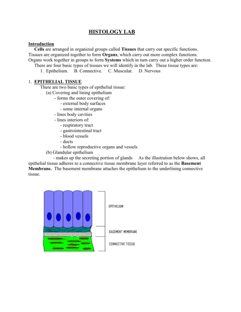
HISTOLOGY LAB Introduction Cells are arranged in organized groups called Tissues that carry out specific functions. Tissues are organized together to form Organs, which carry out more complex functions. Organs work together in groups to form Systems which in turn carry out a higher order function. There are four basic types of tissues we will identify in the lab. These tissue types are: 1. Epithelium. B. Connective. C. Muscular. D. Nervous 1. EPITHELIAL TISSUE There are two basic types of epithelial tissue: (a) Covering and lining epithelium - forms the outer covering of: - external body surfaces - some internal organs - lines body cavities - lines interiors of: - respiratory tract - gastrointestinal tract - blood vessels - ducts - hollow reproductive organs and vessels (b) Glandular epithelium - makes up the secreting portion of glands As the illustration below shows, all epithelial tissue adheres to a connective tissue membrane layer referred to as the Basement Membrane. The basement membrane attaches the epithelium to the underlining connective tissue. The flow chart below illustrates the different types of epithelial tissue Simple Squamous Epithelium As the diagram and photomicrograph below (and on the next page) illustrates, this tissue consists of a single layer of flat cells. The slide you are looking at in lab has only simple squamous epithelium on it, so it is easy to find and identify. Description: A single layer of flat cells. Looks like a platter of fried eggs "sunny-side up". Location: Air sacs (alveoli) of lungs, lines the inside of heart and blood vessels. Function: Filtration, diffusion, osmosis. Drawing of Simple Squamous Epithelium – Superior and lateral aspects Light Micrograph of Simple Squamous Epithelium – Superior aspect (400X) In this view the lumen associated with epithelium cannot be seen as the viewer is looking down on the top (superior aspect) of the tissue. Notice how the nucleus is clearly seen in the center of each cell. Light Micrograph of Simple Squamous Epithelium – Lateral aspect (400X) In this view of the tissue from the side (lateral aspect) the nucleus can still be seen but the overall shape of the cell cannot be discerned ****************************************************************************** Simple Cuboidal Epithelium As the drawings and photomicrographs illustrate below (and on the next page) this tissue consists of a single layer of cube shaped cells. The slide you are looking at in lab is a slide of the kidney. This slide has many different types of tissue on it. Description: A single layer of roughly cube-shaped cells Location: Lines kidney tubules and the small ducts of many glands Function: Secretion and absorption Drawing of Simple Cuboidal Epithelium Notice the central location of the nucleus in this tissue (the distance from the nucleus to the top and bottom of cell is about equal). The drawing gives the impression that this tissue is cube shaped, when in fact it is more polyhedral in shape. The kidney slide has a section of kidney on it that looks like this: In this view of the kidney the papilla can be seen. It is the nipple-like projection extending downward. At this low magnification (40X) the ducts lined with simple Cuboidal epithelium can barely be seen. The same structure should be viewed at a higher magnification (400X) In this view the duct is clearly seen. Note that the simple Cuboidal epithelium lines the lumen (inner space) of the duct. Each Cuboidal cell is roughly cubed shaped with a centrally located nucleus. ***************************************************************************** Simple Columnar Epithelium (non-ciliated) On this slide you must differentiate the liver from the gall bladder in order to locate the Simple Columnar Epithelium that lines the inside of the gall-bladder. Notice that this tissue is often found on finger-like projections of the mucosal lining called villi. Description: A single layer of tall rectangular cells Location: Lines the gall-bladder and most of the G.I. tract Function: Absorption and secretion Drawing of Simple Columnar Epithelium – Superior and Lateral Photomicrograph of Simple Columnar Epithelium (40X) Remember that the slide probably has both liver and gall-bladder on it. To locate the tissue look for the villi as illustrated below (or on the next page) on this slide of a gall-bladder. Notice that the villi is a finger-like projection of the mucosa that extends into the lumen of the gallbladder (40X) Photomicrograph of Simple Columnar Epithelium – from the Gall-bladder (100X) The Simple Columnar Epithelium can be seen in this photomicrograph lining the villi of the gall-bladder. Even at this relatively small magnification the position of the nuclei at the bottom of the cell (close to the basement membrane) can be seen. Photomicrograph of Simple Columnar Epithelium – from the Gall-bladder (400X) The exact shape of the columnar cell is hard to see in this photomicrograph. However, the position of the nuclei in the bottom one-third of each cell is an identifiable characteristic. ***************************************************************************** Stratified Squamous Epithelium As the drawing and photomicrographs below (and on the next page) illustrate this tissue consists of many layers of cells. The cells next to the basement membrane are columnar to Cuboidal in shape and the cells next to the lumen are squamous in shape. Stratified tissues are named according to the shape of the cells next to the lumen. Description: Many layers of cells with squamous cells located next to the lumen Location: Keratiniazed variety located on the outer surface of the epidermis of the skin. The non-keratinized variety lines the mouth, esophagus, and vagina. Function: Protection Drawing of Stratified Squamous Epithelium – Superior and lateral aspect Notice that the cells near the basement membrane are columnar to Cuboidal in shape, and the cells near the surface (lumen) are squamous in shape. This tissue is named according to the shape of the cells near the lumen, so this is Stratified Squamous Epithelium Photomicrograph of Stratified Squamous Epithelium – non-keratinized (40X) At this magnification the Stratified Squamous Epithelium appears as a darkly stained band next to the lumen. This is a slide of the epidermal layer of the vagina. Photomicrograph of Stratified Squamous Epithelium – non-keratinized (200X) At this magnification the Stratified Squamous Epithelium is more obvious (it makes up the Epidermis of the vagina). Photomicrograph of Stratified Squamous Epithelium – non-keratinized (400X) At this magnification of the epidermis of the vagina the squamous shape of the cells next to the lumen can be seen Photomicrograph of Stratified Squamous Epithelium – keratinized (40X) At this magnification the Stratified Squamous Epithelium (Epidermis) appears as two stained bands of cells next to the lumen. The lighter band closer to the lumen is composed of keratinized cells. This type of epithelium is found on the palms of the hands and the soles of the feet. The keratinized layer is the Stratum Corneum. Photomicrograph of Stratified Squamous Epithelium – keratinized (100X) At this magnification the stratified nature of the Stratified Squamous Epithelium can barely be seen. Photomicrograph of Stratified Squamous Epithelium – keratinized (400X) At this magnification the stratified nature of the tissue can be discerned. ***************************************************************************** Pseudostratified Columnar Epithelium (Ciliated) As the drawing and photomicrographs below (and on the following pages) illustrates, this tissue is not really stratified (hence the prefix pseudo). Note that this tissue also has numerous goblet cells and looks a lot like Simple Columnar Epithelium. However, unlike Simple Columnar Epithelium this tissue is not found lining villi. The presence of cilia is an important identification mark as this is the only ciliated tissue you will examine this quarter. Description: Looks like a stratified tissue, but its not. Each cell extends from the basement membrane to the lumen. Location: Lines the upper respiratory tract, the Epididymis, and part of the male urethra. Function: Secretion and movement of mucous by ciliary action Photomicrograph of Pseudostratified Columnar Epithelium (Ciliated) (100X) At this magnification the Pseudostratified Columnar Epithelium (Ciliated) can be seen next to the lumen. The epithelium has a darker stain than the underlying connective tissue. Photomicrograph of Pseudostratifed Columnar Epithelium (Ciliated) (400X) At this magnification the cilia is clearly visible at the apex of the cell (right next to the lumen). Notice that the tissue appears to be stratified due to the presence of nuclei at different levels. ****************************************************************************** Transitional Epithelium As the drawing and photomicrograph below (or on the next page) illustrates this tissue has features common to stratified cuboidal and Stratified Squamous epithelium (hence the name transitional. This tissue can be identified easily from two distinguishing characteristics: 1. The surface cells are scalloped. 2. In the cells next to the lumen the nucleolus can be clearly seen in the nucleus. Description: The appearance of this tissue can vary greatly, that is why it is called transitional epithelium. Location: Lines the urinary bladder and Ureter Function: Permits distention Drawing of Transitional Epithelium – Superior and lateral aspects This drawing illustrates the stratified nature of this tissue. The cells next to the lumen can be squamous, Cuboidal, and/or columnar in shape, hence the name transitional Photomicrograph of Transitional Epithelium (400X) The shape of the cells next to the lumen, the presence of scalloping, and the fact that in the mucleus of the cells next to the lumen the nucleolus can be seen, are all identification clues CONNECTIVE TISSUE The most abundant tissue type in the body. Most connective tissue has a rich blood supply (is highly vascular). The exception to this rule is hyaline cartilage with is avascular. Connective tissue is characterized by widely scattered cells found in an intercellular matrix. The flow chart below illustrates the various types of connective tissue found in the human body. There are three basic elements found in connective tissue. (1) Fibers (such as collagen, reticular, and elastic). (2) Cells (such as fibroblasts, macrophages, and adipose). (3) A ground substance (matrix). All connective tissue (except cartilage) has an extensive nerve and blood supply. The matrix (ground substance) of connective tissue may be: (1) Fluid – as in Vascular Connective Tissue (blood). (2) Semifluid – as in Loose Connective Tissue (Areolar). (3) Gelatinous – as in Mucous Connective Tissue. (4) Fibrous – as in Dense Regular Connective Tissue. (5) Calcified – as Osseous Connective Tissue (bone). Loose (Areolar) Connective Tissue (100X) As the photomicrograph below illustrates this tissue has many collagen and elastic fibers. The nuclei of many cells associated with connective tissue can be seen. Description: Consists of collagen, elastic, and reticular fibers embedded in a semifluid matrix, together with fibroblasts, mast cells, plasma cells and macrophages. Location: Hypodermis (subcutaneous layer of skin), around most body organs. Function: Loosely binds organs together Photomicrograph of Loose (Areolar) Connective Tissue (100X) ****************************************************************************** Vascular Connective Tissue (Blood) Blood is a connective tissue with a fluid matrix called plasma. Three basic types of cells are found suspended in plasma: (1) Erythrocytes (red blood cells). Description: Biconcave cells that are stained pink. The thinner center of the cell is lighter than the rim. Location: Suspended in blood plasma. Function: Transports respiratory gases (oxygen and carbon dioxide). (2) Leukocytes (white blood cells) of which there are five basic types. Description: Stained cells with an obvious (usually multi-lobed) nucleus. Location: Suspended in blood plasma, also found in lymphatic tissues. Function: Involved in immunity. (3) Thrombocytes (or platelets) which are involved in blood clotting. Description: Darkly stained structures much smaller than erythrocytes. Location: Suspended in blood plasma. Function: Involved in blood clotting. Photomicrograph of Vascular Connective Tissue (400X) The erythrocytes (red blood cells) are pink with a light center (where the cell is thinner and the light shines through). The Leukocytes (white blood cells) are stained dark pink to purple and have an obvious darkly stained nucleus. The Thrombocytes appear as small purple specks. ************************************************************************* Adipose Connective Tissue (Fat) As the photomicrograph below illustrates this tissue consists of adipocytes that wrap themselves around lipid droplets. Description: Resembles a chicken-wire fence. The individual adipocytes have a large central storage area where fats and oils are stored. Location: Subcutaneous layer of skin; fatty-capsule of the kidney, yellow bone marrow and around the heart. Function: Energy storage, insulation, protection. Photomicrograph of Adipose Connective Tissue (100X) This is a slide of the hypodermis. Connective tissue fibers, blood vessels, and sweat glands can be seen embedded in the adipose tissue **************************************************************************** Dense Fibrous Connective Tissue (Regular) This tissue contains of numerous collagen fibers oriented in the same direction. This produces great strength in the direction that the fibers run. Description: Many collagen fibers running in the same direction. Individual fibers appear to be "wavy". Fibroblasts can be seen between the fibers. Location: Makes up tendons and ligaments. Function: Attaches muscles to bones (tendons) and bone to bone (ligaments). Dense connective tissue has less flexibility than Areolar connective tissue but is more resistant to stress. The collagen fibers are densely packed together, and run in one direction. There is great strength in the direction of the fibers, but this tissue can be relatively weaker in a direction at right angles to the fiber direction. The fibers and slightly crinkled allowing them to stretch to some degree in the direction of the fibers. Photomicrograph of Dense regular connective tissue (100X) Note that the collagen fibers all run in the same direction (on this view from left to right). The "crinkly" or "wavy" appearance of the fibers is an identifiable characteristic of this tissue. ****************************************************************************** Hyaline Cartilage Connective Tissue This tissue has a matrix that is firm enough to bear a lot of pressure without permanent distortion. Hence, it is found in joints where it functions as a shock-absorber and friction reducing surface for bone articulation. Bones in the embryo start off as hyaline cartilage templates for the later developing bone. This tissue is avascular and has no nerves. Description: Consists of chrondrocytes (cartilage cells) located in spaces called lacuna, surrounded by a bluish matrix interlaced with collagen fibers. Location: Ends of long bones, between the ribs and the sternum, tracheal rings, parts of the larynx, and embryonic skeleton. Function: Reduces friction in joints, support with flexibility. Photomicrograph of Hyaline Cartilage Connective Tissue (100X) At this magnification to ring of Hyaline Cartilage can be seen occupying the center of the wall of the trachea. The individual cells of the cartilage (chondrocytes) are just visible. Photomicrograph of Hyaline Cartilage Connective Tissue (400X) The chondrocytes in the lacuna are obvious at this higher magnification. Notice that the chondrocytes often are found in pairs inside the lacuna. The matrix is blue to bluish-white. **************************************************************************** Osseous Connective Tissue (bone) The slide you will be looking at is a slice of dense bone from the shaft of a long bone, ground down to become extremely thin and translucent (hence the slide name – "ground bone"). Bone tissue has a hard matrix containing ions such as calcium and phosphorus. This matrix is laid down around a dense network of collagen fibers in a layer called lamellae. System of canals containing blood vessels, nerves and lumphatic vessels can be identified. They resemble tree stumps and are known as Haversian systems or Osteons. Bone cells (Osteocytes) can be seen inside their spaces (lacuna). Description: A network of osteons makes ground bone resemble a field of tree stumps. Location: Make up the bones of the body. Function: Protection, support, mineral and fat storage (marrow). Together with skeletal muscle is responsible for movement of the body. Photomicrograph of Osseous Connective Tissue (100X) The Haversian canal is located in the center of the osteon. It contains nerves, lymphatics and blood vessels that run the length of the bone. The wide opening on the left is a Volkmann's canal that transports the same structure from the outside of the bone to the inside of the bone (marrow cavity) and back. ***************************************************************************** MUSCLE TISSUE Muscle tissue is composed of fibers that function to contract and short the muscle for contraction. There are three distinct types of muscle tissue based on location, and structural and/or functional characteristics. Skeletal Muscle Tissue Skeletal muscle tissue is so-called because it attaches to bones and is involved in the movement of bones. It is also referred to as voluntary muscle because of the voluntary control of its contraction, and/or striated muscle because of its striated appearance when you look at it through a light microscope at high magnification. Description: Long, striated fibers with the nuclei located at the edges of the fibers. Location: Attached to bones by tendons. Function: Moves bones, posture, heat production Photomicrograph of Skeletal Muscle Tissue (400X) Notice the striated appearance of this tissue and the location of the nuclei at the edge of the fiber ****************************************************************************** Smooth Muscle Tissue Smooth muscle is composed of elongated cells that are not striated. Each smooth muscle cell is largest at its midpoint and tapered towards its ends. Each cell has a single nucleus located in the middle (or broadest) part of the cell. Description: long tapered fibers with a centrally located nucleus. Location: Walls of hollow internal organs such as the urinary bladder, stomach, intestines, uterus, gall bladder, urinary bladder, blood vessels, and uterus. Function: Movement of the substance inside the hollow organ (example: food in the intestines, blood in the heart etc.). Photomicrograph of Smooth Muscle Tissue (400X) Notice the position of the single nucleus in the center, broadest, portion of the fiber. Notice also that the fibers are nonstriated. ****************************************************************************** Cardiac Muscle Tissue Cardiac muscle is composed of striated fibers that are bifurcated or branched. The nuclei are centrally located. The presence of darkly stained intercalated discs is a good identification characteristis. Description: Branched striated fibers with a centrally located nucleus. Location: Wall of the heart. Function: Pumps blood to all parts of the body. Photomicrograph of Cardiac Muscle Tissue (400X) NERVOUS TISSUE Two basic types of cells are found in nervous tissue. Neurons are large cells that function to conduct impulses from one part of the nervous system to another (example: from the spinal cord to the brain). Neuroglia are small cells that "glue" the neurons together (they have many other functions as well). Description: Large neuron cell bodies with obvious processes extending from them. Many small nuclei of neuroglial cells (the neuroglial cells themselves cannot be seen. Location: Nervous system. Function: Conducts nerve impulse. Nervous Tissue: Ox spinal cord (100X) Notice the large cell body with a conspicuous nucleus. The axon and dendrite (cell processes) are easily identified. The small nuclei of neuroglial cells (astrocytes) are evident.

