AbsorptionSpectrum
advertisement
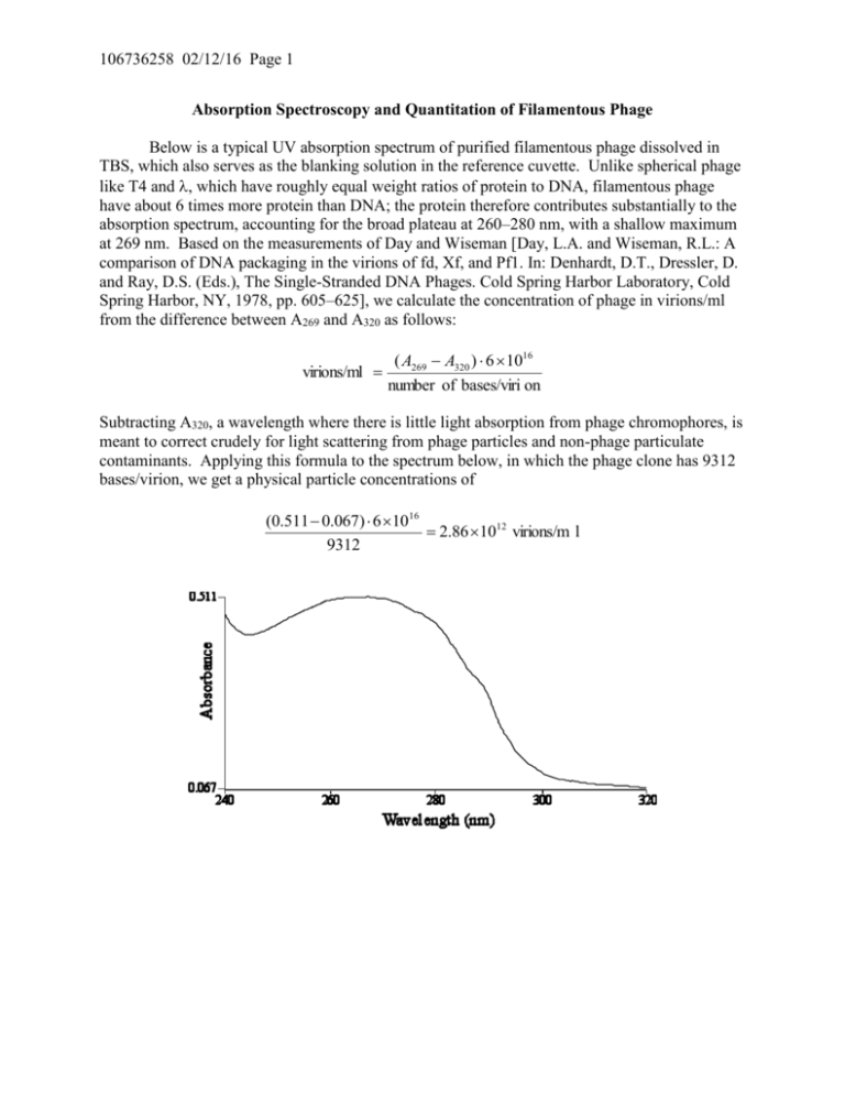
106736258 02/12/16 Page 1 Absorption Spectroscopy and Quantitation of Filamentous Phage Below is a typical UV absorption spectrum of purified filamentous phage dissolved in TBS, which also serves as the blanking solution in the reference cuvette. Unlike spherical phage like T4 and , which have roughly equal weight ratios of protein to DNA, filamentous phage have about 6 times more protein than DNA; the protein therefore contributes substantially to the absorption spectrum, accounting for the broad plateau at 260–280 nm, with a shallow maximum at 269 nm. Based on the measurements of Day and Wiseman [Day, L.A. and Wiseman, R.L.: A comparison of DNA packaging in the virions of fd, Xf, and Pf1. In: Denhardt, D.T., Dressler, D. and Ray, D.S. (Eds.), The Single-Stranded DNA Phages. Cold Spring Harbor Laboratory, Cold Spring Harbor, NY, 1978, pp. 605–625], we calculate the concentration of phage in virions/ml from the difference between A269 and A320 as follows: ( A269 A320 ) 6 1016 virions/ml number of bases/viri on Subtracting A320, a wavelength where there is little light absorption from phage chromophores, is meant to correct crudely for light scattering from phage particles and non-phage particulate contaminants. Applying this formula to the spectrum below, in which the phage clone has 9312 bases/virion, we get a physical particle concentrations of (0.511 0.067) 6 1016 2.86 1012 virions/m l 9312


