Scanning for suspected brain tumours
advertisement
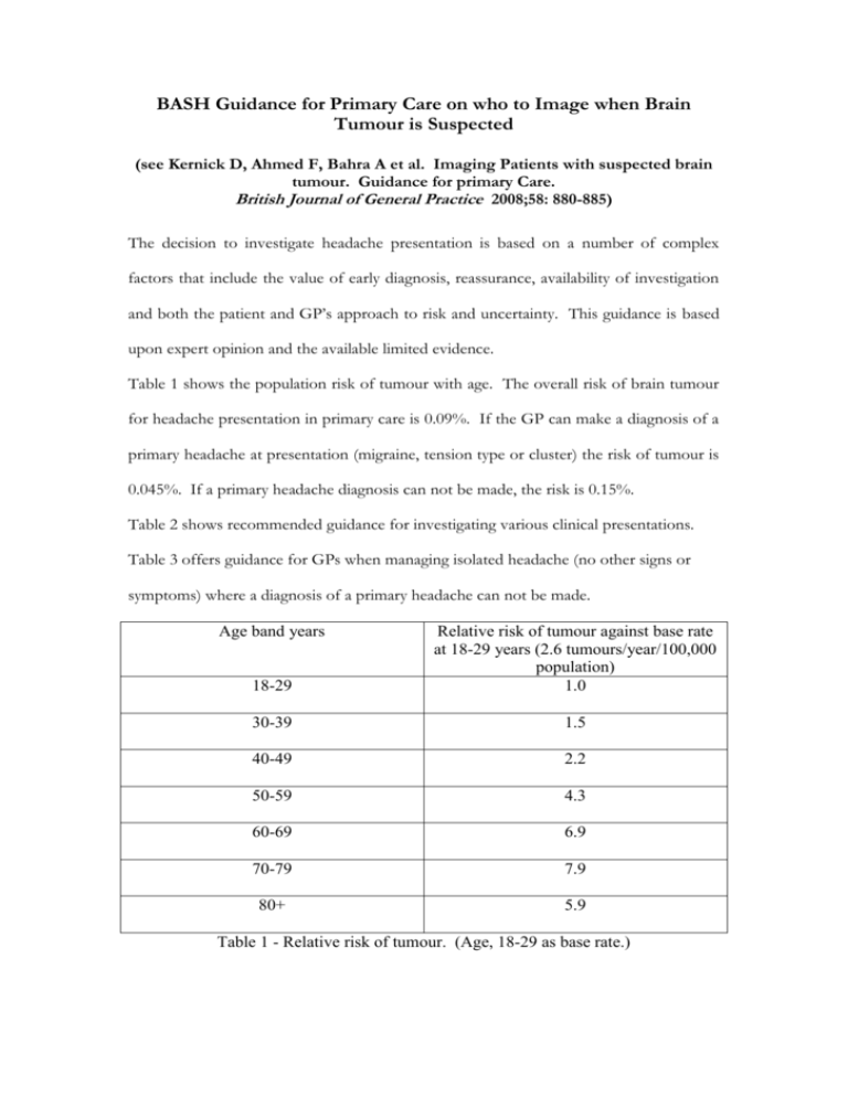
BASH Guidance for Primary Care on who to Image when Brain Tumour is Suspected (see Kernick D, Ahmed F, Bahra A et al. Imaging Patients with suspected brain tumour. Guidance for primary Care. British Journal of General Practice 2008;58: 880-885) The decision to investigate headache presentation is based on a number of complex factors that include the value of early diagnosis, reassurance, availability of investigation and both the patient and GP’s approach to risk and uncertainty. This guidance is based upon expert opinion and the available limited evidence. Table 1 shows the population risk of tumour with age. The overall risk of brain tumour for headache presentation in primary care is 0.09%. If the GP can make a diagnosis of a primary headache at presentation (migraine, tension type or cluster) the risk of tumour is 0.045%. If a primary headache diagnosis can not be made, the risk is 0.15%. Table 2 shows recommended guidance for investigating various clinical presentations. Table 3 offers guidance for GPs when managing isolated headache (no other signs or symptoms) where a diagnosis of a primary headache can not be made. Age band years 18-29 Relative risk of tumour against base rate at 18-29 years (2.6 tumours/year/100,000 population) 1.0 30-39 1.5 40-49 2.2 50-59 4.3 60-69 6.9 70-79 7.9 80+ 5.9 Table 1 - Relative risk of tumour. (Age, 18-29 as base rate.) Red Flags – presentations where the probability of an underlying tumour is likely to be greater than 1%. These warrant urgent investigation. Papilloedema. Significant alterations in consciousness, memory, confusion or co-ordination. New epileptic seizure New onset cluster headache (imaging required but non-urgent). Headache with abnormal findings on neurological examination or other neurological symptoms. Headache with a history of cancer elsewhere particularly breast and lung. Orange Flags - presentations where the probability of an underlying tumour is likely to be between 0.1 and 1%. These need careful monitoring and a low threshold for investigation. New headache where a diagnostic pattern has not emerged after eight weeks from presentation. Headache aggravated by exertion or Valsalva manoeuvre. Headaches associated with vomiting. Headaches that have been present for some time but have changed significantly, particularly a rapid increase in frequency. New headache in a patient over 50. Headaches that wake from sleep. Confusion. Yellow Flags - presentations where the probability of an underlying tumour is likely to be less than 0.1% but above the population rate of 0.01%. These need appropriate management and the need for follow up is not excluded. Diagnosis of migraine or tension type headache. Weakness or motor loss. Memory loss. Personality change. Table 2 – Recommended guidance for investigating for tumour in primary care patient. i) At presentation (Approximately 0.15% risk if a diagnosis of a primary headache is not made) Exclude red flags as below Check blood pressure, fundoscopy and consider ESR if >50years to exclude temporal arteritis. If no diagnosis cannot be made tell patient - “There is no evidence of anything serious underlying your headache but I would like to review you in one month”. ii) Ask patient to keep a headache diary. At one month Exclude urgent features as above. If diagnosis still uncertain: Assess memory and cognitive function during interview. Assess for symptoms that would indicate primary lesion elsewhere Assess for signs that would indicate primary lesion elsewhere (minimum set takes approximately three minutes.) Fundoscopy, pupil responses, visual fields, eye movements, facial movements (raise eyebrows to ceiling, show teeth and grimace), corneal reflexes, protrude tongue, palm drift (outstretched hands, palms uppermost), finger-nose touching with middle or index finger, finger dexterity (play piano), limb and plantar reflexes, standing feet together eyes closed, tandem gait (walk heel to toe in straight line). Consider blood screen to exclude systemic illness or evidence of primary tumour elsewhere - FBC, ESR, CRP, LFT, creatinine, electrolytes, glucose, thyroid function. If diagnosis is still uncertain tell patient - “There is still no evidence of anything serious but I would like to review you again in another month”. iii) At two months Exclude urgent features. Examination as above. If diagnosis is still uncertain tell patient - “There is still no evidence of anything serious underlying your headache but we need to discuss whether it would be appropriate to have a brain scan. The best estimate is that the chances of you having a tumour are between 0.1 and 1%. However, three in every 100 people like you will show an incidental finding that may give rise to unnecessary anxiety and may have implications for future life insurance cover. MRI is a more sensitive investigation and CT can miss up to 10% of lesions. Practical and economic circumstances will inevitably dictate the mode of investigation. A normal investigation does not over-rule the need for further review. Order blood investigations as above if not previously taken in addition to tests dependent on symptoms and history. E.g. VDRL, Lyme titres, antiphospholipids If patient and doctor decide against imaging review again in one month. At this time, 74% of tumours will have become apparent clinically. Table 3 - Clinical guidance for new presentations of isolated non-urgent headache that need a diagnostic pattern to emerge. Reduce the follow up time if orange flag headache features are present.
