answers - Biology Junction
advertisement
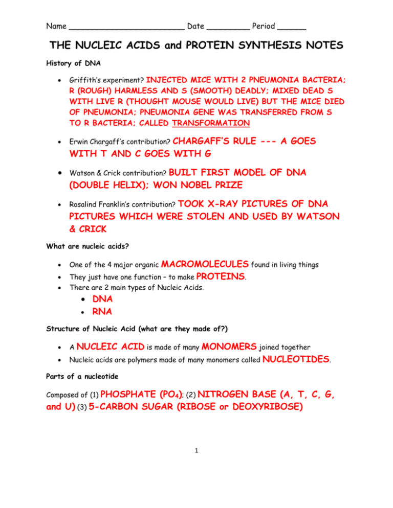
Name ________________________ Date _________ Period ______ THE NUCLEIC ACIDS and PROTEIN SYNTHESIS NOTES History of DNA Griffith’s experiment? INJECTED MICE WITH 2 PNEUMONIA BACTERIA; R (ROUGH) HARMLESS AND S (SMOOTH) DEADLY; MIXED DEAD S WITH LIVE R (THOUGHT MOUSE WOULD LIVE) BUT THE MICE DIED OF PNEUMONIA; PNEUMONIA GENE WAS TRANSFERRED FROM S TO R BACTERIA; CALLED TRANSFORMATION CHARGAFF’S RULE --- A GOES WITH T AND C GOES WITH G Erwin Chargaff’s contribution? Watson & Crick contribution? Rosalind Franklin’s contribution? BUILT FIRST MODEL OF DNA (DOUBLE HELIX); WON NOBEL PRIZE TOOK X-RAY PICTURES OF DNA PICTURES WHICH WERE STOLEN AND USED BY WATSON & CRICK What are nucleic acids? One of the 4 major organic MACROMOLECULES found in living things They just have one function – to make PROTEINS. There are 2 main types of Nucleic Acids. DNA RNA Structure of Nucleic Acid (what are they made of?) A NUCLEIC ACID is made of many MONOMERS joined together Nucleic acids are polymers made of many monomers called NUCLEOTIDES. Parts of a nucleotide Composed of (1) PHOSPHATE (PO4); (2) NITROGEN BASE (A, T, C, G, and U) (3) 5-CARBON SUGAR (RIBOSE or DEOXYRIBOSE) 1 Label a nucleotide and number the carbons on the sugar PHOSPHATE BASE DEOXYRIBOSE SUGAR DNA DEOXYRIBONUCLEIC ACID The structure of DNA is a DOUBLE HELIX (2-stranded spiral) “Deoxy” means ONE oxygen has been WITHOUT OXYGEN Deoxyribose – the type of 5-carbon SUGAR found in DNA (remember sugars end in -OSE). Stands for Where is DNA found? NUCLEUS Can DNA leave the nucleus? NO WHY or WHY NOT? TOO LARGE 2 In the NUCLEUS of the cell, CHROMOSOMES are structures that are made of DNA wrapped tightly around PROTEINS called HISTONES. One Strand of DNA DNA is a DOUBLE stranded molecule. One strand of DNA has millions of nucleotides. The backbone or SIDES of the molecule is alternating SUGARS and PHOSPHATES. In the center, the RUNGS are the nitrogenous BASES Label the DNA strand (phosphate, sugar, nitrogen base) SUGAR PHOSPHATE NITROGEN BASE 3 Four Nitrogenous bases DNA has 4 different bases A T C G - ADENINE - THYMINE - CYTOSINE - GUANINE Two kinds of bases in DNA PYRIMIDINES are the single ring bases PURINES are double ring bases The pyrimidines CYTOSINE and THYMINE each have one ring of carbon and nitrogen atoms The purines ADENINE and GUANINE each have 2 rings of carbon and nitrogen atoms Chargaff’s Rule: ADENINE – A and THYMINE - T always join together CYTOSINE - C and GUANINE - G always join together Two Stranded DNA DNA has 2 strands that fit together something like a The “teeth” or rungs are the NITROGEN together? LADDER or ZIPPER. BASES but why do they stick HYDROPHOBIC CHARGES Hydrogen Bonds The bases attract each other because of 4 HYDROGEN BONDS. Hydrogen bonds are WEAK but there are millions of them in a single molecule of DNA. When making hydrogen bonds, cytosine always pairs up with Adenine always pairs up with TYHMINE. GUANINE. Why do we study DNA? We study DNA for many reasons: Its central importance to all life on earth because it codes for all PROTEINS. Medical benefits such as cures for Better DISEASES. AGRICULTURAL crops. RNA RIBONUCLEIC ACID Composed of NUCLEOTIDES Has only URACIL instead of THYMINE like DNA RNA COPIES the codes from DNA and MAKES the protein. Adenine bonds with URACIL on RNA Is made of ONE strand of nucleotides The 3 types are mRNA, rRNA, and tRNA RNA is involved in the process of PROTEIN SYNTHESIS Stands for RNA Structure Also has 4 nitrogen bases like DNA ADENINE URACIL CYTOSINE GUANINE Has the sugar RIBOSE instead of deoxyribose DNA Replication Occurs in the NUCLEUS during the S or SYNTHESIS stage of interphase. Makes a(n) EXACT copy of DNA before a cell DIVIDES. Uses special proteins called ENZYMES with an –ASE ending 5 Steps in DNA Replication 1. The enzyme HELICASE UNCOILS the DNA strands and then weakens the 2. HYDROGEN bonds between nitrogen bases causing them to separate. DNA POLYMERASE (enzyme) molecules attach to each STRAND of the DNA molecule. 3. Y-shaped RELICATION 4. 5. 6. 7. FORKS forms. DNA polymerase adds NUCLEOTIDES to the 3’ end of each DNA strand. The LEADING strand is synthesized in one piece, while the LAGGING strand is made in pieces called OKAZAKI fragments which must be JOINED or GLUED together by the enzyme LIGASE. HELICASE rejoins the two strands making EXACT copies of the DNA. The two DNAs contain one old and one NEW strand which is known as SEMICONSERVATIVE replication. Steps in Protein Synthesis Step 1: Transcription Location: NUCLEUS Purpose: to copy the DNA code (order of bases) onto mRNA. Events: 1.) DNA is unwound and HELICASE unzips DNA strand. 2.) mRNA reads the complementary base and RNA POLYMERASE adds new nucleotides to the DNA strand. 3.) Editing enzymes clip out INTRONS 4.) (non-coding sections) and leave and rejoin EXONS (coding sections) NEW mRNA is made; it leaves the nucleus to go to ribosome. 6 Step 2: Translation Location: RIBOSOMES in the CYTOPLASM Purpose: to convert the instructions of mRNA into amino acids, to make POLYPEPTIDES or PROTEINS. Events of translation: 1.) The first three bases of mRNA (codon) join the ribosome. Usually (AUG 2.) 3.) – CONSIDERED THE START CODON). tRNA brings the “amino acid” down to the ribosome. The three bases on tRNA called an ANTICODON match the complementary bases on mRNA CODON. Each tRNA has an AMINO ACID, which is determined by its anticodon. Ex: codon (AUG) Amino Acid - methionine 4.) The amino acids are joined by PEPTIDE bonds. 5.) The resulting chain of amino acids is called a Codons & Anticodons CODON – segment of 3 bases on mRNA Start codon: AUG Stop Codons: UAA, UAG, UGA ANTICODON - segment of three bases on tRNA that is complementary to the mRNA codon. 7 POLYPEPTIDE .

