Supplementary Information
advertisement
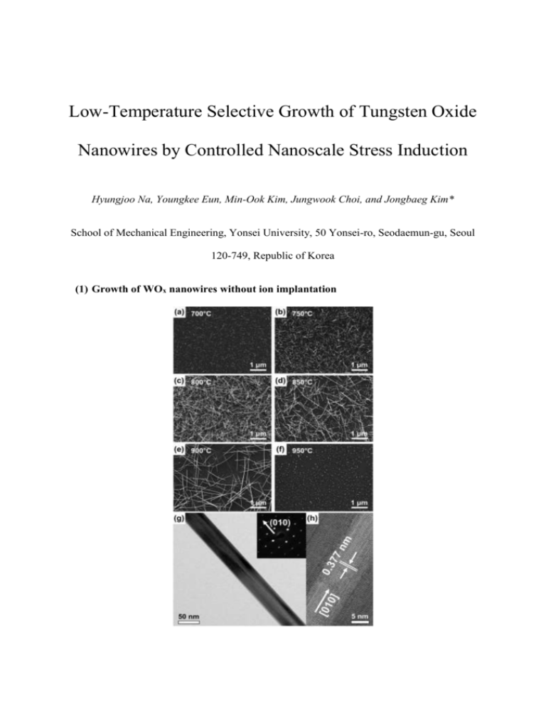
Low-Temperature Selective Growth of Tungsten Oxide Nanowires by Controlled Nanoscale Stress Induction Hyungjoo Na, Youngkee Eun, Min-Ook Kim, Jungwook Choi, and Jongbaeg Kim* School of Mechanical Engineering, Yonsei University, 50 Yonsei-ro, Seodaemun-gu, Seoul 120-749, Republic of Korea (1) Growth of WOx nanowires without ion implantation Supplementary Figure 1. Scanning electron micrographs of WOx nanowires grown without ion implantation on (100) Si substrates at (a) 700°C, (b) 750°C, (c) 800°C, (d) 850°C, (e) 900°C, and (f) 950°C for 20 min in a low-pressure environment with 100 sccm flowing N2 (ca. 11 Torr). (g) Transmission electron micrograph of one of the nanowires in (b), with the corresponding selected area diffraction pattern inset. (h) High-resolution transmission electron micrograph of the nanowire in (g). (2) Nanoscale patterned growth of WOx nanowires using a focused ion beam Supplementary Figure 2 shows the WOx nanowires grown using the same process as for those shown in Fig. 2—that is, with Ga ions implanted at 5 × 10¹⁵ ions/cm2 and 30 keV—but with the samples annealed at 630°C for 20 min (rather than at 600°C for 60 min). Supplementary Figure 2a shows the nanoscale patterns arranged as described for Fig. 2b. Supplementary Figure 2b shows six, four, and a single nanowire respectively in the 100 × 100, 75 × 75, and 50 × 50 nm2 squares of the right-most sample in Supplementary Figure 2a. The red arrows numbered 1, 2, and 3 point towards the nanowires grown on the 100 × 100, 75 × 75, and 50 × 50 nm2 squares, respectively. Supplementary Figure 2. Scanning electron micrographs showing WOx nanowires grown on squares implanted using a 30 keV focused beam of Ga ions. The samples were annealed at 630°C for 20 min. (a) Images of the nanowire patterns obtained with Ga doses of (from left to right) 3 × 10¹⁵, 5 × 10¹⁵, and 7 × 10¹⁵ ions/cm2 on square patterns 1000 × 1000, 500 × 500, 100 × 100, 75 × 75, and 50 × 50 nm2 (from top to bottom) in area. (b) High-magnification image of the region within the red rectangle in (a). The red arrows numbered 1 to 3 indicate the nanowires in the 100 × 100, 75 × 75, and 50 × 50 nm2 squares, respectively. (3) Time-of-flight secondary ion mass spectrometry Supplementary Figure 3 shows the depth profile of As, Ar, and N implanted into WOx thin films on the Si substrate before annealing. Supplementary Figure 3. Concentration profiles of As, Ar, N in WOx thin films as measured by time-of-flight secondary ion mass spectrometry.

