THIN LAYER CHROMATOGRAPHY OF VEGETABLES
advertisement
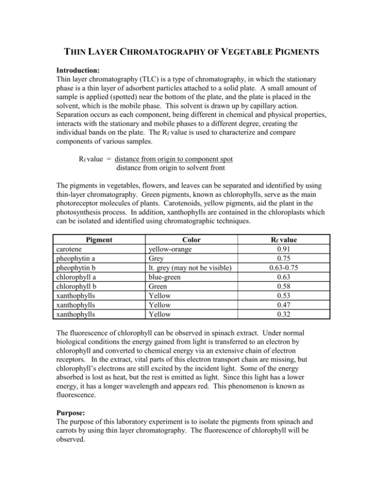
THIN LAYER CHROMATOGRAPHY OF VEGETABLE PIGMENTS Introduction: Thin layer chromatography (TLC) is a type of chromatography, in which the stationary phase is a thin layer of adsorbent particles attached to a solid plate. A small amount of sample is applied (spotted) near the bottom of the plate, and the plate is placed in the solvent, which is the mobile phase. This solvent is drawn up by capillary action. Separation occurs as each component, being different in chemical and physical properties, interacts with the stationary and mobile phases to a different degree, creating the individual bands on the plate. The Rf value is used to characterize and compare components of various samples. Rf value = distance from origin to component spot distance from origin to solvent front The pigments in vegetables, flowers, and leaves can be separated and identified by using thin-layer chromatography. Green pigments, known as chlorophylls, serve as the main photoreceptor molecules of plants. Carotenoids, yellow pigments, aid the plant in the photosynthesis process. In addition, xanthophylls are contained in the chloroplasts which can be isolated and identified using chromatographic techniques. Pigment carotene pheophytin a pheophytin b chlorophyll a chlorophyll b xanthophylls xanthophylls xanthophylls Color yellow-orange Grey lt. grey (may not be visible) blue-green Green Yellow Yellow Yellow Rf value 0.91 0.75 0.63-0.75 0.63 0.58 0.53 0.47 0.32 The fluorescence of chlorophyll can be observed in spinach extract. Under normal biological conditions the energy gained from light is transferred to an electron by chlorophyll and converted to chemical energy via an extensive chain of electron receptors. In the extract, vital parts of this electron transport chain are missing, but chlorophyll’s electrons are still excited by the incident light. Some of the energy absorbed is lost as heat, but the rest is emitted as light. Since this light has a lower energy, it has a longer wavelength and appears red. This phenomenon is known as fluorescence. Purpose: The purpose of this laboratory experiment is to isolate the pigments from spinach and carrots by using thin layer chromatography. The fluorescence of chlorophyll will be observed. Equipment/Materials: TLC silica gel plates developing chambers (jars) spinach shredded carrots disposable Pasteur pipets/bulbs test tube/rack capillary tubes 70:30 hexane-acetone solvent mortar/ pestle hexane acetone flashlight Safety: Always wear safety glasses in the laboratory. Procedure: 1. Obtain a spinach leaf or shredded carrots, and tear the sample into small pieces. Place the pieces in a mortar. 2. Add approximately 5 mL of acetone and carefully grind the sample and acetone together with the pestle. Continue grinding until the acetone becomes dark in color. More acetone may be added to compensate for evaporation. 3. Using a disposable Pasteur pipet, extract the acetone and place it in a small test tube. Be careful not to extract the small pieces of vegetable. 4. If the test tube is not approximately 2/3 full, repeat steps 2 and 3 using the same vegetable remaining in the mortar. 5. Shine a flashlight on the test tube to observe the red fluorescence in any spinach extract. 6. Add approximately 2 mL of hexane to the acetone extract. 7. Mix the contents of the test tube by using a clean disposable Pasteur pipet to repeatedly withdraw a sample and then immediately dispensing the sample back into the test tube. Be careful not to let the test tube overflow. 8. Allow the two layers to separate. 9. With a pencil, draw a line approximately 1 cm from the short edge of the TLC plate. Be careful not to scrape the coating of the plate. Mark the line with two tic marks approximately 1/3 from either side. See diagram. 10. With a capillary tube, draw up a sample of the upper hexane layer from the test tube containing the sample and spot the sample on one of the tic marks on the TLC plate. Be sure to label the spot at the bottom of the TLC plate. 11. Allow the sample to dry. Reapply the sample to the same place at least 3 times, allowing it to dry in between, or until the spot is clearly visible. 12. Repeat steps 10 and 11 using a standard, such as chlorophyll a, chlorophyll b, βcarotene, or xanthophyll. 13. Fill the developing chamber to a depth of approximately 0.5 cm with the 70:30 hexane-acetone mobile phase. (Invert the bottle several times before pouring into the developing chamber.) 14. Place the TLC plate in the chromatography chamber with the sample spot toward the bottom. Be sure the sample spot is above the level of the solvent. Close the chamber. 15. Allow the plate to remain undisturbed until the solvent reaches to within 1 cm of the top. 16. Remove the plate from the chamber and immediately mark the solvent front using a pencil. Trace around the edge of all visible spots. 17. Measure and record the distance from the spotting line (origin) to the center of each spot and from the spotting line to the solvent front. 18. Identify each component (spot). Data: Distance solvent moved from the spotting line (origin) __________________________ Type of Sample:________________________________________________________ Color of spot Distance moved Rf value Identity Standard:______________________________________________________________ Color of spot Distance moved Rf value Identity Calculations: 1. Calculate the Rf value for each spot observed. Questions: 1. Explain the color of the vegetable based on the results of the chromatography. 2. Which pigments, if any, were present in both vegetables? Explain. 3. Why was acetone used? 4. Why was a standard used?
