Word format
advertisement
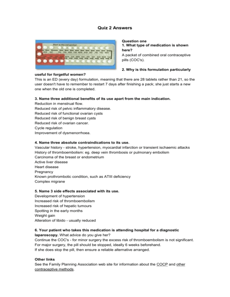
Quiz 2 Answers Question one 1. What type of medication is shown here? A packet of combined oral contraceptive pills (COC's). 2. Why is this formulation particularly useful for forgetful women? This is an ED (every day) formulation, meaning that there are 28 tablets rather than 21, so the user doesn't have to remember to restart 7 days after finishing a pack; she just starts a new one when the old one is completed. 3. Name three additional benefits of its use apart from the main indication. Reduction in menstrual flow. Reduced risk of pelvic inflammatory disease. Reduced risk of functional ovarian cysts Reduced risk of benign breast cysts Reduced risk of ovarian cancer. Cycle regulation Improvement of dysmenorrhoea. 4. Name three absolute contraindications to its use. Vascular history - stroke, hypertension, myocardial infarction or transient ischaemic attacks History of thromboembolism: eg. deep vein thrombosis or pulmonary embolism Carcinoma of the breast or endometrium Active liver disease Heart disease Pregnancy Known prothrombotic condition, such as ATIII deficiency Complex migrane 5. Name 3 side effects associated with its use. Development of hypertension Increased risk of thromboembolism Increased risk of hepatic tumours Spotting in the early months Weight gain Alteration of libido - usually reduced 6. Your patient who takes this medication is attending hospital for a diagnostic laparoscopy. What advice do you give her? Continue the COC's - for minor surgery the excess risk of thromboembolism is not significant. For major surgery, the pill should be stopped, ideally 6 weeks beforehand. If she does stop the pill, then ensure a reliable alternative arranged. Other links See the Family Planning Association web site for information about the COCP and other contraceptive methods. Question two This photograph was taken at diagnostic laparoscopy in a woman who presented with acute pelvic pain. 1. What is the name given to this finding? Fitz-Hugh-Curtis syndrome. 2. With what is is associated? The adhesions are secondary to a perihepatitis due to intraperitoneal spread of a chlamydia pelvic infection. 3. Name three other symptoms that may have been present. Right sided hypochondrial pain Offensive vaginal discharge – although this is less common with chlamydia than the vaginal infections. Patient generally unwell due to systemic infection Nausea or vomiting 4. Name three possible long-term sequelae of the primary condition? Infertility Chronic pelvic pain Dyspareunia Pelvic adhesions Menorrhagia 5.When performing a laparoscopy, what particular risk must the patient be made specifically aware of? Inadvertant perforation of pelvic viscera, such as bowel, bladder & vasculature. Other links A teaching document on PID including some MCQ's to test yourself. Question three A 40 year old lady presents with symptoms of urinary stress incontinence and this treatment is suggested. 1. What is the name of this tool and briefly describe its use? Vaginal cones. The various metal weights go inside the plastic shell. By placing the weighted cones into the vagina & contracting the pelvic floor muscles to hold it in place, pelvic floor tone can be increased. The weight or time held in place is gradually increased. 2. Name 3 surgical alternatives should this fail. Periurethral injectables (Macroplastique) Burch colposuspension Stamey colposuspension Anterior colporrhaphy and buttress to bladder neck (Kelly plication) Sling procedure 3. What 2 investigations are mandatory before embarking on surgery for stress incontinence. Midstream urine culture Urodynamics (cystometry) 4. Name 5 recognised side effects of incontinence surgery. Poor stream UTI Complete urinary retention requiring intermittent self-catheterisation Detrusor instability Enterocele Retropubic haematoma/abscess following Burch colposuspension Infected/fistulous rectus sheath sutures following Stamey 5. List 4 predisposing factors to urinary stress incontinence. Vaginal delivery (especially forceps & prolonged active second stage) Large babies Obesity Smoking Chronic obstructive airways disease (COAD) Menopause Question four 1. What fetal measurement is being made and what is its significance? First trimester nuchal translucency. It is a screening test for Down's Syndrome. The risk of cardiac abnormalities is also increased in chromosomally normal fetuses with a raised NT measurement. 2. Name an alternative method of screening for the target condition. Second trimester serum screening using the double/triple or quadruple test. Amniocentesis offered on the basis of maternal age alone is an alternative, though fewer cases will be detected. 3. What is the main advantage of the test shown as compared to the alternatives? It is carried out in the first trimester, so if an aneuploid fetus is found and termination of pregnancy opted for, a surgical procedure may be safely performed (vacuum termination of pregnancy). 4. What is the usual diagnostic procedure to follow this test if it is positive? Chorion villus sampling (CVS) via either the transcervical or transabdominal route. 5. Name 3 other 'soft markers' that may be detected on a routine anomaly scan. Echogenic bowel Choroid plexus cysts Renal pelvicalyceal dilatation Cardiac ventricular foci (golf-balls) Short femur length Question five This scan is of an ovary of an 18 year old patient who attended your clinic complaining of six month's amenorrhoea. 1. What is the likely diagnosis? Polycystic ovary syndrome (PCOS) 2. Name three appropriate blood tests. Follicular-phase LH/FSH Serum prolactin Serum androgens Sex hormone binding globulin (SHBG) Timed gonadatrophins in the early follicular phase of the cycle are required, due to physiological mid-cycle surges. An abnormally raised LH is indicative of PCOS and a LH:FSH ratio greater than 3 is also suggestive of the condition. Prolactin is mildly raised in 15% of cases of PCOS. Serum androgens are raised and SHBG is depressed, leading to higher levels of free testosterone. 3. Which three other presenting symtoms are common? Hirsutism Subfertility Acne Obesity 4. Given that your patient is not currently trying for pregnancy, does she require treatment? If so, why and what would be appropriate? The combined contraceptive pill would be expected to regulate her menstrual cycle. It is important that she has at least 4 periods per year to ensure protection from endometrial hyperplasia. Oestrogen levels are usually higher than normal due to excess peripheral conversion of androstendione to oestriol in fat. The absence of monthly menstruation means that endometrial hyperplasia is a risk and if untreated can lead to endometrial cancer. 5. Some years later she presents desiring pregnancy. List 4 potential treatments. Clomiphene citrate Laparoscopic ovarian diathermy (drilling) Controlled ovarian stimulation with gonadotrophins Insulin-sensitising drugs such as metformin IVF 6. What are the possible long-term complications of this condition (name 3)? Infertility Increased risk of diabetes Increased risk of hypertension and ischaemic heart disease Other links BMJ review PCOS detailed patient info Question six 1. What type of treatment is shown? Hormone replacement therapy (HRT). From left to right: oestradiol patches, oestradiol implant and sequential oral therapy. 2. Name 4 situations when these may be used. During the climacteric or after the menopause Premature ovarian failure Following bilateral oophorectomy, for example at the time of hysterectomy As add-back therapy with gonadotrophin-releasing agonists (GnRH-a) 3. What is a particular benefit of the medication on the left? Subcutaneous administration avoids first-pass hepatic metabolism and its unfavourable effect on the lipid profile. 4. What are the disadvantages of the middle treatment (name 3)? Requires a surgical procedure Once placed cannot be removed if unsuitable Tachyphylaxis - the phenomenon where increasingly shorter administration intervals are required to provide the same degree of symptom control, leading to supraphysiological levels of oestradiol. 5. Your patient, who has never undergone any surgery, wishes the treatment on the left. What do you also advise, and why? As she has an intact uterus, oestrogen treatment alone will lead to the risk of endometrial hyperplasia and endometrial cancer. Sequential use of a progestogen will bring about a protective withdrawl bleed. An alternative is to use progestogens continuously with the oestrogen (continuous combined therapy) which avoids a withdrawl bleed, but is only suitable for women 12 months after the true menopause (amenorrhoea). In either case the progestogen can be given as a tablet or a patch. 6. If a patient has irregular bleeding on this medication, what investigations are necessary? One or more of the following: outpatient endometrial biopsy (Pipelle), outpatient hysteroscopy, hysteroscopy & endometrial curettage (D&C) under general anaesthesia, transvaginal assessment of endometrial thickness. The last investigation is less useful than in the case of postmenopausal bleeding where no exogenous oestrogens are taken, as one can expect there to be some degree of endometrial proliferation (unless a continuous combined preparation is being taken). Question seven This patient was induced with prostin E2 gel. One hour after administration the cardiotocograph (CTG) shown was recorded. 1. What CTG abnormalities do you see? Reduced baseline variability Late deccelerations Excessive uterine activity (approximately 7 in 10 minutes) 2. What is the diagnosis? Uterine hyperstimulation leading to fetal comprimise 3. What is your management? Move the patient onto her left side to reduce the risk of aortocaval compression. Facial oxygen may improve fetal oxygenation. Use of an intravenous tocolytic agent will reduce the uterine activity. Suitable drugs include salbutamol or ritodrine, both of which are beta-2 agonists. This results in the following trace: Question eight 1. What measurement is being taken? Symphysis-fundal height 2. List 4 causes of an unexpectedly small reading. Incorrect dates Constitutionally small baby Intrauterine growth restriction (IUGR) Oligohydramnios (pathological or ruptured membranes) 3. List 4 causes of an unexpectedly large reading. Incorrect dates Macrosomic baby Polyhydramnios Maternal obesity 4. What is the next step in the case of question 2? Recheck dates using dating from early scan if available Perform ultrasound to check for fetal biometry (abdominal circumference & head circumference) and liquor volume. If fetus is asymmetrically small and/or liquor is reduced, a doppler assessment of flow in the umbilical artery should be made, as well as a biophysical assessment of the baby, which includes: Liquor volume Fetal breathing movements Gross fetal movements Fetal tone Cardiotocography (CTG) 5. The diagnosis of acute polyhydramnios at 28 weeks gestation is made. What 4 further investigations are necessary and what 2 treatments might be employed? Glucose tolerance test TORCH antibody screen Antibody screen Ultrasound of the fetus to check for hydrops and gross abnormalities of the CNS and GI tract Therapeutic amniocentesis can be used to drain off some of the excess liquor and nonsteroidal anti-inflammatory drugs (NSAIDs), such as indomethacin, are sometimes used to reduce the fetal production of liquor. Idiopathic polyhydramnios is the most common aetiology, but others include: diabetes mellitus, hydrops and congenital abnormlities such as hydrocephalus, spina bifida, oesophageal, duodenal or jejunal atresia, abdominal wall defects and diaphragmatic hernia. Hydrops has immune and non-immune causes. Immune hydrops is rarely caused by the D antigen now that the prophylactic use of anti-D is widespread, and usually involves a minor antigen. Non-immune hydrops may be caused by one of the TORCH infections. Question nine Your 34 year old patient presents with a 2 year of primary subfertility and undergoes this investigation. 1. What is the name of the procedure shown, and briefly describe it in patient-friendly terms. Hysterosalpingography (HSG) You will need to come to the x-ray department of the hospital during the first half of your cycle. An examination, like when you have a smear test done is carried out, the doctor passing a speculum into the vagina. A dye is injected through the cervix and x-rays taken to see where the dye goes. It should fill the womb and flow along both fallopian tubes, showing up as a white filling and spillage either side of the uterus. Some women find it a bit uncomfortable, with period-type pains when the dye is injected. See also this page from a US university web site. 2. What is the result of the investigation in this case? Normal filling of the uterus, but no entry into either fallopian tubes. 3. What is the next step? HSG is essentially a screening test and the tubal blockage needs to be confirmed by a formal laparoscopy and dye test. Sometimes a HSG can suggest blockage, but the problem is actually getting enough pressure when injecting the radio-opaque dye. Under anaesthetic you can usually be sure that this isn't a problem. Laparoscopy also allows a better assessment of how the tubes actually look & you can decide whether any blockage is amenable to surgery. 4. If the diagnosis is confirmed, what treatment is advised? In vitro fertilisation (IVF) 5. What are the other 3 essential components of the basic subfertility work-up? Semen analysis Day 2 LH/FSH to check for ovarian reserve (FSH>10 suggests ovarian resistance and raises the suspicion of impending ovarian failure) and the presence of PCO (raised LH) Luteal phase progesterone estimation taken 7 days before expected menses to confirm ovulation Some suggest a routine pelvic ultrasound scan to check for the presence of polycystic ovaries and confirm normal pelvic anatomy. Question ten This baby developed an abnormal fetal heartrate pattern during the latent phase of labour. 1. What method of delivery is shown? Caesarean section 2. List five important risks of the procedure. As usual, categorise these into immediate, early & late: Immediate: primary haemorrhage, damage to bowel, bladder or ureters, anaesthetic risks Early: Infection (chest, urinary, endometritis, wound), Thrombosis: DVT/PE, secondary haemorrhage Late: Increased risk of needing a repeat caesarean, adhesions leading to a greater risk of tubal infertility or chronic pelvic pain, adhesions sticking bladder to the uterus, making repeat sections or future hysterectomy more risky, increased risk of placenta praevia/accreta in the next pregnancy. 3. What would be an alternative course of action if she were later in the first stage? Fetal blood sampling, or assisted delivery by ventouse if the presenting part was very low and full dilatation imminent. Assisted delivery by ventouse in the late first stage should only be carried out by an experienced obstetrician who is sure cephalopelvic disproportion is excluded and when labour is progressing well. 4. What factors determine the likelihood of a successful vaginal birth in her next pregnancy. In general, a non-recurring reason for the first caesarean section suggests that a normal delivery is likely next time. This includes indications such as elective section for breech presentation, pre-eclampsia, placenta praevia, or an emergency section for fetal distress. The implication is that the first section wasn't done for what might have been cephalopelvic disproportion. It used to be that we were less hopeful where the first section was for failure to progress in the late first, or especially the second stage. This was particularly the case when the baby was of moderate size and in a normal position (OA). If the baby was huge, or in a persistent malposition such as OP, then the reason for lack of progress might have, once again, been down to factors to do with that-labour on that-day. Recent studies, however, have found that even where the first section was done for delay in the late first or second stage, the overall successful VBAC (vaginal birth after caesarean) rate is still of the order 70-80%. If a woman has had a previous vaginal delivery before her caesarean section then this would be likely to improve her chances of successful VBAC. Normal delivery after a caesarean is more likely if the next labour begins spontaneously, rather than by induction. 5. Should elective caesarean section be available 'on demand'? Justify your answer with 4 good reasons! I don't mind which way you go on this one, as long as your arguments are satisfactory! Arguments for: Patient autonomy Protection of the pelvic floor & perineum to try & reduce the incidence of third degree tears and incontinence of flatus/faeces. Elective caesarean section is a very safe operation. Timed delivery to suit parents. Perhaps only in selected cases, eg. previous c/s, breech, previous difficult labour or delivery. Arguments against: Although individual risk is small, overall risk of death is still 5 times higher with caesarean section as compared to normal birth. Increased risk of nonfatal thromboses and their incumbent morbidity Increased chance of haemorrhage & needing a blood transfusion, with the risks this carries. See risks of caesarean section, above (particularly late ones). Most women don't get third degree tears and faecal incontinence. Increased risk of baby developing RDS or TTN (transient tachypnoea of the newborn), even when the caesarean section is carried out at 38 weeks. Massive drain on healthcare resources if all or even a significant number of women demanded this. Medicalisation of what is essentially a physiological process for most women. What would we do with all those lovely midwives...!?
