Initiation to Cryo-transmission Electron Microscopy (Cryo
advertisement
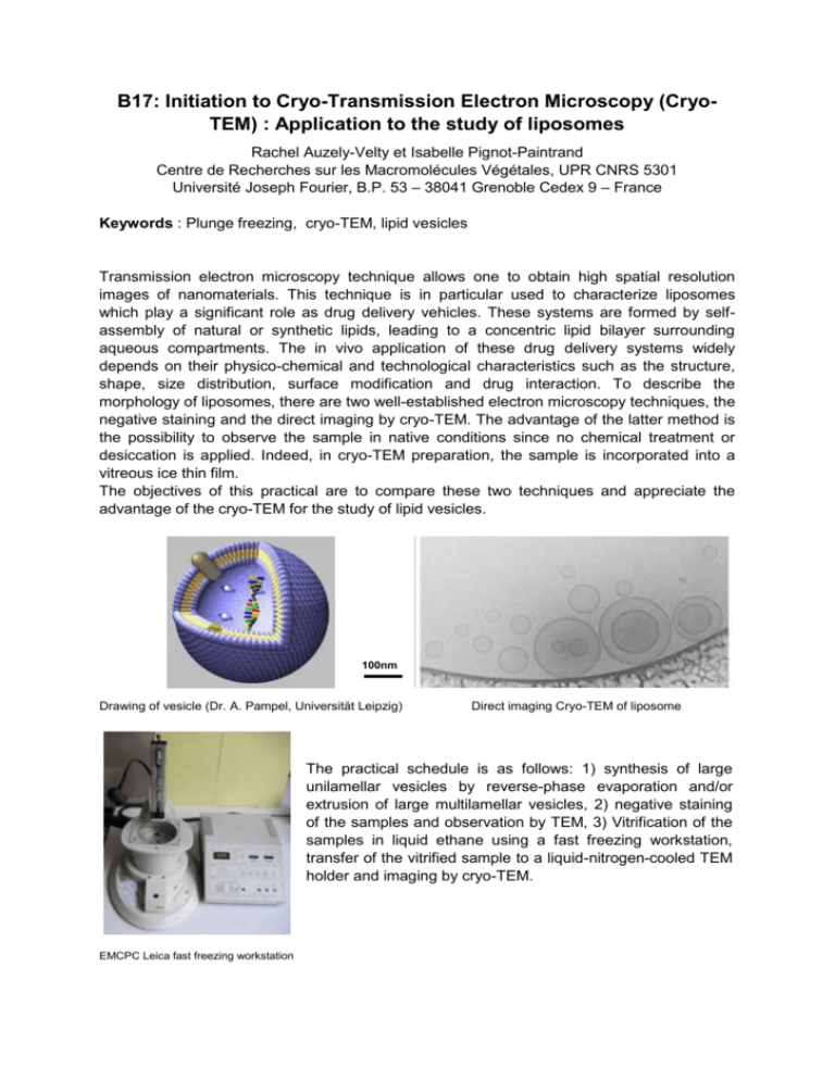
B17: Initiation to Cryo-Transmission Electron Microscopy (CryoTEM) : Application to the study of liposomes Rachel Auzely-Velty et Isabelle Pignot-Paintrand Centre de Recherches sur les Macromolécules Végétales, UPR CNRS 5301 Université Joseph Fourier, B.P. 53 – 38041 Grenoble Cedex 9 – France Keywords : Plunge freezing, cryo-TEM, lipid vesicles Transmission electron microscopy technique allows one to obtain high spatial resolution images of nanomaterials. This technique is in particular used to characterize liposomes which play a significant role as drug delivery vehicles. These systems are formed by selfassembly of natural or synthetic lipids, leading to a concentric lipid bilayer surrounding aqueous compartments. The in vivo application of these drug delivery systems widely depends on their physico-chemical and technological characteristics such as the structure, shape, size distribution, surface modification and drug interaction. To describe the morphology of liposomes, there are two well-established electron microscopy techniques, the negative staining and the direct imaging by cryo-TEM. The advantage of the latter method is the possibility to observe the sample in native conditions since no chemical treatment or desiccation is applied. Indeed, in cryo-TEM preparation, the sample is incorporated into a vitreous ice thin film. The objectives of this practical are to compare these two techniques and appreciate the advantage of the cryo-TEM for the study of lipid vesicles. 100nm Drawing of vesicle (Dr. A. Pampel, Universität Leipzig) Direct imaging Cryo-TEM of liposome The practical schedule is as follows: 1) synthesis of large unilamellar vesicles by reverse-phase evaporation and/or extrusion of large multilamellar vesicles, 2) negative staining of the samples and observation by TEM, 3) Vitrification of the samples in liquid ethane using a fast freezing workstation, transfer of the vitrified sample to a liquid-nitrogen-cooled TEM holder and imaging by cryo-TEM. EMCPC Leica fast freezing workstation
