file - BioMed Central
advertisement
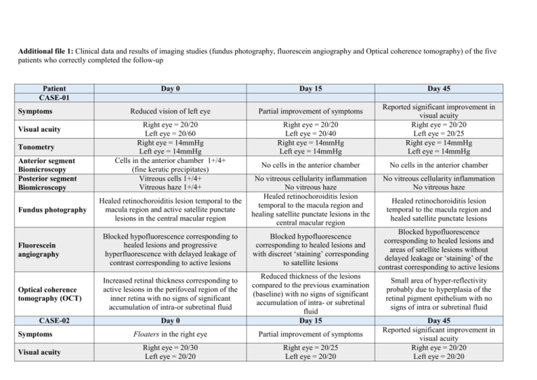
Additional file 1: Clinical data and results of imaging studies (fundus photography, fluorescein angiography and Optical coherence tomography) of the five patients who correctly completed the follow-up Patient CASE-01 Symptoms Visual acuity Tonometry Anterior segment Biomicroscopy Posterior segment Biomicroscopy Fundus photography Day 0 Day 15 Reduced vision of left eye Partial improvement of symptoms Right eye = 20/20 Left eye = 20/60 Right eye = 14mmHg Left eye = 14mmHg Cells in the anterior chamber 1+/4+ (fine keratic precipitates) Vitreous cells 1+/4+ Vitreous haze 1+/4+ Right eye = 20/20 Left eye = 20/40 Right eye = 14mmHg Left eye = 14mmHg Reported significant improvement in visual acuity Right eye = 20/20 Left eye = 20/25 Right eye = 14mmHg Left eye = 14mmHg No cells in the anterior chamber No cells in the anterior chamber No vitreous cellularity inflammation No vitreous haze Healed retinochoroiditis lesion temporal to the macula region and healing satellite punctate lesions in the central macular region No vitreous cellularity inflammation No vitreous haze Healed retinochoroiditis lesion temporal to the macula region and active satellite punctate lesions in the central macular region Fluorescein angiography Blocked hypofluorescence corresponding to healed lesions and progressive hyperfluorescence with delayed leakage of contrast corresponding to active lesions Optical coherence tomography (OCT) Increased retinal thickness corresponding to active lesions in the perifoveal region of the inner retina with no signs of significant accumulation of intra-or subretinal fluid CASE-02 Symptoms Visual acuity Blocked hypofluorescence corresponding to healed lesions and with discreet ‘staining’ corresponding to satellite lesions Day 0 Reduced thickness of the lesions compared to the previous examination (baseline) with no signs of significant accumulation of intra- or subretinal fluid Day 15 Floaters in the right eye Partial improvement of symptoms Right eye = 20/30 Left eye = 20/20 Right eye = 20/25 Left eye = 20/20 Day 45 Healed retinochoroiditis lesion temporal to the macula region and healed satellite punctate lesions Blocked hypofluorescence corresponding to healed lesions and areas of satellite lesions without delayed leakage or ‘staining’ of the contrast corresponding to active lesions Small area of hyper-reflectivity probably due to hyperplasia of the retinal pigment epithelium with no signs of intra or subretinal fluid Day 45 Reported significant improvement in visual acuity Right eye = 20/20 Left eye = 20/20 Tonometry Anterior segment Biomicroscopy Posterior segment Biomicroscopy Fundus photography Right eye = 14mmHg Left eye = 14mmHg Cells in the anterior chamber 1+/4+ (fine keratic precipitates) Vitreous cells 1+/4+ Vitreous haze 1+/4+ Active peridiscal retinochoroiditis lesion in the right eye without signs of macular involvement Fluorescein angiography Progressive hyperfluorescence with delayed leakage of contrast corresponding to active peridiscal lesion Optical coherence tomography (OCT) Increased retinal thickness corresponding to active lesions in the peridiscal region with no signs of significant accumulation of intra-or subretinal fluid CASE-03 Symptoms Visual acuity Tonometry Anterior segment Biomicroscopy Posterior segment Biomicroscopy Fundus photography Fluorescein angiography Optical coherence Day 0 Decreased visual acuity in the left eye Right eye = 20/20 Left eye = 20/40 Right eye = 12mmHg Left eye = 12mmHg Cells in the anterior chamber 1+/4+ (fine keratic precipitates) Vitreous cells 2+/4+ Vitreous haze 1+/4+ Satellite retinochoroiditis lesion in the upper peripheral media of the left eye by a healed retinochoroiditis lesion (old appearance) Progressive hyperfluorescence with delayed leakage of contrast corresponding to active lesion and masked hypofluorescence corresponding to the old scarred lesion Bad quality of OCT examination due to lesion Right eye = 14mmHg Left eye = 14mmHg No cells in the anterior chamber Reduction in keratic precipitates No vitreous cellularity inflammation Vitreous haze 1+/4+ Peridiscal retinochoroiditis lesion in the right eye with signs of healing and without macular involvement Delayed discreet ‘staining’ characteristic of healing of lesion Reduced retinal thickness compared to the previous examination (baseline) with no signs of significant accumulation of intra- or subretinal fluid Day 15 Decreased visual acuity in the left eye Right eye = 20/20 Left eye = 20/40 Right eye = 12mmHg Left eye = 12mmHg Cells in the anterior chamber 1+/4+ (fine keratic precipitates) Vitreous cells 2+/4+ Vitreous haze 1+/4+ Satellite retinochoroiditis lesion in the upper peripheral media of the left eye by a healed retinochoroiditis lesion (old appearance) Progressive hyperfluorescence with delayed leakage of contrast corresponding to active lesion and masked hypofluorescence corresponding to the old scarred lesion Bad quality of OCT examination due Right eye = 14mmHg Left eye = 14mmHg No cells in the anterior chamber No keratic precipitates No vitreous cellularity inflammation No vitreous haze Apparently healed peridiscal retinochoroiditis lesion in the right eye without sings of macular involvement Blocked hypofluorescence corresponding to lesion without areas of delayed leakage or ‘staining’ of the contrast Reduced retinal thickness compared to the previous examination (baseline) with no signs of significant accumulation of intra- or subretinal fluid Day 45 Decreased visual acuity in the left eye Right eye = 20/20 Left eye = 20/40 Right eye = 12mmHg Left eye = 12mmHg Cells in the anterior chamber 1+/4+ (fine keratic precipitates) Vitreous cells 2+/4+ Vitreous haze 1+/4+ Satellite retinochoroiditis lesion in the upper peripheral media of the left eye by a healed retinochoroiditis lesion (old appearance) Progressive hyperfluorescence with delayed leakage of contrast corresponding to active lesion and masked hypofluorescence corresponding to the oldscarred lesion Bad quality of OCT examination due tomography (OCT) CASE-04 Symptoms Visual acuity Tonometry Anterior segment Biomicroscopy Posterior segment Biomicroscopy Fundus photography location Day 0 to lesion location Day 15 Decreased visual acuity in the right eye Partial improvement of symptoms Right eye = 20/40 Left eye = 20/20 Right eye = 12mmHg Left eye = 12mmHg Cells in the anterior chamber 1+/4+ (fine keratic precipitates) Vitreous cells 2+/4+ Vitreous haze 2+/4+ Right eye = 20/25 Left eye = 20/20 Right eye = 12mmHg Left eye = 12mmHg No cells in the anterior chamber Reduction in keratic precipitates Vitreous cells 1+/4+ Vitreous haze 1+/4+ Healing satellite retinochoroiditis lesion in the upper peripheral media of the right eye beside of a healed retinochoroiditis lesion (old appearance) Satellite retinochoroiditis lesion in the upper peripheral media of the right eye beside of a healed retinochoroiditis lesion (old appearance) Progressive hyperfluorescence with delayed leakage of contrast corresponding to active lesion and masked hypofluorescence corresponding to the old scarred lesion Bad quality of OCT examination due to lesion location Day 0 Bad quality of OCT examination due to lesion location Day 15 Decreased visual acuity in the left eye Partial improvement of symptoms Anterior segment Biomicroscopy Right eye = 20/25 Left eye = 20/150 Right eye = 15mmHg Left eye = 14mmHg Cells in the anterior chamber 2+/4+ (granulated keratic precipitates) Right eye = 20/25 Left eye = 20/40 Right eye = 15mmHg Left eye = 15mmHg Cells in the anterior chamber 1+/4+ Reduction in keratic precipitates Posterior segment Biomicroscopy Vitreous cells 3+/4+ Vitreous Haze 3+/4+ Vitreous cells 2+/4+ Vitreous haze 2+/4+ Satellite retinochoroiditis lesion temporal to the macula region of the left eye beside a scarred retinochoroiditis lesion de (old Satellite retinochoroiditis lesion temporal to the macula region of the left eye beside a scarred Fluorescein angiography Optical coherence tomography (OCT) CASE-05 Symptoms Visual acuity Tonometry Fundus photography Delayed discreet ‘staining’ characteristic of healing of lesion to lesion location Day 45 Reported significant improvement in visual acuity Right eye = 20/20 Left eye = 20/20 Right eye = 12mmHg Left eye = 12mmHg No cells in the anterior chamber No keratic precipitates No Vitreous cells No vitreous haze Healed satellite retinochoroiditis lesion in the upper peripheral media of the right eye beside of a healed retinochoroiditis lesion (old appearance) Blocked hypofluorescence corresponding to lesion without areas of delayed leakage or ‘staining’ of the contrast Bad quality of OCT examination due to lesion location Day 45 Reported significant improvement in visual acuity Right eye = 20/25 Left eye = 20/40 Right eye = 15mmHg Left eye = 15mmHg No Cells in the anterior chamber Few precipitates Vitreous cells 1+/4+ Vitreous haze 1+/4+ Retinochoroiditis lesion temporal to the macula region of the left eye beside a scarred retinochoroiditis lesion de appearance) retinochoroiditis lesion (old appearance) (old appearance) Fluorescein angiography Progressive hyperfluorescence with delayed leakage of contrast corresponding to active lesion and masked hypofluorescence corresponding to the old scarred lesion Delayed discreet ‘staining’ characteristic of healing of lesion Blocked hypofluorescence corresponding to lesion without areas of delayed leakage or ‘staining’ of the contrast Optical coherence tomography (OCT) Increased retinal thickness temporal to the macula. Analysis made difficult by the vitreous haze Increased retinal thickness temporal to the macula. Analysis made difficult by the vitreous haze Reduced retinal thickness temporal o the macula

