File - CAPE Biology
advertisement
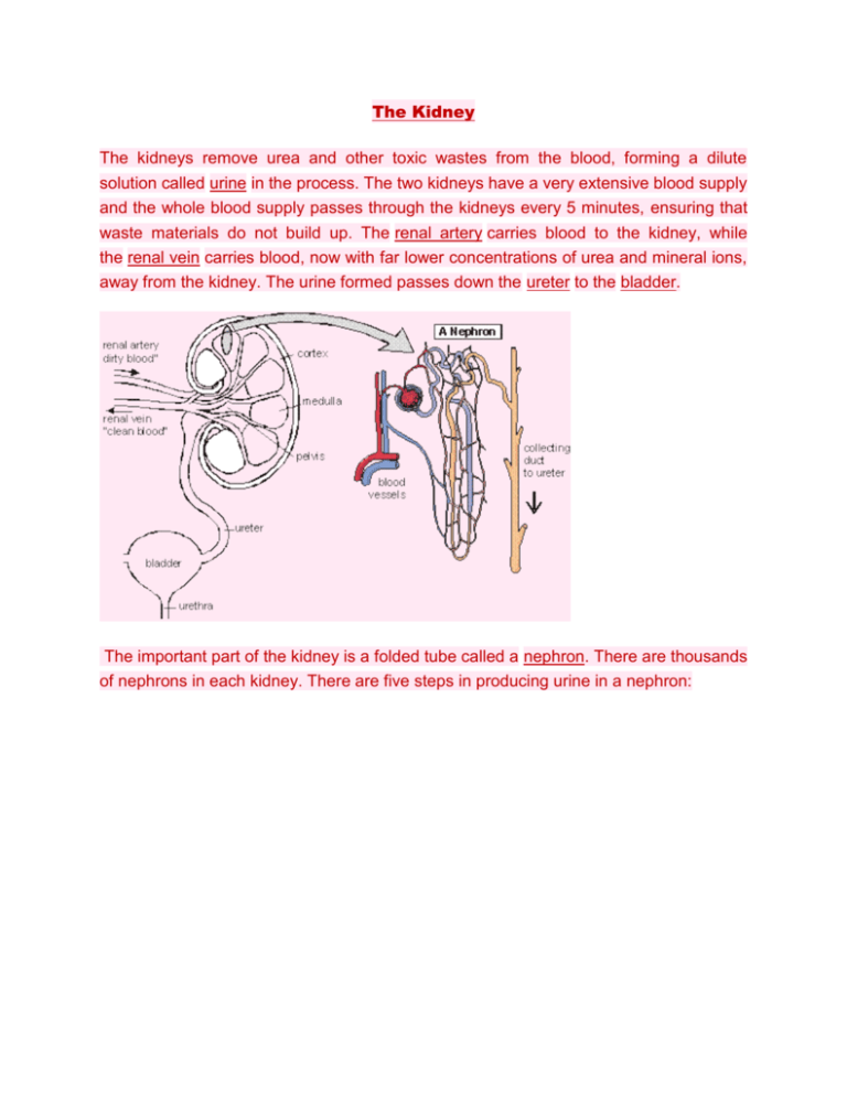
The Kidney The kidneys remove urea and other toxic wastes from the blood, forming a dilute solution called urine in the process. The two kidneys have a very extensive blood supply and the whole blood supply passes through the kidneys every 5 minutes, ensuring that waste materials do not build up. The renal artery carries blood to the kidney, while the renal vein carries blood, now with far lower concentrations of urea and mineral ions, away from the kidney. The urine formed passes down the ureter to the bladder. The important part of the kidney is a folded tube called a nephron. There are thousands of nephrons in each kidney. There are five steps in producing urine in a nephron: 1. Renal capsule – Ultrafiltration The renal artery splits into numerous arterioles, each feeding a nephron. The arteriole splits into numerous capillaries, which form a knot called a glomerulus. The glomerulus is enclosed by the renal capsule (or Bowman’s capsule)- the first part of the nephron. The arteriole leading into the glomerulus (the afferent arteriole) is wider than the one leading out (the efferent arteriole), so there is high blood pressure in the capillaries of the glomerulus. This pressure forces plasma out of the blood by ultrafiltration. Both the capillary walls and the capsule walls are formed from a single layer of flattened cells with gaps between them, so that all molecules with a molecular mass of <70k are squeezed out of the blood to form a filtrate in the renal capsule. Only blood cells and large proteins remain in the blood. 2. Proximal Convoluted Tubule – Reabsorption. The proximal convoluted tubule is the longest (14mm) and widest (60µm) part of the nephron. It is lined with epithelial cells containing microvilli and numerous mitochondria. In this part of the nephron over 80% of the filtrate is reabsorbed into the tissue fluid and then to the blood. This ensures that all the “useful” materials that were filtered out of the blood (such as glucose and amino acids) are now returned to the blood. All glucose, all amino acids and 85% of mineral ions are reabsorbed by active transport from the filtrate to the tissue fluid. They then diffuse into the blood capillaries. Small proteins are reabsorbed by pinocytosis, digested, and the amino acids diffuse into the blood. 80% of the water is reabsorbed to the blood by osmosis. Surprisingly, some urea is reabsorbed to the blood by diffusion. Urea is a small, uncharged molecule, so it can pass through membranes by lipid diffusion and there isn’t much the kidney can do about it. Since this is a passive process, urea diffuses down its concentration gradient until the concentrations of urea in the filtrate and blood are equal. So in each pass through the kidneys half the urea is removed from the blood and half remains in the blood. 3. Loop of Henle – Formation of a Salt Bath. The job of the loop of Henle is to make the tissue fluid in the medulla hypertonic compared to the filtrate in the nephron. The purpose of this “salt bath” is to reabsorb water as explained in step 5. The loop of Henle does this by pumping sodium and chloride ions out of the filtrate into the tissue fluid. The first part of the loop (the descending limb) is impermeable to ions, but some water leaves by osmosis. This makes the filtrate more concentrated as it descends. The second part of the loop (the ascending limb) contains a Na+ and a Cl- pump, so these ions are actively transported out of the filtrate into the surrounding tissue fluid. Water would follow by osmosis, but it can’t, because the ascending limb is impermeable to water. So the tissue fluid becomes more salty (hypertonic) and the filtrate becomes less salty (hypotonic). Since the filtrate is most concentrated at the base of the loop, the tissue fluid is also more concentrated at the base of the medulla, where it is three times more concentrated than seawater. 4. Distal Convoluted tubule – Homeostasis and Secretion In the distal convoluted tubule certain substances are actively transported from the blood into the filtrate, in other words they are secreted. It is relatively short and has a brush border (i.e. microvilli) with numerous membrane pumps for active transport. The important point about this secretion is that it is regulated by hormones, so this is the homeostatic part of the kidney. Substances secreted include H+ (for pH homeostasis), K+ (for salt homeostasis), ethanol, toxins, drugs and other “foreign” substances. 5. Collecting Duct – Concentration As the collecting duct passes through the hypertonic salt bath in the medulla, water leaves the filtrate by osmosis, so concentrating the urine and conserving water. The water leaves through special water channels in the cell membrane called aquaporins. These aquaporin channels can be controlled by the hormone ADH, so allowing the amount of water in the urine to be controlled. More ADH opens the channels, so more water is conserved in the body, and more concentrated urine is produced. This is described in more detail in water homeostasis later. The Bladder The collecting ducts all join together in the pelvis of the kidney to form the ureter, which leads to the bladder. The filtrate, now called urine, is produced continually by each kidney and drips into the bladder for storage. The bladder is an expandable bag, and when it is full, stretch receptors in the elastic walls send impulses to the medulla, which causes the sphincter muscles to relax, causingurination (or micturition). This is an involuntary reflex response that we can learn to control to a certain extent when we are young. Control of Water The control of water in an organism is known as osmoregulation. Hypothalamus Osmoreceptors within the hypothalamus detect when the water content is low since the loss of water reduces their volume which triggers stimulation of nerve cells in the hypothalamus. The hypothalamus are receptors and the effector is the pituitary gland and the walls of the distal convoluted tubule. The stimulated nerve cells produce a chemical known as antidiueretic hormone (ADH), a poly peptide made up of nine amino acids. When nerve cells are stimulated by the osmoreceptors, action potentials travel down them, causing ADH to be released from the blood in the capillaries in the posterior pituitary gland. ADH ADH acts on the kidney cells making up the walls of the collecting ducts, increasing their permeability to water by increasing the amount of water channels in the plasma membrane. It makes the blood less dilute by increasing the concentration of the urine. Negative feedback Again this is a negative feedback system - when blood water content is low, the hypothalamus acts to make the pituitary gland release ADH, and stops once blood water content rises (since the osmoreceptors are no longer being stimulated). Hormones Exocrine/Endocrine An endocrine gland is one that secretes hormones directly into the blood. Exocrine glands are glands that secrete to the outside, into a tube or a duct along which the secretion flows. Structure/Function of Hormones Structure: Relatively small Can be proteins or steroids Usually have a short life in the body and are broken down by enzymes, or lost in urine. Secreted in a very small concentration Function: To carry information from one part of a mammals body to another part Respond to stimulus (such as adrenaline responding to a frightening stimulus) Transported by the blood and have a group of cells which it affects - target cells. These target cells contain receptors specific to the hormone. ADDITONAL NOTES Kidney structure Section through the kidney to show the position of a nephron Scheme of renal tubule and its vascular supply The human body has two kidneys, and each receives blood via a renal artery and returns blood via a renal vein. The ureter carries urine from the kidney to the bladder, which excretes urine to the outside of the body via the urethra. The picture to the right shows a longitudinal section through a kidney, and it's clear that the kidney is covered by a tough capsule, beneath which is the cortex. The central area is the medulla, and where the ureter joins there is an area known as the pelvis. A microscope section shows that the kidney is made up of thousands of tubes known as nephrons. The nephrons structure is shown in the second picture down, and the labelled glomeluar capsule is known also as a renal or Bowman's capsule, and the renal capsules of all nephrons are in the cortex. They then run towards the centre of the kidney, forming a twisted region called the proximal convoluted tubule (PCT), and then a long loop of Henle (in the medulla). This then runs back up toward to cortex where it forms another twisted region called the distal convoluted tubule, before reaching a collecting duct which leads down through the medulla and into the pelvis of the kidney, joining the ureter. Ultrafiltration Urine production is a two-part process, ultrafiltration and reabsorption. The former is done in the renal capsule, where small molecules, including urea are taken out of the blood and into the renal capsule where they take the nephrons route through the kidney. Structure of a renal capsule is as follows; Network of capillaries that come from the afferent arteriole, separated by two cell layers from the renal capsule lumen. The first cell layer is endothelium cells, with thousands of tiny gaps. The second cell layer is the basement membrane formed from epithelial cells which make up the wall of the renal capsule, and have many finger-like projections, and have gaps. They are known as podocytes. The holes in the capillary endothelium and renal epithelium are large enough to allow any dissolved substances in the blood to get through. The basement membrane stops large protein molecules from getting through, as well as blood cells. The rate of ultafiltration is known as the glomerular filtration rate, and is usually around 125cm 3min-1. This is due to a variety of factors. The concentration of solutes in the blood plasma in the capillaries is higher than in the renal capsule, since the plasma protein molecules are too big to get through, creating a water potential that is lower in the blood capillaries. However, the water potential of the blood plasma is raised by the pressure inside the capillaries, causing blood to continue to move down this water potential gradient from the blood to the capsule. Reabsorption Many of the filtered substances need to be kept in the body, and so are selectivley reabsorbed (as some of them do need to be excreted). Most of this process occurs in the proximal convoluted tubule. Proximal Convoluted Tubule Na+/-K+ pump in proximal tubule cell membrane using ATP (made by its numerous mitochondria) to transport sodium ions out of the cell into the blood plasma, lowering the concentration inside the cell, so that they can passively diffuse (using the created concentration gradient) back into it from the proximal tubule. The sodium ions use special transporter proteins, at the same time taking up glucose from the proximal tube and is then taken into the blood. The membrane itself is folded to increases surface area for exchange on the blood plasma side. Glucose, amino acids, vitamins, 65% of the water from the PCT and many sodium/chloride ions are actively reabsorbed here. Some urea is also reabsorbed, around half. The numerous microvilli on the proximal tubule side provide an increased surface area for uptake of solutes. This reduces the volume of liquid remaining from 125cm 3 to around 45cm3 to go to the loop of Henle. Loop of Henle The loop of Henle serves to create a high concentration of salts in the tissue fluid in the medulla of the kidney, allowing a lot of water to be reabsorbed from the fluid in the collecting duct as it flows through, producing very concentrated urine and preserving water for the body. As you can see in the above picture, there is a descending and ascending limb. The method by which it concentrates urine is below; 1. Sodium and Chloride are actively transported out of the ascending limb 2. This raises the concentration of sodium and chloride in the tissue fluid 3. This in turn causes loss of water into the tissue fluid in the descending limb. 4. This loss of water concentrates sodium and chloride in the descending limb 5. The concentrated sodium and chloride ions can then diffuse out of the ascending limb into the tissues. Having the two limbs run side by side allows maximum concentration to be built up both inside and outside the tube at the bottom, and is known as a counter-current multiplier. The fluid then is flowing up the ascending limb of the loop of Henle, losing sodium and chloride as it goes, it continues round into the collecting duct, which dips into the medulla again, passing through the highsolute concentration regions of the tissues. This causes yet more water to move out of the collecting duct via osmosis. Distal Convoluted Tubule The first part of the DCT acts in the same way as the ascending loop of Henle, the second part behaves in the same way as the collecting duct. Functions of second part of the DCT and collecting duct are below; Sodium ions are actively pumped from the fluid in the tubule into the tissue fluid, where they pass into the blood. Potassium ions are actively transported into the tubule
