bit25855-sup-0001-SupData-S1
advertisement
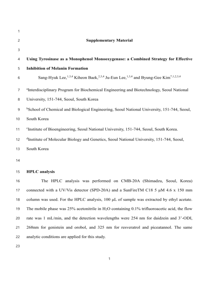
1 2 Supplementary Material 3 4 Using Tyrosinase as a Monophenol Monooxygenase: a Combined Strategy for Effective 5 Inhibition of Melanin Formation 6 Sang-Hyuk Lee,1,3,4 Kiheon Baek,2,3,4 Ju-Eun Lee,1,3,4 and Byung-Gee Kim*,1,2,3,4 7 a 8 University, 151-744, Seoul, South Korea 9 b Interdisciplinary Program for Biochemical Engineering and Biotechnology, Seoul National School of Chemical and Biological Engineering, Seoul National University, 151-744, Seoul, 10 South Korea 11 c 12 d 13 South Korea Institute of Bioengineering, Seoul National University, 151-744, Seoul, South Korea. Institute of Molecular Biology and Genetics, Seoul National University, 151-744, Seoul, 14 15 HPLC analysis 16 The HPLC analysis was performed on CMB-20A (Shimadzu, Seoul, Korea) 17 connected with a UV/Vis detector (SPD-20A) and a SunFireTM C18 5 μM 4.6 x 150 mm 18 column was used. For the HPLC analysis, 100 μL of sample was extracted by ethyl acetate. 19 The mobile phase was 25% acetonitrile in H2O containing 0.1% trifluoroacetic acid, the flow 20 rate was 1 mL/min, and the detection wavelengths were 254 nm for daidzein and 3’-ODI, 21 268nm for genistein and orobol, and 325 nm for resveratrol and piceatannol. The same 22 analytic conditions are applied for this study. 23 1 1 GC analysis 2 After 100 μL of sample was extracted by 400 μL of ethyl acetate, all hydroxyl groups 3 of the product were converted to TMS (trimethylsilyl) derivatives with BSTFA. GC/MS was 4 performed on a Thermo Scientific Trace GC Ultra instrument connected with a Thermo 5 Scientific ITQ1100 MS spectrometer (Rockford, IL, USA) and with a TR-5ms SQC capillary 6 column (30 m × 0.25 mm i.d., 0.25 mM film thickness). The GC/MS analysis of daidzein and 7 resveratrol was carried out as follows: injector temperature, 250 °C; temperature gradient: 8 holding initial temperature at 100 °C for 1 min, gradually increasing temperature up to 9 250 °C by 30 °C/min, holding at 250 °C for 10 min, gradually increasing temperature up to 10 275 °C by 1 °C/min, and holding at 275 °C for 2 min; and MS operating mode: 70 eV 11 electron ionization mode. For genistein: injector temperature, 250 °C; temperature gradient: 12 holding initial temperature at 100 °C for 1 min, gradually increasing temperature up to250 °C 13 by 30 °C/min, holding at 250 °C for 5 min, gradually increasing temperature up to 290 °C by 14 5 °C/min, and holding at 290 °C for 5 min; and MS operating mode: 70 eV electron 15 ionization mode. 16 17 Table S1. Primer list Primer Sequence (5` → 3’) SA_Ty_melC1_F GAA CCATGG AGTTAACCCGGCGT SA_Ty_melC1_R GAC AAGCTT TTAATTAAACGG SA_Ty_melC2_F GAA CCATGG ATGACCGTACGCAA SA_Ty_melC2_R GAC AAGCTT GACGGTGTAGAACG BM_Ty_F GAT GAATTC ATGAGTAACAAGTA BM_Ty_R GCA GTCGAC TGAGGAACGTTTTGA 2 1 Table S2. Reaction conditions. At a reaction temperature of 37℃, 500 mM borate buffer at 2 pH 9 was used. The detailed calculation of the oxygen transfer coefficient of reactors is 3 addressed later in this supporting material. Total reaction volume, mL 5 400 [daidzein]o, mM 5 5 [L-ascorbic acid]o, mM 20 20 Wet cell weight, g 0.1 2 Air flow rate, NL∙min-1 Not controlled 12 0.22 0.467 Oxygen transfer coefficient, -1 min 4 *NL (normal liter): L at 1 atm at 25℃ 5 6 7 8 9 10 11 12 13 14 15 16 17 3 1 Table S3. Kinetic constants of tyrosinase from B. megaterium on daidzein. The total 2 reaction volume was 100 μL, and the range of initial concentration of daidzein was 100 μM 3 to 1 mM. For the reaction, 500 mM borate buffer at pH 9 and 10 mM of NH2OH were used 4 (Yamazaki et al., 2003). The reaction was performed in 1.5 mL tubes at room temperature. 5 The reaction was halted by adding 400 μL of ethyl acetate and 20 μL of 1M HCl. The organic 6 layer was analyzed on HPLC to measure the amount of daidzein left from the reaction. The 7 kinetic constants were calculated based on the triplet sets of the experiment. The average 8 initial velocities of each initial substrate concentration were plotted and the standard errors 9 were obtained by regression wizard of SigmaPlot 10.0. (one site saturation equation was used) 10 [E]0, nM 200 Vmax, μM∙min-1 26.9 ± 2.29 Km, μM 6.11×10² ± 1.05×10² kcat, s-1 8.06×10³ kcat/Km, M-1∙s-1 1.32×10⁷ 11 12 13 14 15 16 17 18 19 4 1 Table S4. The effect of ascorbic acid on the reaction rate of tyrosinase. To measure the 2 first hydroxylation of daidzein, 500 mM of borate was used to make tyrosinase fully focus on 3 phenolase activity as previously studied (Yamazaki et al., 2003). Instead of hydroxylamine, 4 L-ascorbic acid was added for circulating the Ty cycle. The method for measuring k1and k2 is 5 stated in “Calculation of k1 and k2 of BM_Ty on daidzein,” later in this supporting 6 information. 7 [ascorbic acid]0, mM tss, min [B]ss, mM 0 - - 0.5 - - 1 2 5 10 72.29 ± 62.23 ± 83.74 ± 81.39 ± 0.45 0.63 1.22 0.98 0.786 ± 0.896 ± 1± 1± 0.077 0.090 0.0970 0.0851 5.085×10- 2.041×10- 2.687×10- 4.222×10- 4.250×10- 4.378×10k1, min-1 3 2 2 2 2 2 4.901×10- 3.405×10- 1.210×10- 1.241×10k2, min-1 - 3 - 8 5 3 3 3 1 2 Figure S1. The concentration of 3’-ODI produced by tyrosinase from daidzein with 3 various initial concentrations of boric acid. Red lines show the overall concentration of 3’- 4 ODI produced during reaction. The blue lines show the concentration of 3’-ODI that did not 5 form conjugated bonds with borate. Boric acid was added in 100 mM tris-HCl buffer and the 6 pH was maintained at 9 by adding 6N NaOH solution. The conditions for the reaction are as 7 follows: 5 mL reaction, 1 mM daidzein, 10 mM ascorbic acid, 130 nM of enzyme and the 8 temperature was maintained at 37 ℃. 9 6 1 2 3 Figure S2. Auto-oxidation of 3’-ODI in buffer at pH 9. In high pH of solution, diphenols 4 are easily deprotonated since the pka values are generally below 9, which causes oxidation of 5 diphenols to yield unstable quinonic compounds (o-quinones). Since the Ty reaction 6 condition of producing 3’-ODI is held at pH 9, losses due to the auto-oxidation of 3’-ODI 7 were measured. After 1mM of N-methylbenzothiazolone hydrazine (MBTH) was used to 8 detect the amount of o-quinones produced via UV-spectroscopy at 550nm, 500 μM of 3’-ODI 9 with 0.5 v% of DMSO and 1mM of MBTH were added in a glass cuvette containing 1mL of 10 buffer at pH 9, and the absorbance at 550nm of MBTH-o-quinone was measured. In 50 mM 11 of tris-HCl buffer, 3’-ODI was significantly oxidized. After 5 min, the production of MBTH- 12 o-quinone reached a steady state but then increased once air was blown through the pipetting. 13 However, 3’-ODI remained stable in 50 mM borate buffer at pH 9. 14 15 16 7 1 2 Figure S3. Rate changes depending on various concentrations of L-ascorbic acid. The 3 conditions for the reaction are as follows: total reaction volume is 5 mL, 500 mM borate 4 buffer at pH 9, 1 mM daidzein, 130 nM of enzyme and the temperature was maintained at 5 37 ℃. The method for measuring k1and k2 is described in “Calculation of k1 and k2 of 6 BM_Ty on daidzein,” later in this supporting information. All the rates obtained from this 7 graph are listed in Table S2. 8 9 10 11 12 13 8 1 2 Figure S4. Docking simulation of tyrosinase from B. megaterium (3NM8) with ascorbic 3 acid. The crystal structure of tyrosinase from B. megaterium (3NM8) is available at RCSB 4 database. ChemDraw Ultra 8.0 and Chem3D Ultra 8.0 were used for making the PDB file of 5 ascorbic acid. The structure of ascorbic acid was modified with inbuilt minimize energy 6 function of Chem3D. AutoDock Vina 1.1.2 and MGLTools 1.5.6. were used for docking 7 simulation. Grid spacing, exhaustiveness and energy range were set as default, which are 3.75 8 Å, 8 and 3, respectively. X-Score was used to select the best fit conformation. Chimera 1.10rc 9 was used for visualizing the results. 10 11 12 13 9 1 2 Figure S5. The time profile of the concentration of daidzein and 3’-ODI in the 400 mL 3 fed-batch reaction. The reaction conditions are listed as follows: 400 mL total reaction 4 volume in a 1 L bioreactor, 500 mM borate buffer at pH 9, and 3 mM of dadizein. 5 Approximately 2 g of wet cell was used (final OD at 600nm of the reaction mixture was 0.8). 6 After 10 mM ascorbic acid was added initially, 1mM daidzein and 2 mM ascorbic acid were 7 added every 10 min for 70 min. The temperature was maintained at 37℃. The total 8 concentration of daidzein fed was 10 mM and 12 L∙min-1 of air was continuously blown from 9 the bottom of the reactor. The oxygen transfer rate of the reactor was calculated and is further 10 described later in this supporting data, “Calculation of Oxygen transfer rate and coefficient in 11 the 400 mL reaction with the continuous air flow rate of 12 L∙min-1.” The yield of 3’-ODI 12 production for 190 min was 70~75% (7.6 mM 3’-ODI), and 3.2 mM daidzein was left. 10 1 2 11 1 2 3 Figure S6. 1HNMR analysis of the substrate (daidzein) and the product (3’-ODI). Daidzein was dissolved in DMSO-d6 as it was shipped 4 from Sigma-Aldrich. The peaks of 1HNMR of the initial substrate, dadizein, and the product, 3’-ODI, are identical except the peaks a, b, c, d, 5 e, f and g. Product was extracted by diethyl ether from the reaction mixture and the top layer was evaporated. The residual powder and 12 1 daidzein were dissolved in DMSO-d6 and 1H NMR spectra were measured at 400 MHz at 25℃. The 1H NMR spectroscopic data were stated 2 in ppm (δ) from the internal standard (TMS, 0.0 ppm). 1HNMR (DMSO-d6 with 0.05% v/v TMS, 400 MHz) spectrum of 3′-ODI is: δ 10.75 3 (1H, s, C7-OH), 8.98 (1H, s, C3′-OH), 8.94 (1H, s, C4′-OH), 8.25 (1H, s, H2), 7.97 (1H, d, J = 8.8 Hz H5) 7.02 (1H, d, J = 2 Hz, H2′), 6.94 4 (1H, dd, J = 8.8 and 2 Hz, H6), 6.85 (1H, d, J = 2.0 Hz, H5′), 6.82 (1H, dd, J = 8.0 and 2.0 Hz, H6′), 6.77 (1H, d, J = 8.0 Hz, H5′) 5 6 7 8 9 10 11 12 13 13 1 2 3 Figure S7. GC/MS analysis of the product from daidzein. Sampling was started right after the enzyme was added in the reaction: (a) and (c) 4 show the chromatography of the sample, and (b) and (d) show the mass spectra of the peaks (1) and (5), respectively. The peak (1) at 19.08 5 min is daidzein and the peak (2) and (5) which appear at 23.61 min is 3’-ODI. After 60 min of the reaction, peak (1) disappeared and only 14 1 peak (5) was detected. The detected cation for the product, monohydroxylated daidzein, was [M+1]+ ion of m/z 486.86 (6), which 2 corresponds to the summation of three trimethylsilyl groups and 3’-ODI, and [M+1]+ ion of m/z 471.86 (7), which indicates the loss of one 3 phenolic silyl ether group. 15 1 2 16 3 4 5 6 Figure S8. 1HNMR of the hydroxylated product from resveratrol: (a) resveratrol and (b) piceatannol. Product was extracted by diethyl 7 ether from the reaction mixture and the top layer was evaporated. The residual powder and resveratrol were dissolved in DMSO-d6 and 1H 17 8 NMR spectra were measured at 400 MHz at 25℃. Resveratrol was dissolved in DMSO-d6 as it was shipped from Sigma-Aldrich. 1H NMR 9 spectroscopic data are stated in ppm (δ) from the internal standard (TMS, 0.0 ppm). 1HNMR of the product was predicted as piceatannol; (400 10 MHz, 25℃) δ 6.110 (t, 1H, J = 20.), 6.368 (d, 2H, J = 2.0), 6.718 (d, 1H, J = 8.0), 6.728 (d, olefinic H, J = 16.4), 6.841(dd, 1H, J = 2.0, 8.0), 11 6.865(d, olefinic H, J = 16.8), 6.954(d, 1H, J = 2), 9.163(Br s, 4H) 12 13 14 15 16 18 17 18 19 19 20 21 Figure S9. 1HNMR of the hydroxylated product from genistein: (a) genistein and (b) orobol. Samples were extracted by diethyl ether and 22 the top layer was evaporated. The residual powder and genistein were dissolved in DMSO-d6 and 1H NMR spectra were measured at 400 23 MHz at 25℃. Genistein was dissolved in DMSO-d6 as it was shipped from Sigma-Aldrich. 1H NMR spectroscopic data are stated in ppm (δ) 20 24 from the internal standard (TMS, 0.0 ppm). 1HNMR of the product was predicted as 3’,4’,5,7,-tetrahydroxyisogflavone, orobol; (400 MHz, 25 25℃) δ 6.225 (d, 1H, J = 2.0), 6.382 (d, 1H, J = 2.0), 6.772 (d, 1H, J = 8.0), 6.802 (dd, 1H, J = 10, 2.0), 7.004(d, 1H, J =1.6), 8.285(s, 1H), 26 9.011(BRs, 1H), 9.079(BRs, 1H), 10.896(BRs, 1H), 12.999(Br s, 1H) 27 28 29 30 31 32 33 34 35 21 36 37 38 Figure S10. GC/MS anaylsis of the product from resveratrol. Sampling was started right after the enzyme was added in the reaction. (a) 39 and (c) show the chromatography of the sample, and (b) and (d) show the mass spectra of the peaks (1) and (4), respectively. A peak (1) at 22 40 12.60 min is resveratrol, and peaks (2) and (4) which appear at 15.64 min is piceatannol. The detected cation for piceatannol was [M+1]+ ion 41 of m/z 532.96 (5), which corresponds to the summation of four trimethylsilyl groups and piceatannol. 42 43 44 45 46 47 48 49 50 51 23 52 53 54 Figure S11. GC/MS anaylsis of the product from genistein. Sampling was started right after the enzyme was added in the reaction. (a) and 55 (c) show the chromatography of the sample, and (b) and (d) show the mass spectra of peaks (1) and (4), respectively. A peak (1) at 16.61 min 24 56 is genistein and a broad peak (4) at 18.46 is orobol. The detected cation for orobol was [M+1]+ ion of m/z 574.21 but only m/z 502.70 (5) was 57 detected, which corresponds to the summation of three trimethylsilyl groups and orobol. 25 1 Calculation of k1 and k2 of BM_Ty on daidzein 2 The continuous reaction was simplified as described below (steady-state approximation). k1 3 and k2 were obtained by substituting experimental values into the simplified equation, and the 4 ratio of k1 to k2 was calculated. 5 6 7 A, daidzein; B, 3’-ODI; C, o-quinone; [B]ss, concentration of 3’-ODI at steady state; tss, time 8 point at concentration of 3’-ODI reaching at [B]ss; k1, phenolase activity; k2, cateholase 9 activity 10 11 1) Rate constant of phenolase ( k1) : 12 13 14 15 𝑑[𝐴] = −𝑘1 [𝐴] 𝑑𝑡 [𝐴]𝑡 = [𝐴]𝑜 𝑒 −𝑘1 𝑡 ln[𝐴]𝑡 = −𝑘1 𝑡 ln[𝐴]0 16 17 18 19 20 26 − − − (Eq. 1) − − − (Eq. 2) 1 2) Rate constant of diphenolase (k2) : 2 3 At steady state, 4 𝑑[𝐵] = 𝑘1 [𝐴] − 𝑘2 [𝐵] = 0 𝑑𝑡 5 6 8 [𝐵]𝑡 = 𝑘1 𝑘1 [𝐴]𝑡 = [𝐴]0 𝑒 −𝑘1 𝑡 𝑘2 𝑘2 − − − (Eq. 3) Eq. 1 can be inserted into Eq. 3. and then, 𝑘2 = 𝑘1 [𝐴]0 𝑒 −𝑘1 𝑡𝑠𝑠 [𝐵]𝑡 − − − (Eq. 4) 7 9 Below are the time profiles of 1 mM of daidzein consumed and 3’-ODI produced by 130 nM 10 of purified enzyme in 5 mL reaction at 37℃. 500 mM borate buffer at pH 9 and 5 mM of 11 ascorbic acid were used in the reaction. 12 13 14 15 k1, daidzein consumption rate by tyrosinase, can be obtained from Eq. 2, ln[daidzein] vs. time. 16 The slope was obtained through in-built program of Excel (Microsoft). 17 27 1 2 3 k1 = 4.250×10 -2 min -1 4 5 The plot of 3’-ODI (range of 120 min to 360 min of reaction) was drawn separately, and the 6 best fit equation of the graph was obtained by in-built program of Excel, as shown below. 7 8 9 10 [B]ss and tss were calculated through the given equation after finding where the slope of a 11 tangent line become zero. Here, [B]ss is 1 mM and tss is 83.74 min. The values obtained were 12 inserted into Eq. 4 to determine k2, which was 7.26×10 -2 min -1. 13 14 15 28 1 Calculation of oxygen transfer rate and coefficient of the 5 mL reaction in a 50 mL 2 reactor without additional oxygen transfer. 3 4 Rate of oxygen transfer = 𝑑𝐶 𝑑𝑡 = 𝑘𝑣,𝑙 (𝐶𝑠𝑎𝑡 − 𝐶) --------- eq. 1S 5 (𝑘𝑣,𝑙 , overall volumetric oxygen transfer coefficient; C, concentration of oxygen; C sat, 6 saturated concentration of oxygen) 7 8 Integration of eq 1S gives, 𝐶 9 −𝐶 ln 𝐶 𝑠𝑎𝑡−𝐶 = − 𝑘𝑣,𝑙 ∙ 𝑡 𝑠𝑎𝑡 -------- eq. 2S 0 10 11 In 5mL of 50 mM tris-HCl buffer pH 8, N2 was blown to eliminate all oxygen at first, and 12 then D.O. was measured after the N2 nozzle was removed. The reaction was preceded in 50 13 mL Falcon tube with a diameter of 2.8mm 0.5 0.21 ln[(C_sat-C)/(CsatC o ) ] 0.982027183 1.5 1.69 0.834827122 2.5 3.2 0.626597031 3.5 4.32 0.394531426 4.5 5.2 0.098217828 5.5 6.41 - 6.5 6.4 - Time(min) ppm (mg/L) 15 16 14 The overall volumetric oxygen transfer coefficient, 𝒌𝒗,𝒍 = 0.22 min-1 Rate of oxygen dissolving = 𝑑𝑛𝑎𝑞 𝑂2 𝑑𝑡 = 𝑉 𝑑𝐶 𝑚.𝑤.𝑜𝑓 𝑂2 𝑑𝑡 -------- eq. 3S. 17 (V, reactor volume; naq O2 , the moles of oxygen that dissolved in the reaction solution) 18 eq. 1S and eq. 3S can be combined to give, 29 𝑑𝑛𝑎𝑞 𝑂2 1 𝑑𝑡 = 𝑉 𝑚.𝑤.𝑜𝑓 𝑂2 𝑘𝑣,𝑙 (𝐶𝑠𝑎𝑡 − 𝐶) ---------- eq. 4S 2 The rate of oxygen dissolving in 5mL reactor was 0.44 μmol of O2∙min-1 (88 μM of O2∙min-1 3 in the 5 mL solution). 4 During the whole cell reaction (5 mL) with 500 μM daidzein (without shaking or air 5 blowing), the amount of dissolved oxygen (D.O.) was reduced to approximately 50 μM (0.5 6 to 0.8 ppm) but not consumed completely, which means that the oxygen was not deficient. 7 8 9 Calculation of oxygen transfer rate and coefficient of the 400mL reaction in a 1 L 10 reactor with a continuous air flow rate of 12 L∙min-1 . 11 In 400 mL of 500 mM borate buffer pH 9, N2 was blown to eliminate all oxygen at first, and 12 then D.O. was measured with a continuous airflow rate of 12 L∙min-1. The stirring bar (length: 13 5 cm, diameter: 1cm) was spun at 250 rpm. Time (min) 0.0 0.5 1.0 1.5 2.0 2.5 3.0 3.5 4.0 4.5 5.0 5.5 6.0 6.5 7.0 7.5 8.0 ppm (mg∙L-1) 0.23 0.77 1.67 2.38 2.74 3.23 4.10 4.92 5.38 5.60 5.75 5.85 5.93 6.00 6.10 6.15 6.20 ln[(C_sat-C)/(CsatCo)] 0 -0.091589396 -0.265773635 -0.428416935 -0.522235691 -0.66596725 -0.986789715 -1.42765675 -1.799896211 -2.042842389 -2.250481754 -2.417535839 -2.574721422 -2.73598957 -3.023671642 -3.205993199 -3.42913675 30 8.5 9.0 9.5 10.0 10.5 11.0 11.5 12.0 12.5 13.0 13.5 14.0 14.5 15.0 10 6.24 6.27 6.28 6.30 6.32 6.35 6.37 6.37 6.38 6.38 6.39 6.40 6.40 6.40 1 -3.652280302 -3.859919666 2 -3.939962374 -4.122283931 3 -4.345427482 -4.815431111 4 -5.326256735 5 -5.326256735 -5.731721843 6 -5.731721843 7 -6.424869024 8 9 The integrated values (y-axis) can be inserted into eq 2S. and 𝑘𝑣,𝑙 (slope) can be 11 easily determined by trend line equation programmed in Excel. The overall volumetric 12 oxygen transfer coefficient, 𝒌𝒗,𝒍 = 0.4637 min-1. The rate of oxygen dissolving in 400 mL 13 reactor with 12 L/min air flow rate was 74 μmol of O2∙min-1 (185 μM of O2∙min-1 in 400 mL 14 solution) . 15 16 17 Reference 18 Yamazaki S, Itoh S. 2003. Kinetic Evaluation of Phenolase Activity of Tyrosinase Using 19 Simplified Catalytic Reaction System. JACS 125 (43), 13034-13035 31
