MARKER GENE TECHNOLOGIES, Inc
advertisement
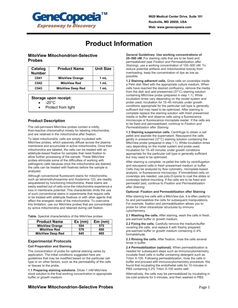
9620 Medical Center Drive, Suite 101 Rockville, MD 20850, USA Web: www.genecopoeia.com Product Information MitoView Mitochondrion-Selective Probes Catalog Number Product Name Unit Size C041 MitoView Orange 1 mL C042 MitoView Red 1 mL C043 MitoView Deep Red 1 mL Storage upon receipt: -20°C Protect from light Product Description The cell-permeant MitoView probes contain a mildly thiol-reactive chloromethyl moiety for labeling mitochondria, and are retained in the mitochondria after fixation. To label mitochondria, cells are simply incubated with MitoView probes, which passively diffuse across the plasma membrane and accumulate in active mitochondria. Once their mitochondria are labeled, the cells can be treated with an aldehyde-based fixative for samples that need fixation to allow further processing of the sample. These MitoView probes eliminate some of the difficulties of working with pathogenic cells because once the mitochondria are stained, the cells can be treated with fixatives before the sample is analyzed. Although conventional fluorescent stains for mitochondria, such as tetramethylrosamine and rhodamine 123, are readily sequestered by functioning mitochondria, these stains are easily washed out of cells once the mitochondria experience a loss in membrane potential. This characteristic limits the use of such conventional stains in experiments that require cells to be treated with aldehyde fixatives or with other agents that affect the energetic state of the mitochondria. To overcome this limitation, use our MitoView probes that are concentrated by active mitochondria and retained during cell fixation. Table. Spectral characteristics of the MitoView probes Product Name Ex (nm) Em (nm) MitoView Orange MitoView Red MitoView Deep Red 554 579 645 575 600 665 Experimental Protocols Cell Preparation and Staining The concentration of probe for optimal staining varies by application. The initial conditions suggested here are guidelines that may be modified based on the particular cell type or on other factors, such as the permeability of the cells or tissues to the probe. 1.1 Preparing staining solutions. Dilute 1 mM MitoView stock solution to the final working concentration in appropriate buffer or growth medium. MitoView Mitochondrion-Selective Probes General Guidelines: Use working concentrations of 25–500 nM. For staining cells that are to be fixed and permeabilized (see Fixation and Permeabilization after Staining), use a working concentration of 100–500 nM. To reduce potential artifacts and mitochondrial toxicity from overloading, keep the concentration of dye as low as possible. 1.2 Staining adherent cells. Grow cells on coverslips inside a Petri dish filled with the appropriate culture medium. When cells have reached the desired confluency, remove the media from the dish and add prewarmed (37°C) staining solution containing MitoView probe (prepared in step 1.1). While incubation times vary depending on the model system and probe used, incubation for 15–45 minutes under growth conditions appropriate for the particular cell type is generally sufficient but may need to be optimized. After staining is complete replace the staining solution with fresh prewarmed media or buffer and observe cells using a fluorescence microscope or fluorescence microplate reader. If the cells are to be fixed and permeabilized, continue to Fixation and Permeabilization after Staining. 1.3 Staining suspension cells. Centrifuge to obtain a cell pellet and aspirate the supernatant. Resuspend the cells gently in prewarmed (37°C) staining solution containing the MitoView probe (prepared in step 1.1). While incubation times vary depending on the model system and probe used, incubation for 15–45 minutes under growth conditions appropriate for the particular cell type is generally sufficient but may need to be optimized. After staining is complete, re-pellet the cells by centrifugation and resuspend cells in fresh prewarmed medium or buffer. Cells may be analyzed by flow cytometry, microplate-based analysis, or fluorescence microscopy. If immobilized cells on coverslips are needed, use poly-D-lysine to coat the slides or coverslips before mounting. If the cells are to be fixed and permeabil ized, continue to Fixation and Permeabilization after Staining. Optional: Fixation and Permeabilization after Staining After staining live cells with a MitoView dye, it is often useful to fix and permeabilize the cells for subsequent manipulations. For example, fixation and permeabilization allows you to probe for other intracellular structures by immuno cytochemistry. 2.1 Washing the cells. After staining, wash the cells in fresh, pre-warmed buffer or growth medium. 2.2 Fixing the cells. Carefully remove the medium/buffer covering the cells, and replace it with freshly prepared, pre-warmed buffer or growth medium containing 2–4% formaldehyde. 2.3 Rinsing the cells. After fixation, rinse the cells several times in buffer. 2.4 Permeabilization (optional). When permeabilization is needed for subsequent steps such as immunocytochemistry, incubate fixed cells in buffer containing detergent such as Triton X-100. Following permeabilization, rinse the cells in buffer and proceed with immunocytochemistry procedure. We found that incubating the endothelial cells for 10 minutes in PBS containing 0.2% Triton X-100 works well. Alternatively, the cells may be permeabilized by incubating in ice-cold acetone for 5 minutes, and then washed in PBS. Page 1
