association of lateral implantation of placenta with development of
advertisement
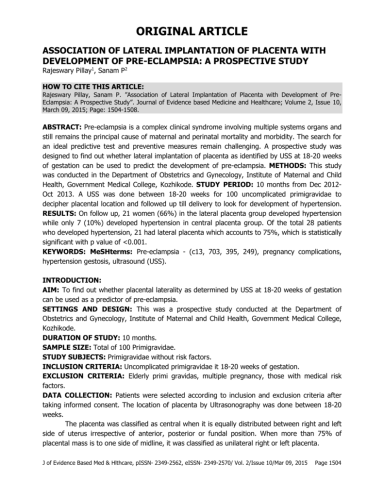
ORIGINAL ARTICLE ASSOCIATION OF LATERAL IMPLANTATION OF PLACENTA WITH DEVELOPMENT OF PRE-ECLAMPSIA: A PROSPECTIVE STUDY Rajeswary Pillay1, Sanam P2 HOW TO CITE THIS ARTICLE: Rajeswary Pillay, Sanam P. ”Association of Lateral Implantation of Placenta with Development of PreEclampsia: A Prospective Study”. Journal of Evidence based Medicine and Healthcare; Volume 2, Issue 10, March 09, 2015; Page: 1504-1508. ABSTRACT: Pre-eclampsia is a complex clinical syndrome involving multiple systems organs and still remains the principal cause of maternal and perinatal mortality and morbidity. The search for an ideal predictive test and preventive measures remain challenging. A prospective study was designed to find out whether lateral implantation of placenta as identified by USS at 18-20 weeks of gestation can be used to predict the development of pre-eclampsia. METHODS: This study was conducted in the Department of Obstetrics and Gynecology, Institute of Maternal and Child Health, Government Medical College, Kozhikode. STUDY PERIOD: 10 months from Dec 2012Oct 2013. A USS was done between 18-20 weeks for 100 uncomplicated primigravidae to decipher placental location and followed up till delivery to look for development of hypertension. RESULTS: On follow up, 21 women (66%) in the lateral placenta group developed hypertension while only 7 (10%) developed hypertension in central placenta group. Of the total 28 patients who developed hypertension, 21 had lateral placenta which accounts to 75%, which is statistically significant with p value of <0.001. KEYWORDS: MeSHterms: Pre-eclampsia - (c13, 703, 395, 249), pregnancy complications, hypertension gestosis, ultrasound (USS). INTRODUCTION: AIM: To find out whether placental laterality as determined by USS at 18-20 weeks of gestation can be used as a predictor of pre-eclampsia. SETTINGS AND DESIGN: This was a prospective study conducted at the Department of Obstetrics and Gynecology, Institute of Maternal and Child Health, Government Medical College, Kozhikode. DURATION OF STUDY: 10 months. SAMPLE SIZE: Total of 100 Primigravidae. STUDY SUBJECTS: Primigravidae without risk factors. INCLUSION CRITERIA: Uncomplicated primigravidae it 18-20 weeks of gestation. EXCLUSION CRITERIA: Elderly primi gravidas, multiple pregnancy, those with medical risk factors. DATA COLLECTION: Patients were selected according to inclusion and exclusion criteria after taking informed consent. The location of placenta by Ultrasonography was done between 18-20 weeks. The placenta was classified as central when it is equally distributed between right and left side of uterus irrespective of anterior, posterior or fundal position. When more than 75% of placental mass is to one side of midline, it was classified as unilateral right or left placenta. J of Evidence Based Med & Hlthcare, pISSN- 2349-2562, eISSN- 2349-2570/ Vol. 2/Issue 10/Mar 09, 2015 Page 1504 ORIGINAL ARTICLE OUTCOME: The end point of study was taken as the development of hypertension as per American College of Obstetrics and Gynecology criteria. The pregnancy outcome was assessed with respect to: • Incidence of pre-eclampsia with lateral location of placenta • Incidence of pre-eclampsia with central location of placenta ANALYSIS: All data collected were coded and entered in Microsoft Excel sheet, rechecked and analysed using SPSS statistical software. Statistical comparison was done using appropriate method and level of significance was estimated with p value <0.05. ETHICAL ASPECTS: Ethical clearance was obtained from Institutional Research committee and Institutional Ethics Committee of Government Medical College, Kozhikode. RESULTS: LATERAL PLACENTA CENTRAL PLACENTA TOTAL TABLE 1: LOCATION OF PLACENTA BY USS (18-20 32 68 100 WEEKS) PLACENTAL POSITION NOT DEVELOPED DEVELOPED TOTAL CENTRAL 61 (84.7%) 7 (25%) 68(68%) LATERAL 11(15.3%) 21(75%) 32(32%) TOTAL 72(100%) 28(100%) 100(100%) TABLE 2: DEVELOPMENT OF HYPERTENSION WITH REGARD TO PLACENTAL LOCATION Out of the 68 patients with central placenta only 7 developed hypertension which accounts for 25% of hypertension, out of 32 with lateral placenta, 21 had hypertension which accounts for 75%, which is statistically significant with p value of <0.001. PLACENTAL POSITION CENTRAL LATERAL TOTAL GESTATIONAL AGE NOT AT 28-32 AT 33-36 WEEKS DEVELOPED WEEKS 61(84.7%) 0(0%) 4(20%) 11(15.3%) 3(100%) 16(80%) 72(100%) 3(100%) 20(100%) TABLE 3: PLACENTAL POSITION AND GESTATIONAL AGE AT WHICH HYPERTENSION DEVELOPED >38 WEEKS 3(60%) 2(40%) 5(100%) In both groups majority of patients developed hypertension at gestational age from 33 to 36 weeks. J of Evidence Based Med & Hlthcare, pISSN- 2349-2562, eISSN- 2349-2570/ Vol. 2/Issue 10/Mar 09, 2015 Page 1505 ORIGINAL ARTICLE PLACENTAL POSITION CENTRAL LATERAL TOTAL IUGR ABSENT PRESENT 57(81.4%) 11(36.7%) 13(18.5%) 19(63.3%) 70(100%) 30(100%) TABLE 4: PLACENTAL POSITION AND IUGR TOTAL 68 32 100 IUGR (intrauterine growth retardation) has a significant association with placental position. In this study 56% of patients with lateral placenta developed IUGR whereas only 16% of those with central placenta had IUGR. In other words, of the 30 patients who had IUGR, 63% were from lateral placenta group and only 37% were from central placenta group, thus showing a statistically significant association with a p value <0.001. BIRTH WEIGHT: Analysis of my study proved a significant association with placental position and birth weight. In those with lateral location 59% had low birth weight (<2500g), whereas only 38% of centrally situated placentae had low birth weight. DISCUSSION: Pre-eclampsia and hypertensive disorders are responsible for up to 10% of maternal and fetal problems.(1) Unfortunately we still are in search of an ideal predictive test for early diagnosis and prevention of this syndrome. Angiotensin sensitivity test and roll-over test are outdated. Since 2004, a new era of serum markers has emerged to predict pre-eclampsia soluble VEGF receptor - 1 (soluble fms- like tyrosine kinase1, sFlt-1), soluble placental growth factor (PIGF) which holds promise for the future.(2, 3) The source of blood supply for the placenta are the two uterine arteries.(4) There is poor evidence of efficient functional collateral anastomosis between the two uterine vessels. So in a centrally located placenta, the blood flow will be abundant from both uterine arteries. But a laterally implanted placenta (which is diagnosed if 75% or more is to one side of the uterine cavity) gets its blood only from one uterine artery which is insufficient for adequate placental perfusion. This is postulated to cause predisposition of uteroplacental insufficiency and IUGR.(5,6,7,8) A deficient perfusion of the placenta will impede proper trophoblastic invasion of spiral arterioles which is the primary inciting factor in development of pre-eclampsia.(9) In the study by Pai et al,(10) out of 426 women, 324 had centrally located placenta and 102 had lateral placenta. A total of 71 women developed pre-eclampsia, of whom 52(74%) had unilaterally located placenta at 20 weeks. This relationship was highly statistically significant (p<0.0001). In this study, the sensitivity is 75%, specificity 85%, positive predictive value is 62% and negative predictive value is 90%. While comparing the performance of placental laterality as a screening test with those of other available ones, it is positively comparable. This test is non-invasive, can be combined with the anomaly scan routinely performed in all antenatal cases and needs to be integrated as an easy predictor of pre-eclampsis and IUGR especially in those cases where these adverse conditions are anticipated. J of Evidence Based Med & Hlthcare, pISSN- 2349-2562, eISSN- 2349-2570/ Vol. 2/Issue 10/Mar 09, 2015 Page 1506 ORIGINAL ARTICLE SUMMARY AND CONCLUSION: This study which followed up primigravidae with placental localisation for development of pre-eclampsia and IUGR, proved that it has a very good specificity of 85% and negative predictive value of 90%. It is cost effective and easy to perform. Placental laterality as detected by USS at 18-20 weeks has significant association with development of pre-eclampsia and can be used as predictor for this condition. BIBLIOGRAPHY: 1. Pregnancy hypertension: Cunningham FG et al. Williams Obstetrics.23rd edition Mc raw Hill, 2010; 706-749. 2. Levine RJ, Maynard SE, Qian C, Lim KH et al. Circulating antigenic factors and the risk for pre-eclampsia. N Engl J Med 2004; 350: 672-683. 3. Thadhani H, Mutter WP, Wolf M Levine RJ et al, First trimester placental growth factor and soluble fms-like tyrosine kinase 1 and the risk for pre-eclampsia. J Clin Endocrinol Metab 2004; 89; 770-775. 4. Fleisher A, Shulman H, Farmakides G et al. Uterine artery doppler velocimetry in pregnant women with hypertension. Am J obstet Gynecol 1986; 154: 806-13. 5. Kofinas AD, Penry M, Swain M et al. Effect of placental laterality on uterine artery resistance and development of pre-eclampsia and IUGR. Am J Obstet Gynecol 1989; 161: 1536-39. 6. Kofinas AD, Penry M, Greiss FC, Meis PJ et al. The effect of placental location on uterine artery flow velocity waveforms. Am J Obstet Gynecol 1988; 159: 1504-08. 7. North RJ, Ferrier C, Long D, Townend K, Kincaid-Smith P. Uterine artery doppler flow velocity waveforms in the second trimester for the prediction of pre-eclampsia and IUGR. Obstet Gynecol 1994; 83: 378-386. 8. Ito Y, Shono H, Muro M, Uchiyama A, Sugimori H. Resistance index of uterine artery and placental location in IUGR. Acta Obstet Gynecol Scand 1998; 77: 385-390. 9. Meekins J W, Prijnenborg R, Hansenns M, McFayden IR, Van Asshe A. A study of placental bed spiral arteries and trophoblastic invasion in normal and severe pre-eclamptic pregnancies. Br J Obstet Gynecol 1994; 101: 669-674. 10. Pai MV, Pillai J. Placental laterality by USS- simple yet reliable predictive test for preeclampsia. J Obstet Gynecol India 2005; 55(5): 431-433. J of Evidence Based Med & Hlthcare, pISSN- 2349-2562, eISSN- 2349-2570/ Vol. 2/Issue 10/Mar 09, 2015 Page 1507 ORIGINAL ARTICLE AUTHORS: 1. Rajeswary Pillay 2. Sanam P. PARTICULARS OF CONTRIBUTORS: 1. Assistant Professor, Department of Obstetrics and Gynaecology, Institute of Maternal and Child Health, Govt. Medical College, Kozhikode. 2. Junior Resident, Department of Obstetrics and Gynaecology, Institute of Medical and Child Health, Govt. Medical College, Kozhikode. NAME ADDRESS EMAIL ID OF THE CORRESPONDING AUTHOR: Dr. Rajeswary Pillay, Assistant Professor, IMCH, Medical College, Kozhikode. E-mail: drsprathap63@yahoo.com Date Date Date Date of of of of Submission: 25/02/2015. Peer Review: 26/02/2015. Acceptance: 04/03/2015. Publishing: 06/03/2015. J of Evidence Based Med & Hlthcare, pISSN- 2349-2562, eISSN- 2349-2570/ Vol. 2/Issue 10/Mar 09, 2015 Page 1508
