I J Pre Clin Dent Res 2015;2(5):1-4
January-March
All rights reserved
International Journal of Preventive &
Clinical Dental Research
Comparative Study Socket Preservation using
PRF and MPM Platelets Concentrates
Abstract
The topical use of platelet concentrates is recent and its efficiency
remains controversial. Several techniques for platelet concentrates are
available; however, their applications have been confusing because each
method leads to a different product with different biology and potential
uses. Pure platelet-rich plasma PRP, Anitua’s and Choukroun’s PRF.
Those are the common types of platelets concentrates. Latest evolution
of platelets concentrates PRP and PRF is the mineralized plasmatic
matrix which is prepared with different ways and has other names like
sticky bone.
Key Words
Extraction; PRF; MPM; socket preservation
INTRODUCTION
Platelet-rich fibrin (PRF) belongs to a new
generation of platelet concentrates, with simplified
processing and without biochemical blood handling,
it is a strictly autologous fibrin matrix containing a
large quantity of platelet and leukocyte cytokines.[1]
The use of platelet gel to improve soft and hard
tissue regeneration is a recent technique in
implantology.[2] The mineralized plasmatic matrix
MPM is a modification of the PRP and the PRF
presented by Prof. Perisse and modified by Dr. El
Moheb. The advantage of the MPM is the
integration of bone grafts particles inside the fibrin
network that is not present in old autologous growth
factors membranes. In fact the bone grafting
materials are prepared and mixed with the
autologous growth factors to produce the MPM.
This offers the MPM the positional stability. This
article focuses on comparing between the healing
patterns of sockets after filling one of them with
PRF and the other with MPM materials. Histologic
evidence is reviewed to provide an in-depth
understanding of the effect of 2 materials on
accelerating the bone healing.
MATERIALS & METHODS
A healthy 46 years man presented with chief
complain badly distracted lower right lateral incisor
and canine and only remaining roots for both teeth
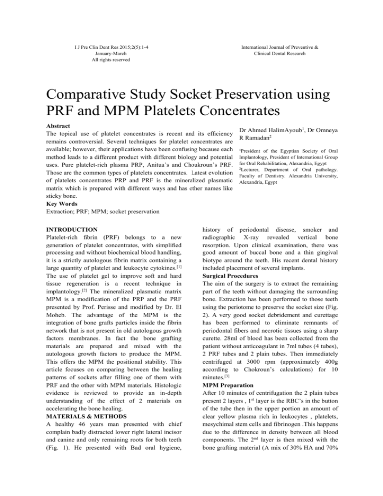
(Fig. 1). He presented with Bad oral hygiene,
Dr Ahmed HalimAyoub1, Dr Omneya
R Ramadan2
1
President of the Egyptian Society of Oral
Implantology, President of International Group
for Oral Rehabilitation, Alexandria, Egypt
2
Lecturer, Department of Oral pathology.
Faculty of Dentistry. Alexandria University,
Alexandria, Egypt
history of periodontal disease, smoker and
radiographic X-ray revealed vertical bone
resorption. Upon clinical examination, there was
good amount of buccal bone and a thin gingival
biotype around the teeth. His recent dental history
included placement of several implants.
Surgical Procedures
The aim of the surgery is to extract the remaining
part of the teeth without damaging the surrounding
bone. Extraction has been performed to those teeth
using the periotome to preserve the socket size (Fig.
2). A very good socket debridement and curettage
has been performed to eliminate remnants of
periodontal fibers and necrotic tissues using a sharp
curette. 28ml of blood has been collected from the
patient without anticoagulant in 7ml tubes (4 tubes),
2 PRF tubes and 2 plain tubes. Then immediately
centrifuged at 3000 rpm (approximately 400g
according to Chokroun’s calculations) for 10
minutes.[3]
MPM Preparation
After 10 minutes of centrifugation the 2 plain tubes
present 2 layers , 1st layer is the RBC’s in the button
of the tube then in the upper portion an amount of
clear yellow plasma rich in leukocytes , platelets,
mesychimal stem cells and fibrinogen .This happens
due to the difference in density between all blood
components. The 2nd layer is then mixed with the
bone grafting material (A mix of 30% HA and 70%
2
PRF and MPM platelets concentrate
Ayoub AH, Ramadan OR
Fig. 1
Fig. 2
Fig. 3
Fig. 4
Fig. 5
Fig. 6
Fig. 7
Fig. 8
Tri calcium phosphate small granules manufactured
by POLYSTOM) and a drop of patient blood from
the extraction socket to provide the thrombin which
will initiate the conversion of insoluble fibrinogen
into soluble fibrin and all mixed together in a sterile
bowel. After a couple of minutes a homogenous
mixture of fibrin network with integrated bone graft
particles inside and the mixture is rich of platelets,
leukocytes and mesynchimal cells (Fig. 3). The
MPM, which has been obtained, is placed in the
extraction socket of the lateral incisor.
PRF Preparation
Within a few minutes, the absence of anticoagulant
allows activation of the majority of platelets
contained in the sample to trigger a coagulation
cascade. In the PRF tubes fibrinogen is at first
concentrated in the upper part of the tube, until the
effect of the circulating thrombin transforms it intoa
fibrin network.[3] Fibrinogen is initially concentrated
in the high part of the tube, before the circulating
thrombin transforms it into fibrin. A fibrin clot is
then obtained in the middle of the tube, just between
the red corpuscles at the bottom and acellular
3
PRF and MPM platelets concentrates
Ayoub AH, Ramadan OR
Fig. 9
Fig. 10
Fig. 11
Fig. 12
Fig. 13
Fig. 14
Fig. 15
plasma at the top (Fig. 8). Platelets are theoretically
trapped massively in the fibrin meshes. The clot is
removed from the tube and the attached red blood
cells scraped off and discarded. The PRF clot is
then placed on the grid in the PRF Box and covered
with the compressor and lid. This produces an
inexpensive autologous fibrin membrane in
approximately one minute. The PRF Box was
devised to produce membranes of constant
thickness that remain Hydrated for several hours
and to recover the serum exudate expressed from
the fibrin clots which is rich in the proteins
vitronectin and fibronectin. The exudate collected at
the bottom of the box may be used to store
autologous grafts.[2] After removing the cover of
PRF box 2 membranes obtained from 2 PRF clots
(Fig. 4). With a specific tweezers the PRF was
inserted in the canine socket. Edges of the mucosa
were approximated to each other and sutured using
3-0 Monocrylsutures. Healing was uneventful, and
the patient was followed up for 4 months
postoperatively, making sure to enforce oral
hygiene and rinsing with chlorhexidine 0.12%. 4
months later bone was harvested from the 2 sockets
using a 3.2mm trephine drill and 2 implants
microdent GN3512 was inserted. The specimen was
sent to histological evaluation.
RESULTS
1st socket MPM histological study showed
decalcified section shows a mass of cancellous bone
with areas of diverse degrees of maturation.
Numerous large lacunae containing osteocytes are a
prominent feature. The bony trabeculae are lined by
numerous osteoblasts with the bony interface
4
PRF and MPM platelets concentrates
Ayoub AH, Ramadan OR
Slide: 1
Slide: 2
Slide: 3
Slide: 4
showing areas of active deposition of woven bone.
Resting lines are seen in some areas. The bone
marrow contains multiple capillaries and lymphatic
vessels with mild inflammatory infiltrate (Fig. 5).
2nd socket PRF histological study showed PRF:
Decalcified section shows a mass of mature
cancellous bone with prominent resting lines. The
bony trabeculae demonstrate multiple osteocytes in
large lacunae. Few viable osteoblasts are seen lining
the bony trabeculae. The bone marrow is clearly
fibrotic with little vascularity and almost no chronic
inflammatory cells infiltrating (Fig. 6).
DISCUSSION
In MPM specimen the ostoeblastic activity is more
prominent and active deposition of woven bone
while in PRF less osteoblastic activity and the bone
marrow is fibrotic with little vascularity. The
plasma obtained after a single spin, is rich in
platelets, fibrinogen and monocytes. The fibrinogen
is necessary for the formation of the MPM. The
Fibrinogen will be transformed into fibrin network
under than action thrombin coming from patient’s
wound,[4] the platelets that will offer the growth
factors and the monocytes once activated by the
interleukine can enhance the production of BMP-2.
The BMP-2 is a bone morphogentique protein that
induces the bone formation. It is a high inductive
protein.[5] The fibrin network that is produced in the
MPM, will link all bone particles together, and also
the platelets and monocytes. This fibrin network is
the scaffold needed by the bone to regenerate, and
also it is the pathway need by the cells to migrate to
heal or to repair the wound. This fibrin network or
the extracellular matrix allows the elasticity of the
MPM, permit the shaping and the remodeling of the
MPM and facilitate the cells migrating inside the
MPM between the particles.
CONCLUSION
From this study, the use of bone filler combined
with fibrin, platelets and leukocytes in the form of
MPM has shown a better histologic evidence of
bone formation after 4 months than the use of PRF
as sole filling material for the extraction socket.
REFERENCES
1. David M, Ehrenfest D, Rasmusson L,
Albrektsson T. Classification of platelet
concentrates: from pure platelet-rich plasma
(P-PRP) to leucocyte- and platelet-rich fibrin
(L-PRF). Trends in Biotechnology. 27(3).
2. Choukroun J, Adda F, Schoeffler C, Vervelle
A. Une opportunite en paro-implantologie: le
PRF. Implantodontie. 2000;42:55-62.
3. Oral Surg Oral Med Oral Pathol Oral Radiol
Endod. 2006;101(3):45-50.
4. Mazzoni L, Périssé J. Apports de la
microscopie électro-nique à balayage pour la
Matrice Plasmatique Minéralisée. La Lettre de
la Stomatologie. 2011;51.
5. Biomaterials. 2011;32:8190-204
doi:
10.1016/j.biomaterials.2011.07.055.
Epub
2011 Aug 10.


