Corneal Topography Testing – Patient informaton 2013
advertisement
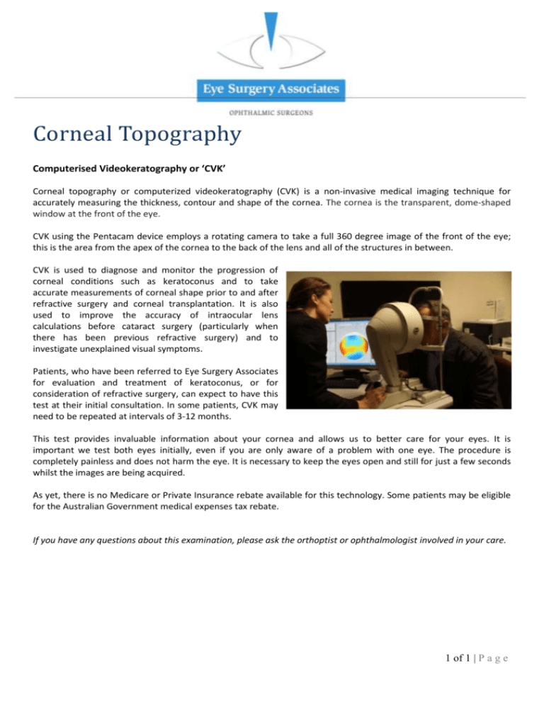
Corneal Topography Computerised Videokeratography or ‘CVK’ Corneal topography or computerized videokeratography (CVK) is a non-invasive medical imaging technique for accurately measuring the thickness, contour and shape of the cornea. The cornea is the transparent, dome-shaped window at the front of the eye. CVK using the Pentacam device employs a rotating camera to take a full 360 degree image of the front of the eye; this is the area from the apex of the cornea to the back of the lens and all of the structures in between. CVK is used to diagnose and monitor the progression of corneal conditions such as keratoconus and to take accurate measurements of corneal shape prior to and after refractive surgery and corneal transplantation. It is also used to improve the accuracy of intraocular lens calculations before cataract surgery (particularly when there has been previous refractive surgery) and to investigate unexplained visual symptoms. Patients, who have been referred to Eye Surgery Associates for evaluation and treatment of keratoconus, or for consideration of refractive surgery, can expect to have this test at their initial consultation. In some patients, CVK may need to be repeated at intervals of 3-12 months. This test provides invaluable information about your cornea and allows us to better care for your eyes. It is important we test both eyes initially, even if you are only aware of a problem with one eye. The procedure is completely painless and does not harm the eye. It is necessary to keep the eyes open and still for just a few seconds whilst the images are being acquired. As yet, there is no Medicare or Private Insurance rebate available for this technology. Some patients may be eligible for the Australian Government medical expenses tax rebate. If you have any questions about this examination, please ask the orthoptist or ophthalmologist involved in your care. 1 of 1 | P a g e

