Statechart Modeling and Experimental Validation
advertisement
Tactile modulation of whisking via the brainstem loop: Statechart modeling and
experimental validation
Supporting information
Dana Sherman1,2, Tess Oram1, Dudi Deutsch1, Goren Gordon1, Ehud Ahissar*1 and David Harel*2
1
Department of Neurobiology, Weizmann Institute of Science, Rehovot 76100, Israel
Department of Computer Science and Applied Mathematics, Weizmann Institute of Science, Rehovot 76100, Israel
*, equal contribution
2
Model specifications
Number of elements in the model
The brainstem loop model contained five rows of whiskers, with two rows of four whiskers and three rows of seven
whiskers, corresponding to rows A-B and C-E in the rat. Each whisker was innervated by a separate pool of neurons that
closed the loop between that whisker and the muscles attached to it, as shown in Figure S1.
Model parameters
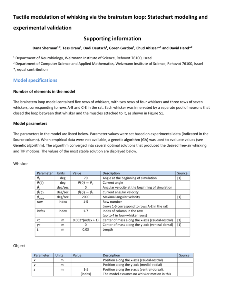
The parameters in the model are listed below. Parameter values were set based on experimental data (indicated in the
Source column). When empirical data were not available, a genetic algorithm (GA) was used to evaluate values (see
Genetic algorithm). The algorithm converged into several optimal solutions that produced the desired free-air whisking
and TIP motions. The values of the most stable solution are displayed below.
Whisker
Parameter
𝜃0
𝜃(𝑡)
𝜃̇0
𝜃̇(𝑡)
𝜃̇𝑚𝑎𝑥
row
index
xc
yc
L
Units
deg
deg
deg/sec
deg/sec
deg/sec
index
Value
70
𝜃(0) = 𝜃0
0
̇
𝜃(0) = 𝜃̇0
2000
1-5
index
1-7
m
m
m
0.002*(index + 1)
0
0.03
Description
Angle at the beginning of simulation
Current angle
Angular velocity at the beginning of simulation
Current angular velocity
Maximal angular velocity
Row number
(rows 1-5 correspond to rows A-E in the rat)
Index of column in the row
(up to 4 in four-whisker rows)
Center of mass along the x-axis (caudal-rostral)
Center of mass along the y-axis (ventral-dorsal)
Length
Source
[1]
[1]
[1]
[1]
Object
Parameter
x
y
z
Units
m
m
m
Value
1-5
(index)
Description
Position along the x-axis (caudal-rostral)
Position along the y-axis (medial-radial)
Position along the z-axis (ventral-dorsal).
The model assumes no whisker motion in this
Source
present
logical
True/False
axis, thus z value simply indicates the row
number in which the object is present
True when object is present
Whisking cells (SN1_W)
Parameter
𝑃𝑊 (𝜃, 𝜃̇ )
Units
-
value
0-1
Description
Firing probability: the current probability of the cell to fire.
𝜃
−|𝜃−𝜃𝑓 |
𝜃̇
𝑃𝑊 (𝜃, 𝜃̇ ) = 𝐾1 ∙ 𝑟𝑎𝑛𝑔𝑒
+ 𝐾2 ∙
𝜃𝑟𝑎𝑛𝑔𝑒
threshold
-
0
k1
k2
index
𝜃𝑓
index
deg
0.84
0.16
0-72
50 + index
𝜃𝑟𝑎𝑛𝑔𝑒
deg
5.6
𝑡𝑟𝑒𝑙𝑎𝑦
msec
2
ARP
RRP
msec
msec
1
3
Source
𝜃̇𝑚𝑎𝑥
Set PW (θ, θ̇ ) = 0: (1) when 𝑃𝑊 (𝜃, 𝜃̇ ) < 0, (2) upon cell’s firing
The threshold to be crossed in order to successfully fire (𝑃𝑊
must be > 0)
A relative-weight constant
A relative-weight constant; k2=1-k1
Each of the whisker's 73 SN1_Ws takes a different index
Favorite angle: its whisker's angle to which the cell is most
sensitive, i.e., fires with highest probability
The range of angles of its whisker to which the cell responds.
𝜃
𝜃
A cell responds to the range: [𝜃𝑓 − 𝑟𝑎𝑛𝑔𝑒
, 𝜃𝑓 + 𝑟𝑎𝑛𝑔𝑒
]
2
2
Conductance time of the stimulus along the cell's axon to its
post-synaptic target
Absolute refractory period duration
Relative refractory period duration
GA
GA
GA
[2]
[3]
Na+ channels
recovery time
Contact (SN1_C)
Parameter
𝑡𝑟𝑒𝑠𝑝𝑜𝑛𝑠𝑒
Units
msec
𝑡𝑟𝑒𝑙𝑎𝑦
msec
value
2-5 (2)
[range (median)]
1
ARP
RRP
msec
msec
1
3
index
index
0-32
Description
Time to generate action potential in
response to contact stimulus
Conductance time of the stimulus along
the cell's axon to its post-synaptic target
Absolute refractory period duration
Relative refractory period duration
Source
[4]
[2]
[3]
Na+ channels
recovery time
Each of the whisker's 33 SN1_Cs takes a
different index
Pressure (SN1_P)
Parameter
𝑡𝑟𝑒𝑠𝑝𝑜𝑛𝑠𝑒 (𝑥, 𝑦, 𝐿)
Units
msec
value
1-34
Description
Time to generate action potential in response to contact stimulus.
Changes for every cell according to the radial distance of contact
between its whisker and an object.
𝑡𝑟𝑒𝑠𝑝𝑜𝑛𝑠𝑒 = 𝑡𝑑𝑒𝑙𝑎𝑦 ∗ 𝐹(𝑥, 𝑦)
𝑡𝑑𝑒𝑙𝑎𝑦 – "basic" delay of a cell; 8-34 (16) [range (median)].
F(x,y) – change in 𝑡𝑑𝑒𝑙𝑎𝑦 as a function of object's radial distance,
measured from whisker's base (
𝑡𝑟𝑒𝑙𝑎𝑦
msec
1
ARP
RRP
msec
msec
1
3
index
index
0-27
√(𝑥−𝑥𝑐)2 +(𝑦−𝑦𝑐)2
𝐿
).
Conductance time of the stimulus along the cell's axon to its postsynaptic target
Absolute refractory period duration
Relative refractory period duration
Each of the whisker's 28 SN1_Ps takes a different index
Source
[4,5]
[2]
[3]
Na+ channels
recovery time
Detach (SN1_D)
Parameter
𝑡𝑟𝑒𝑠𝑝𝑜𝑛𝑠𝑒
Units
msec
Value
1
𝑡𝑟𝑒𝑙𝑎𝑦
msec
1
ARP
RRP
msec
msec
1
3
Description
Time to generate action potential in response to
detachment of its whisker from an object
Conductance time of the stimulus along the cell's axon
to its post-synaptic target
Absolute refractory period duration
Relative refractory period duration
index
index
0-27
Each of the whisker's 28 SN1_Ds takes a different index
Source
[4]
[2]
[3]
Na+ channels
recovery time
SN2 (W,C,P,D)
Parameter
type
Units
-
Value
W,C,P,D
Description
Cell type: whisking (W), contact (C), pressure (P), or
detach (D)
index
index
0-T
𝑡𝑟𝑒𝑠𝑝𝑜𝑛𝑠𝑒
msec
1
𝑡𝑟𝑒𝑙𝑎𝑦
msec
1-4
ARP
RRP
msec
msec
2
3
Each of the whisker's SN2s of each type takes a
different index: T = 69,29,24,24 for SN2 of type
W,C,P,D
Time to generate action potential in response to
stimuli from its pre-synaptic SN1s.
Not a parameter; results from simulation dynamics*
Conductance time of the stimulus along the cell's
axon to its post-synaptic target
Absolute refractory period duration
Relative refractory period duration
Source
These SN2s also project to the
thalamus, where the different
types of information are relayed
separately [6].
GA
[7]
Na+ channels recovery time
* The delay in firing of an SN2 following pre-synaptic stimuli (𝑡𝑟𝑒𝑠𝑝𝑜𝑛𝑠𝑒 ) depended on the time it took the cell to
cross a threshold. The time for crossing the threshold depended on the threshold (increased in the “RRP” state
relative to “Rest”; value evaluated by GA), strength of the pre-synaptic stimuli and time interval between stimuli:
𝑇
𝐴𝑆𝑁2 (𝑡) = 𝐴𝑆𝑁2 (𝑡 − 1) +
∑ 𝐸𝑆𝑁1_𝑋𝑖𝑛𝑑𝑒𝑥 − 𝐶
𝑖𝑛𝑑𝑒𝑥=1
where 𝑡 is the elapsed time from the moment the SN2 entered the “GenerateAP” state,
𝐴𝑆𝑁2 (𝑡 − 1) is the SN2 excitability value (arbitrary units) at time t-1,
𝑋 is the type of information the SN2 relayed, i.e., whisking (W), contact (C), pressure (P) or detach (D),
𝐸𝑆𝑁1_𝑋𝑖𝑛𝑑𝑒𝑥 is the effect of a pre-synaptic cell "index" of the corresponding type (𝑋), on its SN2 excitability,
𝑇 is the number of primary afferents of type 𝑋 activated at time 𝑡,
𝐶 is a constant.
Once an SN2 crossed threshold, its excitability values were zeroed (𝑡 = 0; 𝐴𝑆𝑁2 (0) = 0).
Thus the effect of an SN1 firing on its SN2 excitability depended on the synaptic strength (𝐸𝑆𝑁1_𝑋𝑖𝑛𝑑𝑒𝑥 ) and
decreased with time, as moving away from the firing moment of the SN1 (embodied in 𝐶 ∙ (𝑡 − 1)).
𝐴𝑆𝑁2 (𝑡) value was not changed if the SN2 was at the "absolute refractory period" state.
MN (ExtP, Int, ExtR)
Parameter
type
𝑡𝑟𝑒𝑠𝑝𝑜𝑛𝑠𝑒
Units
msec
Value
ExtP, Int, ExtR
1-4
𝑡𝑟𝑒𝑙𝑎𝑦
msec
1
ARP
RRP
msec
msec
2
3
Description
MN type: extrinsic protractor, intrinsic, or extrinsic retractor
Time to generate action potential in response to stimuli from
its pre-synaptic sources (SN2s, CPG).
Not a parameter; results from simulation dynamics*
Conductance time of the stimulus along the cell's axon to its
post-synaptic target
Absolute refractory period duration
Relative refractory period duration
Source
[8,9]
GA
[10,11]
[10]
Na+ channels
recovery time
* The delay in firing of an MN following pre-synaptic stimuli (𝑡𝑟𝑒𝑠𝑝𝑜𝑛𝑠𝑒 ) depended on the time it took the cell to
cross a threshold. The time for crossing the threshold depended on the threshold (increased in the “RRP” state
relative to “Rest”; value evaluated by GA), strength of the pre-synaptic stimuli and time interval between stimuli:
𝑇
𝐴𝑀𝑁 (𝑡) = 𝐴𝑀𝑁 (𝑡 − 1) + 𝐸𝐶𝑃𝐺 +
∑ 𝐸𝑆𝑁2_𝑋𝑖𝑛𝑑𝑒𝑥 − 𝐶
𝑖𝑛𝑑𝑒𝑥=1
where 𝑡 is the elapsed time from the moment the MN entered the “GenerateAP” state,
𝐴𝑀𝑁 (𝑡 − 1) is the MN excitability value (arbitrary units) at time t-1,
𝐸𝐶𝑃𝐺 is the effect of the pre-synaptic CPG on its MN excitability,
𝑋 is the type of information the SN2 relayed, i.e., whisking (W), contact (C), pressure (P) or detach (D),
𝐸𝑆𝑁2_𝑋𝑖𝑛𝑑𝑒𝑥 is the effect of a pre-synaptic SN2_X number "index" on its MN excitability,
𝑇 is the number of secondary afferents of type 𝑋 activated at time 𝑡,
𝐶 is a constant.
Once an MN crossed threshold, its excitability values were zeroed (t = 0; AMN (0) = 0).
Thus the effect of a pre-synaptic source (CPG or SN2) firing on its MN excitability depended on the synaptic
strength (𝐸𝐶𝑃𝐺 or 𝐸𝑆𝑁2_𝑋𝑖𝑛𝑑𝑒𝑥 ) and decreased with time, as moving away from the firing moment of the presynaptic source (embodied in 𝐶 ∙ (𝑡 − 1)).
𝐴𝑀𝑁 (𝑡) value was not changed if the MN was at the "absolute refractory period" state.
Muscle (ExtP, Int, ExtR)
Parameter
type
Units
-
𝐶𝑎0
𝐶𝑎(𝑡)
M
M
𝑟0
𝑟(𝑡)
-
t
msec
𝜏𝑐
𝜏𝑟
A
stimuliNum
threshold
msec
msec
index
Value
ExtP, Int, ExtR
10-7
𝑟0 𝜏𝑐
[𝑒
𝜏𝑐 −𝜏𝑟
−𝑡
𝜏𝑐
−𝑡
𝜏𝑟
− 𝑒 ] + 𝐶𝑎(𝑡 − 1)𝑒
−𝑡
𝜏𝑐
2.55
𝑒
−𝑡
𝜏𝑟
7.4
5
*
0-(size of innervating MN pool)
1
of the innervating MN pool
3
Description
Muscle type: extrinsic protractor, intrinsic, or extrinsic
retractor
Concentration of Ca2+ ions in muscle cytoplasm at rest
Concentration of Ca2+ ions released from the SR to the
cytoplasm; 𝐶𝑎(0) = 𝐶𝑎0
A constant
The fraction of open ryanodine receptors (RyRs);
𝑟(𝑡) = 𝑟0
Elapsed time following MN stimulation. Initialized to
zero at stimulation onset time
Time constant of the decay of intracellular Ca+2 ions
Decay time constant of r
Force scaling factor
Number of MN stimuli in the last 4 msec
Minimal number of stimuliNum, during a timewindow of 4 msec, required for muscle contraction
Source
[12]
[1]
[1]
[1]
[1]
[1]
[1]
GA
* Muscles force scaling factor:
Muscle type
Intrinsic
Pseudo
intrinsic
Extrinsic
protractor
Extrinsic
retractor
Value
0.30
0.83
Description
0.05, CPG − induced activation
𝐴={
0.3, sensory feedback − induced activation
0.05, CPG − induced activation
𝐴={
𝑓(𝑥), sensory feedback − induced activation
i.e., muscle's ExtP_MNs triggerend by ExtP_CGP.
i.e., muscle's ExtP_MNs triggerend by SN2_D.
i.e., ExtR_MNs triggerend by ExtR_CGP.
i.e., ExtR_MNs triggerend by SN2_C,P.
When activated by SN2_C,P, A=f(x), is a function
of the activated number of SN2_C,P (x) in
response to whisker-object contact.
f(x) ~ x (size principle of motor units recruitment);
0.3 ≤ f(x) ≤ 0.5
Once a muscle successfully crosses its threshold, its parameters’ updated values are passed to a Matlab function,
which calculates muscle’s force and transforms it into whisker motion. The calculation beyond the scope of this
study and is describes in detail in [1]. Briefly, total muscle force, F, is composed of two components: 𝐹 = 𝐹𝑐 + 𝐹𝑙 ,
4
𝐶𝑎(𝑡)
where the 𝐹𝑐 component depends on 𝐶𝑎(𝑡) and A as follows: 𝐹𝑐 = 𝐴1+𝐶𝑎(𝑡)
4 , and the 𝐹𝑙 component depends on
muscle's length (See formulas (6)-(7) in [1]).
CPG (ExtP, Int, ExtR)
Parameter
type
cycleDuration
Units
msec
Value
ExtP, Int, ExtR
4
currentCycle
index
0-cyclesNum
cyclesNum
index
10,17,18
silenceDuration
msec
110,82,78
Description
CPG type: extrinsic protractor, intrinsic, or extrinsic retractor
Time interval between successive stimuli of the corresponding
type of MNs.
Corresponds to CPG’s firing rate.
Current cycle’s number.
Only at initialization, currentCycle gets a non-positive value*:
0,-2,-21 for ExtP, Int, ExtR CPGs
Number of successive stimuli by ExtP, Int, and ExtR CPGs.
Corresponds to “Activate” state’s duration**
for the ExtP, Int, and ExtR CPG, respectively.
silenceDuration = 150 - cycleDuration * cyclesNum
(150 msec is whisk cycle duration, i.e., protraction + retraction)
Source
[13]
GA
[13]
[13]
* At the beginning of the simulation currentCycle gets a non-positive value, resulting in a delayed activation of the
CPG (delayed by |currentCycle|*cycleDuration, i.e., by 0,8,84 msec for the ExtP, Int, ExtR CPGs [13]).
** Model execution results in the following periods of activity of the different CPGs throughout a whisking cycle:
CPG type
ExtP
Int
ExtR
Active period [msec]
0-40
8-80
84-150
Muscle forces & whisker motion
A major part of our model was based on another study done in Ahissar’s group, which constructed a transformation
function that converts an MN’s spikes to whisker movement. The transformation is done in two parts: motor neurons
spiking are first converted to intrinsic muscle force production, and muscle force is then translated into motion of the
whiskers. Calculation in each part is composed of several steps, specified in great detail in [1].
The transformation function was developed and written in a Matlab environment, and underwent several changes in
order to fit our model.
The Matlab function is called many times during model execution (approximately every 1 msec), to recalculate the
anticipated whiskers’ movement each time a motor neuron fires a spike. Function accuracy depends on whisking
amplitude, since the viscoelastic tissue is modeled as a system of linear springs and dampers, an approximation
appropriate for small protraction angles [See Discussion in 1]. Thus, amplitudes <15° give very high accuracy while
amplitudes >15° allow a less accurate calculation of whisker movement.
Original code is available at: http://senselab.med.yale.edu/ModelDB/ShowModel.asp?model=127512
Since the underlying code in our model is written in java, a java-Matlab linking tool was implemented, allowing to
directly use the complex transformation function code that already exists in Matlab without converting it to Java code,
an action that has a high probability of introducing bugs.
This tool, consisting of two parts – a server part and a client part, makes it possible to call Matlab functions from Java
programs. The server can be run inside the Matlab installation on a network host. The client calls the server by using
remote method invocation (RMI) and allows for calling Matlab function on the fly, without saving results to a temporary
file.
Code for this tool is available at: http://jamal.sourceforge.net/about.shtml
Genetic algorithm
In order to find the most fitted values for parameters whose empirical data were not available, a genetic algorithm was
used (a parametric analysis technique often used in optimization problems).
In genetic algorithms, a set of parameters, which encode candidate solutions (called individuals) to an optimization
problem, evolves toward better solutions. The evolution starts from a population of randomly generated individuals and
happens in generations. In each generation, the fitness of every individual in the population is evaluated, multiple
individuals are stochastically selected from the current population (based on their fitness), and modified to form a new
population. The new population is then used in the next iteration of the algorithm. Commonly, the algorithm terminates
when either a maximum number of generations has been produced, or a satisfactory fitness level has been reached for
the population.
The parameters indicated by “GA” in the above tables were used. The algorithm was executed eight times, starting each
time from 50 randomly generated individuals and converging to a satisfactory solution that accurately simulated whisker
motion. Out of the eight solutions, four were found to be stable, where the stability of a solution is defined as follows:
changing the value of each one of the evaluated parameters (while keeping the other parameters constant), still results
in the desired motion. The larger the change that would still result in the optimal solution is, the more stable/robust the
solution is. The most stable solution of the four stable solutions is presented here (for each parameter, a change of up to
±(5-10)% of its possible range still resulted in the desired motion).
Model behavior in statecharts
Neurons
Since the behavior of all types of neurons in the model is very similar, the behavior of a generic neuron is described. The
statechart that defines the behavior of a generic neuron is displayed in paper Figure 3B and explained in the text. In the
model, each type of neuron has its own statechart, which is very similar to the diagram in Figure 3B, but with two major
differences between the different types of neurons:
(1) Type of stimulus that triggers a neuron to fire:
Cell type
SN1_W
SN1_C
SN1_P
SN1_D
SN2s, MNs
Firing depends on…
Whisker’s angle and angular velocity
Contact time - upon touch
(1) Contact time - upon and during touch period
(2) Radial distance of contact (stimulus strength)
Detachment time - upon detachment
Strength of stimulus - must reach threshold
The firing probability of the above cells is specified in detail in Model parameters.
(2) Post-synaptic target:
Pre-synaptic cell
Primary afferents*
Secondary afferents
CPG
Motor neurons
Post-synaptic target
Secondary afferents*
Motor neurons
Muscles
* Each type of primary afferents (i.e., W, C, P or D) innervates SN2s of the corresponding type – that relay the same
type of information
CPG, Muscle and Whisker
The statecharts of these elements are displayed in paper Figures 3A,C-D and are explained in the text.
Obstacle
The obstacle statechart has two states (Figure S2):
(1) “No obstacle” – in which no obstacle is present.
(2) “Obstacle” - in which an obstacle is present.
At the beginning of the simulation, no obstacle is present (present = “false”) and the obstacle is in the “No Obstacle”
state. When a “Put obstacle” event is sent, the obstacle moves to the “Obstacle” state and an obstacle is added to the
simulation at (x,y,z). The obstacle stays in this state until a “Remove obstacle” event is sent, setting present to “true”.
Manager
The Manager’s behavior is not described here, as it basically helps overcome technical issues, such as synchronization
between instances. This component does not have any equivalent biological entity.
References
1. Simony E, Bagdasarian K, Herfst L, Brecht M, Ahissar E, et al. (2010) Temporal and spatial characteristics of vibrissa
responses to motor commands. J Neurosci 30: 8935-8952.
2. Descheˆnes M, Timofeeva E, Lavalle´e P (2003) The Relay of High-Frequency Sensory Signals in the Whisker-toBarreloid Pathway. The Journal of Neuroscience 23: 6778–6787.
3. Leiser SC, Moxon KA (2007) Responses of trigeminal ganglion neurons during natural whisking behaviors in the awake
rat. Neuron 53: 117-133.
4. Szwed M, Bagdasarian K, Ahissar E (2003) Encoding of Vibrissal Active Touch. Neuron 40: 621–630.
5. Szwed M, Bagdasarian K, Blumenfeld B, Barak O, Derdikman D, et al. (2006) Responses of trigeminal ganglion neurons
to the radial distance of contact during active vibrissal touch. J Neurophysiol 95: 791-802.
6. Yu C, Derdikman D, Haidarliu S, Ahissar E (2006) Parallel Thalamic Pathways for Whisking and Touch Signals in the Rat.
PLoS Biology 4: 819-824.
7. Jacquin MF, Mooney RD, Rhoades RW (1986) Morphology, response properties, and collateral projections of
trigeminothalamic neurons in brainstem subnucleus interpolaris of rat. Exp Brain Res 61: 457-468.
8. Klein BG, Rhoades RW (1985) Representation of Whisker Follicle Intrinsic Musculature in the Facial Motor Nucleus of
the Rat. The Journal of Comparative Neurology 232: 55-69.
9. Herfst LJ, Brecht M (2008) Whisker movements evoked by stimulation of single motor neurons in the facial nucleus of
the rat. J Neurophysiol 99: 2821-2832.
10. Martin MR, Biscoe TJ (1977) Physiological studies on facial reflexes in the rat. Quarterly Journal of Experimental
Physiology 62: 209-221.
11. Yetiser S, Kahraman E, Satar B, Karahatay S, Akcam T Anatomy of the Extratemporal Facial Nerve in Rats. The
Mediterranean Journal of Otology.
12. Haidarliu S, Simony E, Golomb D, Ahissar E (2010) Muscle architecture in the mystacial pad of the rat. Anat Rec
(Hoboken) 293: 1192-1206.
13. Hill DN, Bermejo R, Zeigler HP, Kleinfeld D (2008) Biomechanics of the vibrissa motor plant in rat: rhythmic whisking
consists of triphasic neuromuscular activity. J Neurosci 28: 3438-3455.
