anesthetic considerations in chronic obstructive pulmonary disease
advertisement
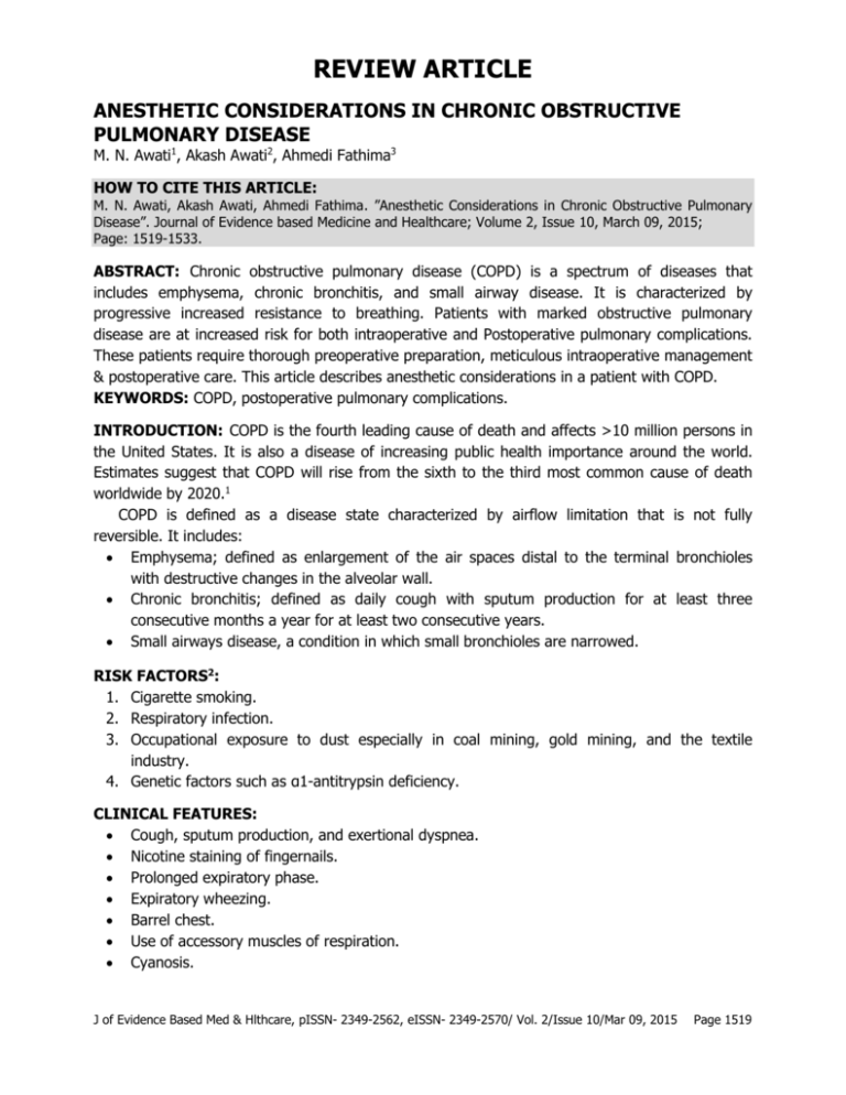
REVIEW ARTICLE ANESTHETIC CONSIDERATIONS IN CHRONIC OBSTRUCTIVE PULMONARY DISEASE M. N. Awati1, Akash Awati2, Ahmedi Fathima3 HOW TO CITE THIS ARTICLE: M. N. Awati, Akash Awati, Ahmedi Fathima. ”Anesthetic Considerations in Chronic Obstructive Pulmonary Disease”. Journal of Evidence based Medicine and Healthcare; Volume 2, Issue 10, March 09, 2015; Page: 1519-1533. ABSTRACT: Chronic obstructive pulmonary disease (COPD) is a spectrum of diseases that includes emphysema, chronic bronchitis, and small airway disease. It is characterized by progressive increased resistance to breathing. Patients with marked obstructive pulmonary disease are at increased risk for both intraoperative and Postoperative pulmonary complications. These patients require thorough preoperative preparation, meticulous intraoperative management & postoperative care. This article describes anesthetic considerations in a patient with COPD. KEYWORDS: COPD, postoperative pulmonary complications. INTRODUCTION: COPD is the fourth leading cause of death and affects >10 million persons in the United States. It is also a disease of increasing public health importance around the world. Estimates suggest that COPD will rise from the sixth to the third most common cause of death worldwide by 2020.1 COPD is defined as a disease state characterized by airflow limitation that is not fully reversible. It includes: Emphysema; defined as enlargement of the air spaces distal to the terminal bronchioles with destructive changes in the alveolar wall. Chronic bronchitis; defined as daily cough with sputum production for at least three consecutive months a year for at least two consecutive years. Small airways disease, a condition in which small bronchioles are narrowed. RISK FACTORS2: 1. Cigarette smoking. 2. Respiratory infection. 3. Occupational exposure to dust especially in coal mining, gold mining, and the textile industry. 4. Genetic factors such as α1-antitrypsin deficiency. CLINICAL FEATURES: Cough, sputum production, and exertional dyspnea. Nicotine staining of fingernails. Prolonged expiratory phase. Expiratory wheezing. Barrel chest. Use of accessory muscles of respiration. Cyanosis. J of Evidence Based Med & Hlthcare, pISSN- 2349-2562, eISSN- 2349-2570/ Vol. 2/Issue 10/Mar 09, 2015 Page 1519 REVIEW ARTICLE Dyspnea on walking uphill I Mild Walks more slowly than people of the same age on the level II Moderate Stops for breath after walking about 100 yards /after a few mins on the level III Severe Too breathless to leave the house/ unable to dress or undress IV Very severe Table 1: MRC grading of dyspnea Features Chronic bronchitis Emphysema Mechanism of airway obstruction Decreased lumen due to mucus & inflammation Loss of elastic recoil Dyspnea Moderate Severe FEV1 Decreased Decreased PaO2 Marked decrease (Blue bloaters) Modest decrease (pink puffers) PaCO2 Increased Normal to decreased Diffusing capacity Normal Decreased Haematocrit Increased Normal Cor pulmonale Marked Mild Prognosis Poor Good Table 2: Differentiating features between chronic bronchitis & emphysema1 LABORATORY INVESTIGATIONS: 1. Pulmonary Function Testing: Shows airflow obstruction with a reduction in FEV1 and FEV1/FVC. With worsening disease severity, increase in total lung capacity, functional residual capacity, and residual volume. In patients with emphysema, the diffusing capacity may be reduced. Fig 1: Spirometry normal & in COPD J of Evidence Based Med & Hlthcare, pISSN- 2349-2562, eISSN- 2349-2570/ Vol. 2/Issue 10/Mar 09, 2015 Page 1520 REVIEW ARTICLE 2. Arterial blood gases: Provide information about resting or exertional hypoxemia, alveolar ventilation & acidbase status. 3. Radiographic studies: Bullae, paucity of parenchymal markings or hyperlucency suggests the presence of emphysema. Flattening of the diaphragm suggest hyperinflation. Computed tomography (CT) scan is the definitive test for establishing the presence or absence of emphysema. GOLD Stage Severity Symptoms Spirometry 0 At Risk Chronic cough, sputum production Normal I Mild With or without chronic cough or sputum production FEV1/FVC <0. 7 and FEV1 80% predicted II Moderate With or without chronic cough or sputum production FEV1/FVC <0. 7 and 50% FEV1 <80% predicted III Severe With or without chronic cough or sputum production FEV1/FVC <0. 7 and 30% FEV1 <50% predicted With or without chronic cough or sputum production FEV1/FVC <0. 7 and FEV1 <30% predicted Or FEV1 <50% predicted with respiratory failure or signs of right heart failure IV Very severe Table 3: GOLD grading for COPD severity3 TREATMENT: 1. Smoking Cessation: Crucial for stopping progression of the disease. 2. Inhaled Bronchodilator therapy: Beta agonists. Anticholinergics. 3. Inhaled steroids. 4. Mucolytics: N-acetyl cysteine. 5. Chest physiotherapy. 6. Theophylline. 7. Supplemental oxygen: For patients with resting hypoxemia (resting O2 saturation 88% or <90% with signs of pulmonary hypertension or right heart failure).1 8. Hydration & nutrition. 9. Antitussives. J of Evidence Based Med & Hlthcare, pISSN- 2349-2562, eISSN- 2349-2570/ Vol. 2/Issue 10/Mar 09, 2015 Page 1521 REVIEW ARTICLE Beta-Adrenergic agonists: albuterol, metaproterenol, fenoterol, terbutaline, epinephrine Increases adenylate cyclase, increasing cAMP and decreasing smooth muscle tone (bronchodilation); short-acting b-adrenergic agonists (e. g., albuterol, terbutaline, and epinephrine) are the agents of choice for acute exacerbations Methylxanthines: aminophylline, Theophylline Phosphodiesterase inhibition increases cAMP; potentiates endogenous catecholamines; improves diaphragmatic contractility; central respiratory stimulant Corticosteroids: methylprednisolone, dexamethasone, prednisone, cortisol Anti-inflammatory and membrane stabilizing; inhibits histamine release; potentiates beta-agonists Anticholinergics: atropine, glycopyrrolate, ipratropium Blocks acetylcholine at postganglionic receptors, decreasing cGMP, relaxing airway smooth muscle Cromolyn sodium Also a membrane stabilizer, preventing mast cell degranulation, but must be given prophylactically Antileukotrienes: zileuton, montelukast Inhibition of leukotriene production and/or zafirlukast, leukotriene antagonism; antiinflammatory; used in addition to corticosteroids; however, may be considered first-line antiinflammatory therapy for patients who cannot or will not use corticosteroids Table 4: Drugs used in COPD4 Non-pharmacologic Therapies1: 1. Lung Volume Reduction Surgery (LVRS): Patients with upper lobe–predominant emphysema and a low post rehabilitation exercise capacity are most likely to benefit. Exclusion criteria: 1. Significant pleural disease 2. Pulmonary artery systolic pressure >45 mmHg 3. Congestive heart failure or other severe comorbid conditions. 4. FEV1 <20% of predicted. 5. Diffusing capacity of lung for carbon monoxide (DLCO) <20% of predicted 2. Lung transplantation: Current recommendations are that candidates for lung transplantation: Should be <65 years, Should have severe disability despite maximal medical therapy and Should be free of comorbid conditions such as liver, renal, or cardiac disease. 3. General Medical Care: Patients with COPD should receive the influenza vaccine annually. Polyvalent pneumococcal vaccine is also recommended. J of Evidence Based Med & Hlthcare, pISSN- 2349-2562, eISSN- 2349-2570/ Vol. 2/Issue 10/Mar 09, 2015 Page 1522 REVIEW ARTICLE 4. Pulmonary Rehabilitation: ACUTE EXACERBATION OF COPD. Sustained worsening of patients symptoms from their usual stable state, which is beyond normal day-to-day variations and is acute in onset. Clinical features: Increased shortness of breath. Increased sputum production and/or change in colour. Increased cough. Increased wheeze/tightness. Decreased exercise tolerance. Increased fatigue. Confusion. Treatment: 1. Inhaled short-acting beta2-agonists: Albuterol (agent of choice) 2. Inhaled short-acting anticholinergics: Ipratoprium bromide 3. Antibiotics: If the patient has symptoms and signs suggesting infection; Choice of antibiotics should be based on the severity. 4. Glucocorticoids: 30–40 mg of oral prednisolone or its equivalent for a period of 10–14 days. 5. Oxygen Supplemental O2 should be supplied to keep arterial saturations 90%. Mechanical Ventilatory Support: 1. Non-invasive positive-pressure ventilation (NIPPV)5,6,7: A temporary measure for ventilatory support. Improves blood gases and pH, reduces in-hospital mortality, need for intubation, complications of therapy, hospital length of stay & decreases infection. BIPAP is best. Improvement should be apparent in the first 30-120 minutes. If no improvement, proceed to endotracheal intubation. Monitor: Level of consciousness, vital signs, and ABG’s. Contraindications8: a. Cardiovascular instability. b. Impaired mental status. c. Inability to cooperate. d. Copious secretions or the inability to clear secretions. e. Craniofacial abnormalities or trauma precluding effective fitting of mask. f. Extreme obesity. g. Significant burns. J of Evidence Based Med & Hlthcare, pISSN- 2349-2562, eISSN- 2349-2570/ Vol. 2/Issue 10/Mar 09, 2015 Page 1523 REVIEW ARTICLE 2. Invasive mechanical ventilation: Indications: a. patients with severe respiratory distress despite initial therapy, life-threatening hypoxemia, b. severe hypercarbia and/or acidosis, c. markedly impaired mental status, d. respiratory arrest, e. Hemodynamic instability or other complications. Ventilatory strategies2: Ventilatory setting: maximize alveolar gas emptying and avoid dynamic hyperinflation & auto PEEP. Low respiratory frequency (8-10). Low I: E ratio (1:3 or more). Appropriate VT (8-10ml/Kg) to maintain oxygenation and ventilation (ET CO2). Airway pressure<35 cm of H2O. Fig. 2: Therapy at each stage MANAGEMENT OF ANESTHESIA: Preoperative3: Smoking history: number of packs per day and duration in years Dyspnea, wheezing, productive cough, and exercise tolerance Hospitalizations for COPD, including the need for intubation and mechanical ventilation J of Evidence Based Med & Hlthcare, pISSN- 2349-2562, eISSN- 2349-2570/ Vol. 2/Issue 10/Mar 09, 2015 Page 1524 REVIEW ARTICLE Medications, including home oxygen therapy and steroid use, either systemic or inhaled. Recent pulmonary infections, exacerbations, or change in character of sputum Weight loss that may be caused by end-stage pulmonary disease or lung cancer Symptoms of right-sided heart failure. Bedside Pulmonary Function Tests9: 1. Cough test: ask he patient to cough once after deep inspiration, test is positive if it leads to recurrent coughing & is suggestive of bronchitis. 2. Wheeze test: ask patient to take 5 deep breaths, auscultate for wheeze in the interscapular region. 3. Maximum laryngeal height: if the distance between top of thyroid cartilage & suprasternal notch at the end of expiration is <4cm, it is an accurate sign of obstructive airway disease. 4. Debono`s Whistle test: patient is asked to exhale as forcefully as possible into the tube. It indirectly estimates PEFR upto 300litre/min. 5. Breath holding test (Sabrasez): Patient is asked to take a deep breath and hold it for as long as possible. Duration of more than 40 sec is considered normal. <15 sec indicates reduced VC. It roughly estimates cardiopulmonary and ventilatory status. 6. Snider`s Match Blowing test: lighted match stick is held at 15cms away from patient’s mouth and then patient is asked to blow out the match. Maximum breathing capacity <50L/min if distance is <7. 5cms. 7. Watch and Stethoscope test: sound is heard over the trachea during a forced expiration. Normal value is 3-4 sec, >6sec indicates obstructive disease. 8. Hand Held Spirometry: to assess FEV1 and PEFR, here the patient is asked to blow forcefully into a hand held spirometer after a maximal inspiration. 9. Single breath count test: patient is asked to count out loud numbers from 1 onwards after maximal inspiration. <15 indicate severe impairment of VC. INVESTIGATIONS: Complete blood count ECG 2D ECHO: In patients with features of heart failure Chest X-RAY PA view PFT: to grade the severity of the disease ABG in severe COPD Flow Volume Loops:In patients with COPD, there is a decrease in the expiratory flow rate at any given lung volume. The expiratory curve is concave upward due to uniform emptying of the airways. The residual volume is increased due to air trapping. Indications for a preoperative pulmonary evaluation (consultation with a pulmonologist &/or PFT)2: 1. Hypoxemia on room air or the need for home oxygen therapy without a known etiology. 2. Bicarbonate more than 33 mEq/L or PCO2 more than 50 mm Hg in a patient whose pulmonary disease has not been previously evaluated. J of Evidence Based Med & Hlthcare, pISSN- 2349-2562, eISSN- 2349-2570/ Vol. 2/Issue 10/Mar 09, 2015 Page 1525 REVIEW ARTICLE 3. 4. 5. 6. 7. 8. 9. A history of respiratory failure due to a problem that still exists. Severe shortness of breath attributed to respiratory disease. Planned pneumonectomy. Difficulty assessing pulmonary function by clinical signs. Distinguishing among potential etiologies of significant respiratory compromise. Determining the response to bronchodilators, and Suspected pulmonary hypertension. Preoperative preparation: 1. Cessation of smoking: Effects of Smoking on Different Organ Systems10: Cardiovascular system: 1. Smoking is a risk factor for development of cardiovascular disease. 2. Carbon monoxide decreases oxygen delivery and increases myocardial work. 3. Smoking releases catecholamines and causes coronary vasoconstriction. 4. Smoking decreases exercise capacity. Respiratory system: 1. Smoking is the major risk factor for development of chronic pulmonary disease. 2. Smoking decreases mucociliary activity. 3. Smoking results in hyper-reactive airways. Other effects: Decreased immnunity & delayed wound healing. 12-24 hrs Decreases CO & nicotine levels 48-72 hrs COHb level normalized, ciliary function improves 1-2 wks Decreased sputum production 4-6wks PFTs improve 6-8 wks Immune function & metabolism normalizes 8-12wks Decreases post op morbidity & mortality Table 5: Beneficial effects of smoking cessation & time course10 2. Treatment of chest infections. 3. Bronchodilator therapy. 4. Steroids cover if patient is on steroids. J of Evidence Based Med & Hlthcare, pISSN- 2349-2562, eISSN- 2349-2570/ Vol. 2/Issue 10/Mar 09, 2015 Page 1526 REVIEW ARTICLE Dose Surgery Recommendations <10mg/day Minor/moderate/major Additional steroid dose not required >10mg/kg Minor 25mg hydrocortisone at induction & usual medications postoperative >10mg/kg Moderate Usual dose preop & 25mg hydrocortisone at induction then 25mg iv TID for 1day, then recommence preop dosage >10mg/kg Major Usual dose preop & 100mg hydrocortisone at induction then 100mg iv TID for 2-3day, then recommence preop dosage Table 6: Recommendations for steroids11 5. Hydration & mucolytic therapy to loosen the secretions. 6. Chest physiotherapy & postural drainage of secretions. INTRAOPERATIVE2: Regional anesthesia is suitable for operations that do not invade the peritoneum and for surgical procedures performed on the extremities. General anesthesia is the usual choice for upper abdominal and intrathoracic surgery. The choice of anesthetic technique does not seem to alter the incidence of postoperative pulmonary complications. Regional anesthesia: Advantages: Avoiding airway instrumentation & hence, bronchospasm. Reduced incidence of postoperative atelectasis & acute respiratory failure Cardiac complications are also low. Reduced incidence of thromboembolism. Early ambulation due to better analgesia. Disadvantages: Inadequate muscle relaxation. Hypotension. Higher levels of blockade decreases inspiratory and expiratory capacity. Need for heavy sedation. May reduce effective coughing secondary to abdominal muscle dysfunction, leading to decreased sputum clearance and atelectasis. General anesthesia Advantages: Good control of airway & ventilation. Adequate muscle relaxation. Complications of spinal/epidural avoided J of Evidence Based Med & Hlthcare, pISSN- 2349-2562, eISSN- 2349-2570/ Vol. 2/Issue 10/Mar 09, 2015 Page 1527 REVIEW ARTICLE Disadvantages: Increased incidence of postoperative respiratory failure. GA+ RA: Reduction in dosage of anesthetic drugs. Faster recovery of bowel motility. Early mobilization. Excellent analgesia. Reduced incidence of POPC. Volatile anesthetics: Volatile anesthetics are more useful because of the ability of these drugs to be rapidly eliminated, thereby reducing residual ventilatory depression during the early postoperative period. Sevoflurane is the best choice. Desflurane must be avoided as it may irritate airway. Furthermore, volatile anesthetics produce bronchodilation. (Halothane> Enflurane> isoflurane> sevoflurane) Provide humidified gases. Intravenous agents: Propofol is the first choice in hemodynamically stable patients with reactive airways. Thiopentone may provoke bronchospasm. Ketamine or Etomidate in hemodynamic instability. Ketamine is also preferred in actively wheezing patient. Muscle relaxants: Vec /roc/ cis-atracurium. Histamine releasing muscle relaxants must be avoided. Avoid long acting NMBDs. Opioids: prolonged ventilatory depression as a result of their slow rate of metabolism or elimination. Avoid nitrous oxide in the presence of bullae or pulmonary hypertension. Ventilation strategies: Large tidal volumes (8-10mL/kg). Slow respiratory rates (8to 10 breaths per minute). Low I: E ratio (1:3 or more). Advantages: Maintain optimal ventilation-to-perfusion matching. Provide sufficient time for complete exhalation to occur, thereby preventing auto PEEP. Sufficient time for venous return and are less likely to be associated with undesirable degrees of hyperventilation. Intraoperative complications: 1. Bronchospasm 2. Tension pneumothorax 3. Auto PEEP J of Evidence Based Med & Hlthcare, pISSN- 2349-2562, eISSN- 2349-2570/ Vol. 2/Issue 10/Mar 09, 2015 Page 1528 REVIEW ARTICLE Intraoperative bronchospasm Causes: Lighter planes. Kinked ETT. Secretions /blood. Pulmonary edema. Aspiration. Endobronchial intubation. Tension pneumothorax. Diagnosis: Extensive rhonchi. Increase in peak and plateau pressure. Decrease in slope of expired CO2. Hypoxia, hypercarbia & hypotension due to dynamic hyperinflation and development of autopeep. Treatment: 100% oxygen. Deepen the plane of anesthesia. Stop the surgical stimulation. beta 2 agonist: nebulization. IV corticosteroid. Prevention: Optimize preoperative symptom control. Adequate preoperative anxiolysis. Inhaled β2 agonists or muscarinic antagonists immediately before induction. Regional anesthesia if feasible. Minimize airway instrumentation as feasible. Induction with propofol, ketamine, or volatile agent. Use volatile anesthetics early and often (avoid desflurane). Adequate depth of anesthesia before airway instrumentation. Intravenous lidocaine and opioids as adjuncts for endotracheal intubation. Auto-PEEP: Air trapping is known as auto-PEEP and results from “stacking” of breaths when full exhalation is not allowed to occur. It results in impairment of oxygenation and ventilation and hemodynamic compromise by decreasing preload and increasing pulmonary vascular resistance. Can be prevented by increasing the expiratory phase of ventilation and decreasing the respiratory rate. For intra-operative diagnosis of auto-PEEP, “apnea test” is done. Hypotension may develop due to hyperinflation and autopeep, disconnect the patient from the ventilator for 30 mins. If it leads to normalization of blood pressure, it confirms the diagnosis of auto peep. J of Evidence Based Med & Hlthcare, pISSN- 2349-2562, eISSN- 2349-2570/ Vol. 2/Issue 10/Mar 09, 2015 Page 1529 REVIEW ARTICLE EXTUBATION: Should be slow and smooth, after complete reversal of residual neuromuscular blockade. Neostigmine can cause bronchospasm, therefore, should be preceeded by anticholinergic agents. Preferably under deep anaesthesia. Decrease the risk of bronchospasm, by administration of I. V. lidocaine or with inhaled bronchodilators. POST-OPERATIVE CARE: Supplemental oxygen. Judicious fluid therapy. Adequate analgesia-multimodal approach. Early ambulation. Early postoperative respiratory therapy & drainage of secretions, along with nebulization, mucolytics and bronchodilators. Postoperative neuraxial analgesia with opioids may permit early tracheal extubation. The sympathetic blockade, muscle weakness, and loss of proprioception that are produced by local anesthetics are not produced by neuraxial opioids. Therefore, early ambulation is possible which serves to increase FRC and improve oxygenation, presumably by improving ventilation-to-perfusion matching. Neuraxial opioids may be especially useful after intrathoracic and upper abdominal surgery. Sedation may accompany neuraxial opioid administration and delayed respiratory depression can be seen. Lung expansion maneuvers: These techniques decrease the risk of atelectasis by increasing lung volumes, thereby decreasing the frequency of postoperative pulmonary complications. Deep breathing exercises Incentive spirometry Chest physiotherapy Positive-pressure breathing techniques Post-operative mechanical ventilation: Continued mechanical ventilation during the immediate postoperative period may be necessary in patients with severe COPD who have undergone major abdominal or intrathoracic surgery. Patients with preoperative FEV1/FVC ratios less than 0. 5 or with a preoperative PaCO2 of more than 50 mm Hg are likely to need some postoperative mechanical ventilation. If the PaCO2 has been chronically increased, it is important not to correct the hypercarbia too quickly because this will result in a metabolic alkalosis that can be associated with cardiac dysrhythmias and central nervous system irritability and even seizures. J of Evidence Based Med & Hlthcare, pISSN- 2349-2562, eISSN- 2349-2570/ Vol. 2/Issue 10/Mar 09, 2015 Page 1530 REVIEW ARTICLE When continued mechanical ventilation is necessary, FIO2 and ventilator settings should be adjusted to maintain the PaO2 between 60 and 100 mm Hg and the PaCO2 in a range that maintains the pH at 7. 35 to 7. 45. The decision to discontinue mechanical support of ventilation and to perform tracheal extubation is based on the patient's clinical status and indices of pulmonary function. POSTOPERATIVE COMPLICATIONS Pneumonia. Atelectasis. Bronchospasm. Acute respiratory failure requiring mechanical ventilation. DVT/ Pulmonary embolism. Risk factors for POPC12: Patient-Related: Age ≥60 yr. ASA-PS class ≥2. Heart failure. Partial or total functional dependence. Chronic obstructive pulmonary disease. Weight loss. Delirium. Cigarette smoking. Alcohol use. Abnormal chest examination findings. Procedure-Related: Thoracic surgery. Abdominal surgery. Neurosurgery. Head and neck surgery. Emergency surgery. Vascular surgery. Use of general anaesthesia. Blood product transfusion. Laboratory Test–Related: Albumin level <35 g/L Abnormal chest radiograph BUN level >7. 5 mmol/L (>21 mg/dL) J of Evidence Based Med & Hlthcare, pISSN- 2349-2562, eISSN- 2349-2570/ Vol. 2/Issue 10/Mar 09, 2015 Page 1531 REVIEW ARTICLE Criteria to predict POPC: Nunn & Milledge criteria: a. FEV1< 1L, normal PaO2 & PaCO2- low risk. b. FEV1< 1L, low PaO2 & PaCO2- patient will need prolonged O2. c. FEV1< 1L, lowPaO2 & PaCO2- may need postoperative ventilation. Based on spirometry: a. Predicted FVC<50%. b. Predicted FEV1<50% or 2L. c. Predicted MVV <50% or <5oL/min. d. Predicted DLCO <50%. e. Predicted RV/TLC >50%. Risk reduction strategies to decrease the incidence of postoperative pulmonary complications13: Pre-operative: Encourage cessation of smoking for at least 6weeks. Treat evidence of expiratory airflow obstruction. Treat respiratory infection with antibiotics. Initiate patient education regarding lung volume expansion maneuvers. Intra-operative: Use minimally invasive surgery (endoscopic) techniques when possible. Consider use of regional anesthesia. Avoid surgical procedures likely to require more than 3hours. Post-operative: Institute lung volume expansion maneuvers (voluntary deep breathing, incentive spirometry, continuous positive pressure) Maximize analgesia (neuraxial opioids, intercostals nerve blocks, patient-controlled analgesia). Role of LMA in COPD patients: Reduced manipulation of airway. Decreased incidence of bronchospasm. Decreased sympathetic stimulation. Proseal LMA is recommended. CONCLUSION: Pulmonary complications remain a major cause of morbidity and mortality for patients undergoing surgery and anesthesia. Patients with COPD have fewer postoperative pulmonary complications when a perioperative program of intensive chest physiotherapy is initiated preoperatively. J of Evidence Based Med & Hlthcare, pISSN- 2349-2562, eISSN- 2349-2570/ Vol. 2/Issue 10/Mar 09, 2015 Page 1532 REVIEW ARTICLE REFERENCES: 1. Chronic Obstructive Pulmonary Disease, Harrison’s principles of internal medicine, 18th Edition; 2012. 2. Sharif al-ruzzeh, viji kurup, Respiratory diseases. Hines & Marschall: Stoelting's Anesthesia and Co-Existing Disease; 5th edition: 10; 189-195. 3. Global initiative for chronic obstructive Lung Disease: 2011 update. 4. Howard j miller, Chronic Obstructive Pulmonary Disease. Anesthesia secrets; 4th edition: 40; 278-84. 5. Mehta S, Micholos S Hill, Noninvasive ventilation Am J Raspir Crit Care Med 2001; 163: 540577. 6. Bochard L, Mancebo J, Wysocki M et al. Noninvasive ventilation for acute exacerbation of chronic obstructive pulmonary disease. N Engl J Med 1995; 333: 817-22. 7. Foglio C, Vitacca M, Quadri A, et al. Acute exacerbations in severe COPD patients: treatment using positive pressure ventilation by nasal mask. Chest 1992; 101: 1533-38. 8. Chandrasekhar TR. Noninvasive ventilation. In proceedings of “Mehanical ventilation – Basics and beyond”, Bangalore; 2004. p 167-190. 9. Straus SE, et al, for the CARE-COAD1 group. The accuracy of patient history, wheezing & laryngeal measurements in diagnosing obstructive airway diseases. JAMA. 2000; 283: 185367. 10. Chandra Rodrigo. The effects of cigarette smoking on anesthesia. Anaesth prog. 2000; 47: 143-50. 11. Marik, P. E. & Varon, J. Requirement of perioperative stress doses of corticosteroids: a systematic review of the literature. Archives of Surgery 143, 1222 (2008). 12. Smetana GW, Lawrence VA, Cornell JE, et al: Preoperative pulmonary risk stratification for noncardiothoracic surgery: systematic review for the American College of Physicians, Ann Intern Med 144: 581-595, 2006. 13. Smetana GW. Preoperative pulmonary evaluation. N Engl J Med. 1999; 340: 937-944. Copyright 1999 Massachusetts Medical Society. AUTHORS: 1. M. N. Awati 2. Akash Awati 3. Ahmedi Fathima PARTICULARS OF CONTRIBUTORS: 1. Professor & HOD, Department of Anaesthesiology, M. R. Medical College, Gulbarga, Karnataka. 2. DM, Department of Neurology, M. R. Medical College, Gulbarga, Karnataka. 3. Post Graduate, Department of Anaesthesiology, M. R. Medical College, Gulbarga, Karnataka. NAME ADDRESS EMAIL ID OF THE CORRESPONDING AUTHOR: Dr. M. N. Awati, #10/105/39, Next to ‘DIJA’ Science Centre, Gulbarga-585105. E-mail: hodanaesthesiology@gmail.com Date Date Date Date of of of of Submission: 22/02/2015. Peer Review: 23/02/2015. Acceptance: 04/03/2015. Publishing: 06/03/2015. J of Evidence Based Med & Hlthcare, pISSN- 2349-2562, eISSN- 2349-2570/ Vol. 2/Issue 10/Mar 09, 2015 Page 1533
