volumes
advertisement
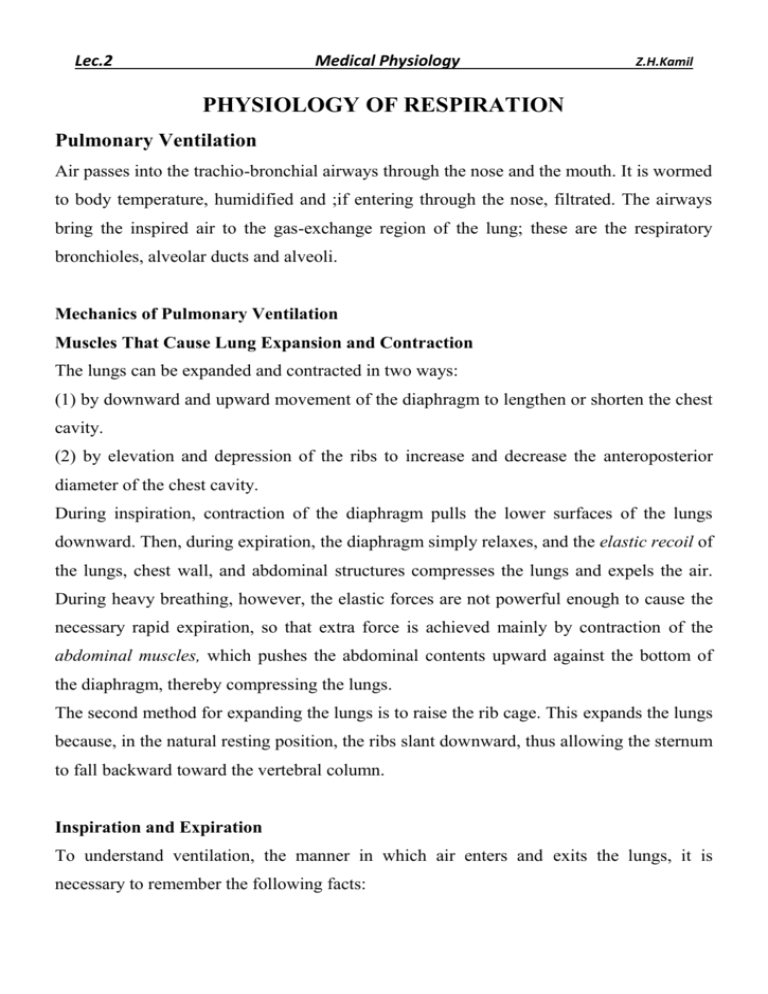
Lec.2 Medical Physiology Z.H.Kamil PHYSIOLOGY OF RESPIRATION Pulmonary Ventilation Air passes into the trachio-bronchial airways through the nose and the mouth. It is wormed to body temperature, humidified and ;if entering through the nose, filtrated. The airways bring the inspired air to the gas-exchange region of the lung; these are the respiratory bronchioles, alveolar ducts and alveoli. Mechanics of Pulmonary Ventilation Muscles That Cause Lung Expansion and Contraction The lungs can be expanded and contracted in two ways: (1) by downward and upward movement of the diaphragm to lengthen or shorten the chest cavity. (2) by elevation and depression of the ribs to increase and decrease the anteroposterior diameter of the chest cavity. During inspiration, contraction of the diaphragm pulls the lower surfaces of the lungs downward. Then, during expiration, the diaphragm simply relaxes, and the elastic recoil of the lungs, chest wall, and abdominal structures compresses the lungs and expels the air. During heavy breathing, however, the elastic forces are not powerful enough to cause the necessary rapid expiration, so that extra force is achieved mainly by contraction of the abdominal muscles, which pushes the abdominal contents upward against the bottom of the diaphragm, thereby compressing the lungs. The second method for expanding the lungs is to raise the rib cage. This expands the lungs because, in the natural resting position, the ribs slant downward, thus allowing the sternum to fall backward toward the vertebral column. Inspiration and Expiration To understand ventilation, the manner in which air enters and exits the lungs, it is necessary to remember the following facts: 1. Normally, there is a continuous column of air from the pharynx to the alveoli of the lungs. 2. The lungs lie within the sealed-off thoracic cavity. The rib cage, consisting of the ribs joined to the vertebral column posteriorly and to the sternum anteriorly, forms the top and sides of the thoracic cavity. The intercostal muscles lie between the ribs. The diaphragm and connective tissue form the floor of the thoracic cavity. 3. The lungs adhere to the thoracic wall by way of the pleura. Any space between the two pleurae is minimal due to the surface tension of the fluid between them. Inspiration Inspiration is the active phase of ventilation because this is the phase in which the diaphragm and the external intercostal muscles contract (Fig. 1-a). In its relaxed state, the diaphragm is dome-shaped; during deep inspiration, it contracts and lowers. Also, the external intercostals muscles contract, and the rib cage moves upward and outward. Following contraction of the diaphragm and the external intercostal muscles, the volume of the thoracic cavity will be larger than it was before. As the thoracic volume increases, the lungs expand. Now the air pressure within the alveoli decreases, creating a partial vacuum. Because alveolar pressure is now less than atmospheric pressure (air pressure outside the lungs), air naturally flows from outside the body into the respiratory passages and into the alveoli. It is important to realize that air comes into the lungs because they have already opened up; air does not force the lungs open. This is why it is sometimes said that humans inhale by negative pressure. The creation of a partial vacuum in the alveoli causes air to enter the lungs. While inspiration is the active phase of breathing, the actual flow of air into the alveoli is passive. Expiration Usually, expiration is the passive phase of breathing, and no effort is required to bring it about. During expiration, the elastic properties of the thoracic wall and lungs cause them to recoil. In addition, the lungs recoil because the surface tension of the fluid lining the alveoli tends to draw them closed. During expiration, the abdominal organs press up against the diaphragm, and the rib cage moves down and inward (Fig. 1-b). What keeps the alveoli from collapsing as a part of expiration? Recall that the presence of surfactant lowers the surface tension within the alveoli. Also, as the lungs recoil, the pressure between the pleura decreases, and this tends to make the alveoli stay open. The importance of the reduced intrapleural pressure is demonstrated when, by design or accident, air enters the intrapleural space. Now the lung collapses. The diaphragm and external intercostal muscles are usually relaxed when expiration occurs. However, when breathing is deeper and/or more rapid, expiration can be active. Contraction of the internal intercostal muscles can force the rib cage to move downward and inward. Also, when the abdominal wall muscles contract, they push on the viscera, which push against the diaphragm, and the increased pressure in the thoracic cavity helps expel air. Figure 1: Inspiration versus expiration. Movement of Air In and Out of the Lungs the Pressures That Cause the Movement Pleural Pressure and Its Changes During Respiration Pleural pressure is the pressure of the fluid in the thin space between the lung pleura and the chest wall pleura. As noted earlier, this is normally a slight suction, which means a slightly negative pressure. The normal pleural pressure at the beginning of inspiration is about –5 centimeters of water, which is the amount of suction required to hold the lungs open to their resting level. Then, during normal inspiration, expansion of the chest cage pulls outward on the lungs with greater force and creates more negative pressure, to an average of about –7.5 centimeters of water. These relationships between pleural pressure and changing lung volume are demonstrated in Figure 2, showing in the lower panel the increasing negativity of the pleural pressure from –5 to –7.5 during inspiration and in the upper panel an increase in lung volume of 0.5 liter. Then, during expiration, the events are essentially reversed. Alveolar Pressure Alveolar pressure is the pressure of the air inside the lung alveoli. When the glottis is open and no air is flowing into or out of the lungs, the pressures in all parts of the respiratory tree, all the way to the alveoli, are equal to atmospheric pressure, which is considered to be zero reference pressure in the airways—that is, 0 centimeters water pressure. To cause inward flow of air into the alveoli during inspiration, the pressure in the alveoli must fall to a value slightly below atmospheric pressure (below 0). The second curve (labeled “alveolar pressure”) of Figure 2 demonstrates that during normal inspiration, alveolar pressure decreases to about –1 centimeter of water. This slight negative pressure is enough to pull 0.5 liter of air into the lungs in the 2 seconds required for normal quiet inspiration. During expiration, opposite pressures occur: The alveolar pressure rises to about +1 centimeter of water, and this forces the 0.5 liter of inspired air out of the lungs during the 2 to 3 seconds of expiration. Transpulmonary Pressure Finally, note in Figure 2 the difference between the alveolar pressure and the pleural pressure. This is called the transpulmonary pressure. It is the pressure difference between that in the alveoli and that on the outer surfaces of the lungs, and it is a measure of the elastic forces in the lungs that tend to collapse the lungs at each instant of respiration, called the recoil pressure. Figure 2: Changes in lung volume, alveolar pressure, pleural pressure, and transpulmonary pressure during normal breathing. Non-respiratory Air Movements Many situations other than breathing move air into or out of the lung and may modify the normal respiratory rhythm. These movements are: 1. Cough: taking a deep breath, closing glottis and forcing air superiorly from lungs against glottis. Then the glottis opens suddenly and a blast of air rushes upward. Coughs act to clear the lower respiratory passageways. 2. Sneeze: similar to cough except that the expelled air is directed through the nasal cavities instead of through oral cavity. The uvula, a tag of tissue hanging from the soft palate, becomes depressed and closes oral cavity off from pharynx, routing the air through nasal cavities, sneezes clear upper respiratory passageways. 3. Crying: inspiration followed by release of air in a number of short breaths. Primarily an emotionally induced mechanism. 4. Laughing: essentially same as crying in terms of the air movements produced. Also an emotionally induced response. 5. Hiccups: sudden inspiration resulting from spasms of diaphragm; initiated by irritation of diaphragm or phrenic nerves, which serve diaphragm. The sound occurs when inspired air hits vocal folds of closed glottis. 6. Yawn: very deep inspiration, take with jaws wide open. Formerly believed to be triggered by need to increase amount of oxygen in blood but this theory is now being questioned, ventilates all alveoli (this is not the case in normal quiet breathing). Pulmonary Volumes and Capacities A simple method for studying pulmonary ventilation is to record the volume movement of air into and out of the lungs, a process called spirometry. A spirogram indicating changes in lung volume under different conditions of breathing. For ease in describing the events of pulmonary ventilation, the air in the lungs has been subdivided in this diagram into four volumes and four capacities, which are the average for a young adult man. Pulmonary Volumes In Figure 3 there are listed four pulmonary lung volumes that, when added together, equal the maximum volume to which the lungs can be expanded. The significance of each of these volumes is the following: 1. The tidal volume is the volume of air inspired or expired with each normal breath; it amounts to about 500 milliliters in the adult male. 2. The inspiratory reserve volume is the extra volume of air that can be inspired over and above the normal tidal volume when the person inspires with full force; it is usually equal to about 3000 milliliters. 3. The expiratory reserve volume is the maximum extra volume of air that can be expired by forceful expiration after the end of a normal tidal expiration; this normally amounts to about 1100 milliliters. 4. The residual volume is the volume of air remaining in the lungs after the most forceful expiration; this volume averages about 1200 milliliters. Pulmonary Capacities In describing events in the pulmonary cycle, it is sometimes desirable to consider two or more of the volumes together. Such combinations are called pulmonary capacities. The important pulmonary capacities can be described as follows: 1. The inspiratory capacity equals the tidal volume plus the inspiratory reserve volume. This is the amount of air (about 3500 milliliters) a person can breathe in, beginning at the normal expiratory level and distending the lungs to the maximum amount. 2. The functional residual capacity equals the expiratory reserve volume plus the residual volume. This is the amount of air that remains in the lungs at the end of normal expiration (about 2300 milliliters). 3. The vital capacity equals the inspiratory reserve volume plus the tidal volume plus the expiratory reserve volume. This is the maximum amount of air a person can expel from the lungs after first filling the lungs to their maximum extent and then expiring to the maximum extent (about 4600 milliliters). 4. The total lung capacity is the maximum volume to which the lungs can be expanded with the greatest possible effort (about 5800 milliliters); it is equal to the vital capacity plus the residual volume. All pulmonary volumes and capacities are about 20 to 25 per cent less in women than in men, and they are greater in large and athletic people than in small and asthenic people. Figure 3: Diagram showing respiratory excursions during normal breathing and during maximal inspiration and maximal expiration. Respiratory sounds As air flows in and out of respiratory tree, it produces two recognizable sounds that can be picked up with a stethoscope. Bronchial sounds are produced by air rushing through the large respiratory passageways (trachea and bronchi) Vesicular breathing sounds occurs as air fills the alveoli. The vesicular sounds are soft and resemble a muffled breeze.
