Leaf Pigment Chromatography Lab: High School Biology
advertisement
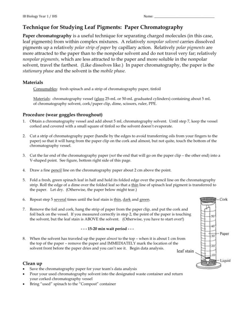
IB Biology Year 1 / IHS Name: ______________________________________________ Technique for Studying Leaf Pigments: Paper Chromatography Paper chromatography is a useful technique for separating charged molecules (in this case, leaf pigments) from within complex mixtures. A relatively nonpolar solvent carries dissolved pigments up a relatively polar strip of paper by capillary action. Relatively polar pigments are more attracted to the paper than to the nonpolar solvent and do not travel very far; relatively nonpolar pigments, which are less attracted to the paper and more soluble in the nonpolar solvent, travel the farthest. (Like dissolves like.) In paper chromatography, the paper is the stationary phase and the solvent is the mobile phase. Materials Consumables: fresh spinach and a strip of chromatography paper, tinfoil Materials: chromatography vessel (glass 25-mL or 50-mL graduated cylinders) containing about 5 mL of chromatography solvent, cork/paper clip, dime, scissors, ruler, PPE. Procedure (wear goggles throughout) 1. Obtain a chromatography vessel and add about 5 mL chromatography solvent. Until step 7, keep the vessel corked and covered with a small square of tinfoil so the solvent doesn’t evaporate. 2. Cut a strip of chromatography paper (handle by the edges to avoid transferring oils from your fingers to the paper) so that it will hang from the paper clip on the cork and almost, but not quite, touch the bottom of the chromatography vessel. 3. Cut the far end of the chromatography paper (not the end that will go on the paper clip – the other end) into a V-shaped point. See figure, bottom right side of this page. 4. Draw a fine pencil line on the chromatography paper about 2 cm above the point. 5. Fold a fresh, green spinach leaf in half and hold its folded edge over the pencil line on the chromatography strip. Roll the edge of a dime over the folded leaf so that a thin line of spinach leaf pigment is transferred to the paper. Let dry. (Otherwise, the paper below might tear.) 6. Repeat step 5 several times until the leaf stain is thin, dark and green. 7. Remove the foil and cork, hang the strip of paper from the paper clip, and put the cork and foil back on the vessel. If you measured correctly in step 2, the point of the paper is touching the solvent, but the leaf stain is ABOVE the solvent. (Otherwise, you have to start over!) - - - 15-20 min wait period - - - 8. When the solvent has traveled up the paper almost to the top – when it is about 1 cm from the top of the paper – remove the paper and IMMEDIATELY mark the location of the solvent front before the paper dries and you can’t see it. Begin data analysis. leaf stain Clean up Save the chromatography paper for your team’s data analysis Pour your used chromatography solvent into the designated waste container and return your corked chromatography vessel Bring “used” spinach to the “Compost” container Data Analysis 1. Either draw and color the team’s chromatogram using colored pencils, photograph it (close up) and print with a color printer, or make a color photocopy. (The original chromatogram will fade and be useless in a few days.) Turn in with this handout! 2. Calculate the Retention factor (Rf) of each pigment and record in Table 1 below. We usually see 4 - 5 pigments. a. First, look at the chromatography strip and measure the distance from the original green leaf stain to the final point the solvent traveled. This is the solvent distance measurement. b. Next, measure the distance from the original green leaf stain to the highest point the first pigment traveled up the strip. This is the pigment distance measurement. c. Continue doing this for each pigment on your chromatogram. d. Calculate Rf using the formula below. Both distance measurements must be in the same units. pigment distance measurement Rf = solvent distance measurement Table 1. Spinach leaf chromatography data Pigment Number (1 = closest to original green leaf stain) Pigment distance measurement Solvent distance measurement (same for all pigments) Rf value* (display as a decimal) Hypothesis: identity of pigment (see # 3 below) 1 2 3 4 5 Space to show one Rf calculation: 2 3. Based on their colors, relative Rf values and molecular structures (next page) identify each pigment in your chromatogram. Write their identities in the table! 3 4 (a carotenoid) (a carotenoid) 5
