Additional file 1: Figure S1.
advertisement
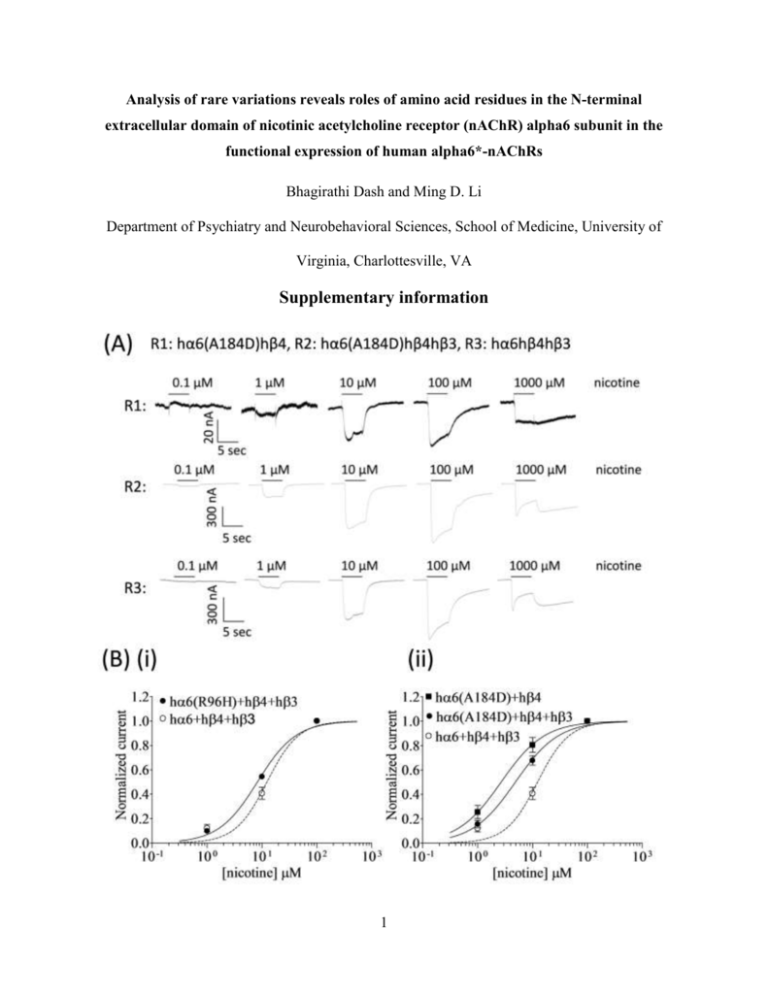
Analysis of rare variations reveals roles of amino acid residues in the N-terminal extracellular domain of nicotinic acetylcholine receptor (nAChR) alpha6 subunit in the functional expression of human alpha6*-nAChRs Bhagirathi Dash and Ming D. Li Department of Psychiatry and Neurobehavioral Sciences, School of Medicine, University of Virginia, Charlottesville, VA Supplementary information 1 Figure S1. Variations in nAChR hα6 subunit influence the nicotine sensitivity of hα6hβ4*nAChRs. (A) Representative traces are shown for current responses from oocytes (voltage clamped at -70 mV) responding to the application of indicated concentrations of nicotine (shown with the duration of drug exposure as black bars above the traces) and expressing indicated nAChR (i.e., R1: hα6A184Dhβ4-nAChR, R2: hα6A184Dhβ4hβ3-nAChR, R3: hα6hβ4hβ3-nAChR). (B) Results averaged across experiments were used to produce concentration-response (CR) curves (ordinate-mean normalized current ± SEM; abscissa - ligand concentration in log M) for inward current responses to nicotine as indicated for the nAChR expressed in oocytes and voltage clamped at -70 mV. Current amplitudes are represented as a fraction of the peak inward current amplitude in response to the most efficacious concentration of nicotine. Leftward shifts in nicotine CR curves for hα6R96Hhβ4hβ3-() [(B) (i)], hα6A184Dhβ4hβ3-() [(B) (ii)], hα6D199Yhβ4hβ3-() [(B) (iii)], or hα6S233Chβ4hβ3-() [(B) (iv)] nAChR are evident relative to that of hα6hβ4hβ3-nAChR (). Furthermore nicotine curves for hα6A184Dhβ4-() [(B) (ii)], hα6D199Yhβ4-() [(B) (iii)], or hα6S233Chβ4-() [(B) (iv)] nAChR are shifted leftward relative to those nAChR containing the same subunits but in the additional presence of hβ3 subunits. See Table S1 for parameters of nicotine action. 2 Table S1. Parameters for nicotine action at WT or variant hα6hβ4*- nAChRs Potencies [micromolar EC50 values with 95% confidence intervals (CI)], Hill coefficients (nH ± SE) and concentrations (M) where maximal peak current amplitudes (Imax) achieved are provided for nicotine acting at nAChR composed of the indicated subunits and from the indicated number of independent experiments (n) based on studies as shown in Fig S1. or indicate a significant (p<0.05) increase or decrease in indicated parameter at the indicated nAChR subtype relative to nAChR containing the same subunits but in the presence of the WT hα6 subunit (i.e., hα6hβ4- vs. variant-hα6hβ4- nAChR; hα6hβ4hβ3- vs. variant-hα6hβ4hβ3nAChR). or indicate a significant (p<0.05) increase or decrease, respectively, in indicated parameter at the indicated nAChR subtype relative to nAChR containing the same subunits but in the absence WT β3 subunits (i.e., hα6hβ4- vs. hα6hβ4hβ3- nAChR; varinat-hα6hβ4- vs. varianthα6hβ4hβ3- nAChR). ‘-‘ indicates that inconsistent functional responses in two electrode voltage clamp studies precluded determination of the parameter of interest. nAChR subunit combinations hα6+hβ4 hα6(R96H)+hβ4 hα6(A184D)+hβ4 hα6(D199Y)+hβ4 hα6(S233C)+hβ4 n hα6+hβ4+hβ3 hα6(R96H)+hβ4+hβ3 hα6(A184D)+hβ4+hβ3 hα6(D199Y)+hβ4+hβ3 hα6(S233C)+hβ4+hβ3 1 nH ± SE nH 3 3 3 3 EC50 (M) (95 % CI) 7.11 2.7 (1.8-3.9) 4.4 (2.4-8) 6.2 (5.2-7.4) 1.1±0.15 1.1±0.26 1.4±0.13 Imax conc. (µM) 100 100 100 100 100 4 3 3 3 3 12 (9.2-17) 8.2 (6.9-9.8) 4.9 (3.8-6.3) 11 (8.7-15) 6.9 (6-7.9) 1.5±0.45 1.2±0.13 1.1±0.11 1.5±0.42 1.4±0.12 100 100 100 100 100 From Kuryatov et al (2000) 3
