BMED2801 Lecture 2 Bacterial structure: General features Learning
advertisement
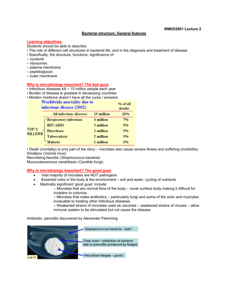
BMED2801 Lecture 2 Bacterial structure: General features Learning objectives Students should be able to describe: • The role of different cell structures in bacterial life, and in the diagnosis and treatment of disease • Specifically, the structure, functions, significance of: – nucleoid – ribosomes – plasma membrane – peptidoglycan – outer membrane Why is microbiology important? The bad guys • Infectious diseases kill ~ 15 million people each year • Burden of disease is greatest in developing countries • Modern medicine doesn’t have all the cures / answers • Death (mortality) is only part of the story – microbes also cause severe illness and suffering (morbidity) Smallpox (Variola virus) Necrotising fasciitis (Streptococcus bacteria) Mucocutaneonous candidiasis (Candida fungi) Why is microbiology important? The good guys Vast majority of microbes are NOT pathogens Essential roles in the body & the environment – soil and water, cycling of nutrients Medically significant ‘good guys’ include: – Microbes that are normal flora of the body – cover surface body making it difficult for invaders to colonize. – Microbes that make antibiotics – particularly fungi and some of the actin and myocytes invaluable to treating other infectious diseases. – Weakened strains of microbes used as vaccines – weakened strains of viruses – allow immune system to be stimulated but not cause the disease Antibiotic- penicillin discovered by Alexander Flemming What are microbes? • Viruses: very small + simple, have genes and enzymes but no metabolism, require a host cell to replicate • Bacteria (Prokaryotes): intermediate size + complexity, smallest free-living organisms, have own metabolism (living organism) • Fungi and protists (Eukaryotes): largest + most complex microbes, have nucleus and membrane-bound organelles Bacterial cell morphology: a diversity of shapes A lot of the filamentous one’s are important in making antibiotics e.g. streptomyces (largest antibiotic producing genus) Bacterial morphology: clinical relevance • Morphology is useful for identification and classification e.g. Staphylococcus & Streptococcus: Both cocci, but differ in arrangement: clusters vs. chains (this is quite distinctive – can distinguish the two apart) e.g. Spirochaetes are rare pathogens. Cell shape v.useful for diagnosis ((because this uncommon morphology is specific and unique) eg. spirochaetes observed in genital sores = syphilis e.g. Gastric ulcers + spirilla = Helicobacter pylori (is a Gram-negative, microaerophilic bacterium that inhabits various areas of the stomach and duodenum. It causes a chronic low-level inflammation of the stomach lining and is strongly linked to the development of duodenal and gastric ulcers and stomach cancer) Previously – antacids thought to treat stomach ulcers but now know that antibiotics is more effective. e.g. Respiratory distress + irregular rods in palisades (parallel rods) = Diphtheria Bacterial/prokaryotic cell structure, function and significance The Nucleoid Eukaryotic cells have two or more chromosomes contained within a membrane-delimited nucleus = TURE NUCLEUS Prokaryotes lack a membrane-delimited nucleus = NUCLEOID no membrane result in irregular structure – fingerlike projections into cytoplasm --> reason for irregular shape – some parts of nucleoid being transcribed more actively than others so DNA is being used and read and made into protein and these actively described parts extend out into cytoplasm to contact other bits of machinery required for transcription. • Irregularly shaped region called nucleoid Where the chromosome is housed • Bacteria usually have a single circular double stranded DNA arranged chromosome • Chromosome (d.s. DNA) + structural proteins (histones) together comprise the nucleoid • Bacterial nucleoid is NOT membrane bound •it is not the same as a true ‘nucleus’ (eg. eukaryotes) The nucleoid: not the only DNA in bacteria • Bacteria often contain accessory DNA elements that can replicate independently of the chromosome • Plasmids: - made of circular or linear d.s DNA - no protein coat - Many prokaryotes contain extrachromosomal DNA molecules apart from the nucleoid. - they replicate autonomously - plasmids play many impt. roles in the lives of organism that have them they also have proved invaluable to microbiologists/molecular geneticists in constructing and transferring new genetic combinations and in cloning genes. - e.g. types of plasmids: o resistance factors (R factors. R plasmids) – confer antibiotic resitance on the cells that contain them. have genes that code for enzymes capable of destroying/modifying antibiotics. • Bacteriophage (phage): - diverse genome either - made of linear d.s or s.s DNA or RNA - AND have protective protein coat (a virus that infects bacteria and may integrate into the genetic material of its host cell. Bacteriophages are used as vectors in gene cloning and have other biotechnological uses.) e.g. lambda ‘phage – live in ecoli – head filled DNA and tail – tail attaches to bacterial cell injecting DNA from head into cell – page invade cell and reproduce more phage inside cell until cell burst. The nucleoid: functions and significance • Nucleoid is the core genome of the cell (Plasmids & phage: accessory functions or parasites) • Information flows from nucleoid to messenger RNA (via RNA polymerase) transcription to protein (via ribosomes) translation • Antibiotics can target the nucleoid: ciprofloxacin inhibits DNA synthesis (Its mode of action depends upon blocking bacterial DNA replication by binding itself to an enzyme called DNA gyrase, thereby preventing the enzyme's ability to untwist the DNA double helix, which is required for DNA replication) rifampin inhibits RNA synthesis (Rifampin inhibits DNA-dependent RNA poymerase which leads to suppression of RNA synthesis in susceptible bacteria. The site of action appears to be the b subunit of the enzyme.) Ribosomes Ribosomes: protein synthesis factories • Small (~20 nm) note cell = 1μm and very numerous (~20,000/cell) – this is testament to importance of ribosomes to make proteins and keep making proteins as environmental conditions change. • Complex structure, made of many 2 different subunits: both ribosomal RNA (rRNA) (has a structural role only) and ribosomal proteins • pocaryotic ribosomes are called 70S ribosomes – constructed of a 50S and 30S subunit. • The 16S rRNA (most highly conserved part of ribosome) – sequence found in the 30S subunit is strongly conserved in all bacteria can use PCR to amplify little bits of ribosomal sequence present in all bacteria which is conveniently used to diagnose disease or identifying bacteria in particular environments furthermore, allows construction of - phylogeny (the evolutionary history of a species, genus, or group, as contrasted with the development of an individual ontogeny) reflect evolution really closely because it’s such a conserved molecule. Ribosome function and significance Function: Translation of mRNA into protein they are the site of protein synthesis – cytoplasmic ribosomes synthesise proteins destined to remain within the cell, whereas plasma membrane ribosomes make protein for transport outside the cell. • Proteins (enyzmes) are central to all aspects of life (metabolism, structural functions, sensors and regulatory function) translation is a critical cellular function hence, if we can disrupt this function we can kill the bacteria • Many different antibiotics inhibit ribosome function eg. streptomycin (bacteria- streptomyces), chloramphenicol, tetracycline The Cell Envelope Intro to the cell envelope • Cell envelope surrounds cell. Primary components are the cell wall and cell membrane(s) • Overall function: protective barrier against environment, separates ordered cytoplasm from chaotic exterior • Differences in cell envelope provide basis for dividing bacteria into Gram-negative and Gram-positive The Gram stain differentiates Gram-negative bacteria (red) from Gram-positive bacteria (purple) Outer membrane – lipid + Lipoplysacchairde (impt. for pathogens for tying to invade human body) Porin- traverse outer membrane – hollow channel – allow substrate and solutes e.g. nutrients to get into cytoplasm. Plasma membrane Phospholipid bilayer: detailed structure Bilayer self-assembles to thermodynamically favourable configuration: hydrophobic (water hating) inside, hydrophilic (water loving) outside Other plasma membrane components Proteins: eg. ATP synthase uses proton gradient across plasma membrane to generate energy (ATP) – relies on presence of membrane to maintain proton gradient to create ATP. Steroids: Prokaryote membranes contain hopanoids to give rigidity. (eukaryotes contain cholesterol) Plasma membrane functions • Selective permeability – the cell has to be able to protect itself from environment but need let: influx of nutrients, efflux of waste; Selectivity critical: can’t let cytoplasm out or toxins in • Metabolic processes eg. electron transport, respiration, and lipid biosynthesis, photosynthesis all occur in the membrane • Detection of environmental signals (signal has to get from outside of cell to inside of cell) : membrane receptors switch on regulatory proteins activate transcription Plasma membrane: clinical & other significance • Many antimicrobials disrupt membrane structure high pressure cell leakage of cytoplasm cell death • Eg. detergents (eg. SDS), antiseptics (eg. benzalkonium), and antibiotics (eg. polymyxin) • Membrane-disrupting toxins are used by bacteria to kill other bacteria eg. lantibiotics made by Lactobacillus lantibiotic toxin forms channels in cell membrane allowing interest to medicine- potential to use this to kill lactobacillus strains. Peptidoglycan - is unique to bacteria- impt. target for antibiotic. The cell wall contains peptidoglycan • Cell wall definition depends on bacterial type: Gram + Bacteria: peptidoglycan + teichoic acids Gram - Bacteria: peptidoglycan + outer membrane • Peptidoglycan (PG): structural polymer made of sugars and amino acids that is unique to bacteria • PG backbone made of a repeating di-saccharide unit; N-acetylglucosamine(NAG) + N-acetylmuramic acid (NAM) • PG cross-linked by peptide bridges = rigidity and structural strength. Peptidoglycan function + significance protect cell form bursting – high osmotic pressure • Function is osmotic protection. Cytoplasm very salty high pressure; membrane alone can’t resist this • Destruction of PG (eg. by lysozyme) leads to swelling, lysis, and death of bacteria • Many antibiotics target PG biosynthesis eg. penicillin, cephalosporins, vancomycin (used for front line of defence against golden staph which has become resistant to most antibiotics) • Thickness of PG determines Gram reaction PG is a useful diagnostic tool AND an excellent drug target Outer membrane structure (Gram negatives only) Outer membrane (OM) function & significance • Protective barrier against toxins eg. antibiotics, bile salts --> Deoxycholate is a bile salt: functions in intestine as emulsifier and antimicrobial properties selects types of bacteria in gut – generally gram neg. bacteria. • Presence of many sugars in LPS defines bacterial surface properties: hydrophilic, negative charge (due to phosphate in sugar) • O-antigen is highly variable, allows Gram neg. bacteria to rapidly change surface by changing sugar components valuable to bacteria appear different to immune system hence able to evade immune system because all it can see is surface of cell. e.g e-coli one species - there hundreds of O-antigen variants known. • Lipid A component is also known as endotoxin released into blood = fever, general malaise (depression) systemic toxic effects in Gram neg. infections Summary: structures / functions / significance • Cell morphology: diagnosis via microscopy • Nucleoid: information archive / cipro. / rif. • Ribosomes: protein factories killed by streptomycin • Plasma membrane: selective barrier attacked with detergents • Peptidoglycan: osmotic protection / penicillin • Outer membrane: protection- toxins, immune system

