- The Annual Congress of Tanta Faculty of Medicine
advertisement
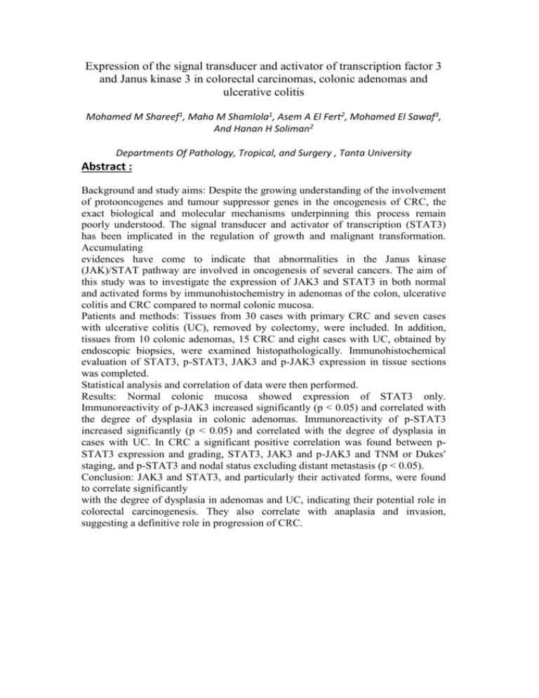
Expression of the signal transducer and activator of transcription factor 3 and Janus kinase 3 in colorectal carcinomas, colonic adenomas and ulcerative colitis Mohamed M Shareef1, Maha M Shamlola1, Asem A El Fert2, Mohamed El Sawaf3, And Hanan H Soliman2 Departments Of Pathology, Tropical, and Surgery , Tanta University Abstract : Background and study aims: Despite the growing understanding of the involvement of protooncogenes and tumour suppressor genes in the oncogenesis of CRC, the exact biological and molecular mechanisms underpinning this process remain poorly understood. The signal transducer and activator of transcription (STAT3) has been implicated in the regulation of growth and malignant transformation. Accumulating evidences have come to indicate that abnormalities in the Janus kinase (JAK)/STAT pathway are involved in oncogenesis of several cancers. The aim of this study was to investigate the expression of JAK3 and STAT3 in both normal and activated forms by immunohistochemistry in adenomas of the colon, ulcerative colitis and CRC compared to normal colonic mucosa. Patients and methods: Tissues from 30 cases with primary CRC and seven cases with ulcerative colitis (UC), removed by colectomy, were included. In addition, tissues from 10 colonic adenomas, 15 CRC and eight cases with UC, obtained by endoscopic biopsies, were examined histopathologically. Immunohistochemical evaluation of STAT3, p-STAT3, JAK3 and p-JAK3 expression in tissue sections was completed. Statistical analysis and correlation of data were then performed. Results: Normal colonic mucosa showed expression of STAT3 only. Immunoreactivity of p-JAK3 increased significantly (p < 0.05) and correlated with the degree of dysplasia in colonic adenomas. Immunoreactivity of p-STAT3 increased significantly (p < 0.05) and correlated with the degree of dysplasia in cases with UC. In CRC a significant positive correlation was found between pSTAT3 expression and grading, STAT3, JAK3 and p-JAK3 and TNM or Dukes’ staging, and p-STAT3 and nodal status excluding distant metastasis (p < 0.05). Conclusion: JAK3 and STAT3, and particularly their activated forms, were found to correlate significantly with the degree of dysplasia in adenomas and UC, indicating their potential role in colorectal carcinogenesis. They also correlate with anaplasia and invasion, suggesting a definitive role in progression of CRC. Probiotic Lactobacilus Casei (L.Casei) Can Support Intestinal Barrier through Immune Modulation in Experimental Cirrhosis Hanan H. Soliman1, Abdel-Razek A. Sheta2 & Omayma K. Afifi3 Departments of Tropical medicine & infectious diseases1, Anatomy2 & Histology3, Faculty of medicine, Tanta University, Egypt ABSTRACT Background/Aim: Liver cirrhosis induces intestinal mucosal barrier dysfunction and impairs mucosal defenses against luminal bacteria. This work aims to study the histological changes in the intestine of cirrhotic rats and to determine the ability of probiotic lactobacilus casei (L.casei) to immune-modulate or prevent cirrhosis-induced intestinal changes. Patients & Methods: Twenty seven adult rats were used in this study and classified into, 9 control, 9 injected with Ccl4 to induce cirrhosis and 9 animals received L.casei in addition to Ccl4. Sera from all animals were tested for TNF-α and INF-γ, and intestinal segments were examined histologically by light and electron microscopes. Results: In cirrhotic rats, TNF-α increased significantly while INF-γ showed non-significant increase compared to control. Histologically, intestinal mucosa showed degenerated lining epithelium and submucosal thickening. The intestinal wall appeared thin and atrophied in severely affected animals with complete damage of the lining epithelium. PAS stained sections showed apparently decreased goblet cells number and reactivity of the brush border. Ultrastructurally, enterocytes showed degenerated microvilli, wide intercellular spaces, dilated rER, swollen mitochondria, small irregular nuclei and many secondary lysosomes. In group III, TNF-α increased non-significantly, while IFN-γ showed a significant increase. Histologically, apparent normal villi with intact lining epithelium were noticed except few areas that were slightly affected. The brush border and goblet cells maintain their normal PAS-positive reaction. Ultrastructurally, Except for mild rER dilatation, most of enterocytes organelles appeared normal. Conclusion: L.casei is a good probiotic in preventing intestinal mucosal damage associating cirrhosis. These effects may be mediated by immunomodulation through TNF-α and IFN-γ. Key words: cirrhosis, intestinal barrier, probiotic, L.casei Corresponding Author: Hanan H. Soliman M.D. Department of Tropical Medicine, Faculty of Medicine, Tanta University, Egypt. E-mail: hanansol@gmail.com The Impact of H. pylori Eradication on Response to Oral Iron Therapy in Patients with Iron Deficiency Anemia 1Nesreen A Kotb, 1Mohamed M Bedewy, 2Hanan H Soliman, 3 Hala M Nagy, 4 Eman A Hasby Departments of 1Internal Medicine, 2 Tropical and Infectious Disease, 3 Clinical Pathology and 4Pathology, Faculty of Medicine, Tanta University, Tanta, Egypt. Abstract Infection with Helicobacter pylori has been associated with Iron deficiency anemia (IDA). We assessed the effect of eradication of H. pylori infection on response to oral iron treatment. Twenty patients with IDA of no obvious cause, unresponsive to oral iron therapy with positive gastric biopsy for H. pylori infection received sequential eradication therapy for 10 days followed by oral iron therapy. Treated patients were followed up at 6 weeks and 12 weeks post eradication to assess dynamic changes in hemoglobin, serum ferritin and transferrin saturation. While 65% of anemic H. pylori infected cases had pangastritis, 35% had antral gastritis. In the antrum severe and moderate gastritis were found in 40% and 45% of cases, respectively. Hemoglobin and serum ferritin level correlated inversely with grade of gastritis (P <0.001). Improvement in hematological parameters and serum iron profile was observed 6 weeks and 12 weeks post successful H.pylori eradication oral iron therapy. In conclusion, eradication of H. pylori infection enhances the response to oral iron supplementation in patients with refractory IDA. Screening and eradication of H. pylori should be a standard procedure for patients with IDA. Assessment of the Role of L-Carnitine in Improving Hepatic Encephalopathy Using MR Spectroscopy Hanan H. Soliman1, Dina H. Ziada1, Mohamed Hefeda2, Manal Hamisa2 and Samy A. Khodeir3 Tropical Medicine and Infectious Diseases1, Radiology2, Internal Medicine3, Departments, Faculty of Medicine Tanta University-Egypt Abstract: Background and aim: Hepatic encephalopathy (HE) is related to the presence of abnormal cerebral metabolites. MR Spectroscopy (MRS) can demonstrate the neurometabolite changes associated with therapy .The aim of this study was to evaluate the influence of L-carnitine on mental conditions, serum ammonia and neurometabolite levels in patients with HE using MRS. Patients and methods: Ten control subjects and 54 patients with grades II to III HE, were randomized into (GI) receiving lactulose 30ml/t.d.s as standard therapy and (GII) receiving L-Carnitine 350mg t.d.s in addition to the lactulose. Clinical assessment, fasting serum ammonia levels, and measurement of neurometabolite levels using proton MRS were recorded and compared at base line and after one week. Results: After one week, 25% of HE patients were reversed in group I versus 42.3% in group II. Fasting ammonia levels were significantly decreased in both groups compared to pretreatment levels and significantly lower in the L-carnitine and lactulose treated group compared to the lactulose group(P=0.041). Neurometabolites mI/Cre, Cho/Cre, Gx/Cre, and (Cho+mI)/Gx ratios were significantly improved in both groups compared to pre treatment levels, but the L Carnitine added group(II), showed a significant increase in mI/Cre, and (Cho+mI)/Glx ratios and decrease in Glx/Cre ratio in comparison to the lactulose group(p=0.002-p=0.003-p=0.002 respectively). Conclusion: Adding L Carnitine to lactulose for treatment of hepatic encephalopathy hastened the clinical improvement and was associated with significant improvements in serum ammonia and neurometabolites specially mI/Cre, and (Cho+ mI)/Glx and Glx/Cre ratios. Key words: Hepatic encephalopathy, Neurometabolites, Magnetic Resonance Spectroscopy, L-Carnitine. Abbreviations: (HE) hepatic encephalopathy, (MRS) Magnetic Resonance Spectroscopy, (Cho) Choline, (Cre) creatine, (NAA) N-acetylaspartate, (Glx)glutamine+glutamate, and(mI) myo-inositol Comparison of Partial Splenic embolisation versus Splenic Irradiation as Treatment of Hypersplenism in Advanced Cirrhosis Dina H. Ziada, Hanan H. Soliman, Amr Al-Badery*, Nehal El Mashd** Tropical Medicine and Infectious Diseases, Radiology *, Oncology and Nuclear Medicine**Departments- Tanta University-Egypt. Abstract: Hypersplenism in advanced cirrhotic patient is a conflicting issue as surgical splenectomy carries high perioperative morbidity and mortality rates. Partial splenic embolization (PSE) and low dose splenic irradiation (LDSI) are alternative procedures available in Egypt. The aim of this prospective controlled study was to compare (PSE) versus (LDSI) for ablation of spleen as treatment of hypersplenism in advanced cirrhosis. Patients and methods: Seventy one cirrhotic patients suffering hypersplenism underwent abdominal ultrasonography, liver functions, INR and bone marrow examination to confirm the diagnosis. They were enrolled in 3 groups. Group (I):26patients treated by (LDSI), Group (II):25patients treated by (PSE) and GroupIII:20 patients received conventional therapy as control group then we followed up these patients for 6months. Results: LDSI group showed steady progressive increase in all CBC elements. A significant increase in platelets started by week 2 While that of Hb and WBCs started by first month. While these increases started by the first week in PSE group which reached its peak in second week then gradually decreased but still significantly higher than pre treatment and control levels. Both procedures were well tolerated by the patients but more complications were reported with PSE. Both LDSI and PSE didn't affect liver functions. Conclusion: Both LDSI and PSE are safe and effective in treating hypersplenism in decompensated cirrhosis. They induce improvement of cytopenia and splenomegaly. The rapid onset and higher efficacy of PSE should be weighed against the wide safety and moderate efficacy of Splenic irradiation. Keywords: hypersplenism, advanced cirrhosis, Partial splenic embolization (PSE) and low dose splenic irradiation (LDSI). Leukocyte esterase and nitrate strip as a bedside test to diagnose Spontaneous Bacterial Peritonitis in Egyptian cirrhotic patients Hanan H. Soliman, Dina H. Ziada, and Naglaa Ghoneim* Tropical Medicine and Infectious Diseases, and microbiology* Departments- Tanta University-Egypt . ABSTRACT: Background and aim: Rapid diagnosis of Spontaneous Bacterial Peritonitis (SBP) is essential to reduce its mortality. The aim of this study was to evaluate the usefulness of Leukocyte esterase tests either alone or combined with nitrate tests in same dipstick as a bedside tool for diagnosis of SBP in Egyptian cirrhotic patients Patients and methods: Two hundred consecutive ascitic fluid samples were collected from 103 cirrhotic patients. Chylus, Bloody, serosangounus or bile stained samples were excluded. ,The routine and bacteriologic examination of ascitic samples were performed and ascitic sample tested using dipstick (Urocolor Test™ SD Standard diagnostics, Inc, Korea) for granulocyte esterase and nitrates designed for urine analysis. The procedure was the same as what the manufacturer described for urine. Results: By comparing the results of the routine and bacteriologic examination of ascitic and dipstick test, Leucocytic esterase (LE) test had sensitivity (sens.), specificity (sp.), positive predictive value(PPV), and negative predictive value(NPV) of 93.6%, 98.7% , 95.7%, and 98.7% respectively at colorimetric level 2+ ; and 74.5%, 99.3%, 97.2%, and 92.2% respectively at colorimetric level 3+. The nitrite test had sens., sp., PPV, and NPV of 12.7%, 85.6%, 21.4%, and 85.6% respectively. While combining both testes reveilles; 11%, 98.7%, 71%, and 78.2% respectively. LE test was superior to nitrite test in diagnosis of SBP Conclusion: Leukocyte esterase is more reliable than nitrate test in diagnosis of SBP. Uriscan SD dipstick test is better to be interpreted at colorimetric +2 which has high NPV(98.7%) allow to rollout the diagnosis of SBP in most cases. Uriscan SD dipstick test is rapid, cheap, and available everywhere, so it can be used as bedside test to screen SBP in Egyptian cirrhotic patients. Key words: Leukocyte esterase, nitrate test, Spontaneous Bacterial Peritonitis

