The Title Goes Here
advertisement
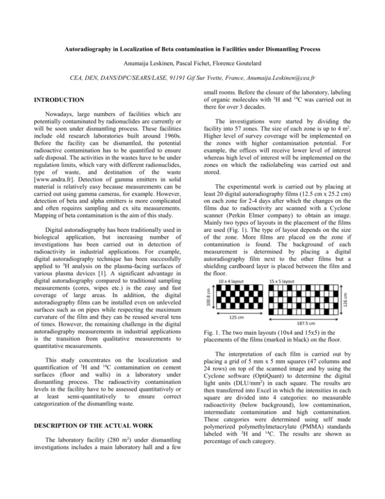
Autoradiography in Localization of Beta contamination in Facilities under Dismantling Process Anumaija Leskinen, Pascal Fichet, Florence Goutelard CEA, DEN, DANS/DPC/SEARS/LASE, 91191 Gif Sur Yvette, France, Anumaija.Leskinen@cea.fr INTRODUCTION Nowadays, large numbers of facilities which are potentially contaminated by radionuclides are currently or will be soon under dismantling process. These facilities include old research laboratories built around 1960s. Before the facility can be dismantled, the potential radioactive contamination has to be quantified to ensure safe disposal. The activities in the wastes have to be under regulation limits, which vary with different radionuclides, type of waste, and destination of the waste [www.andra.fr]. Detection of gamma emitters in solid material is relatively easy because measurements can be carried out using gamma cameras, for example. However, detection of beta and alpha emitters is more complicated and often requires sampling and ex situ measurements. Mapping of beta contamination is the aim of this study. Digital autoradiography has been traditionally used in biological application, but increasing number of investigations has been carried out in detection of radioactivity in industrial applications. For example, digital autoradiography technique has been successfully applied to 3H analysis on the plasma-facing surfaces of various plasma devices [1]. A significant advantage in digital autoradiography compared to traditional sampling measurements (cores, wipes etc.) is the easy and fast coverage of large areas. In addition, the digital autoradiography films can be installed even on unleveled surfaces such as on pipes while respecting the maximum curvature of the film and they can be reused several tens of times. However, the remaining challenge in the digital autoradiography measurements in industrial applications is the transition from qualitative measurements to quantitative measurements. This study concentrates on the localization and quantification of 3H and 14C contamination on cement surfaces (floor and walls) in a laboratory under dismantling process. The radioactivity contamination levels in the facility have to be assessed quantitatively or at least semi-quantitatively to ensure correct categorization of the dismantling waste. DESCRIPTION OF THE ACTUAL WORK The laboratory facility (280 m2) under dismantling investigations includes a main laboratory hall and a few small rooms. Before the closure of the laboratory, labeling of organic molecules with 3H and 14C was carried out in there for over 3 decades. The investigations were started by dividing the facility into 57 zones. The size of each zone is up to 4 m2. Higher level of survey coverage will be implemented on the zones with higher contamination potential. For example, the offices will receive lower level of interest whereas high level of interest will be implemented on the zones on which the radiolabeling was carried out and stored. The experimental work is carried out by placing at least 20 digital autoradiography films (12.5 cm x 25.2 cm) on each zone for 2-4 days after which the changes on the films due to radioactivity are scanned with a Cyclone scanner (Perkin Elmer company) to obtain an image. Mainly two types of layouts in the placement of the films are used (Fig. 1). The type of layout depends on the size of the zone. More films are placed on the zone if contamination is found. The background of each measurement is determined by placing a digital autoradiography film next to the other films but a shielding cardboard layer is placed between the film and the floor. Fig. 1. The two main layouts (10x4 and 15x5) in the placements of the films (marked in black) on the floor. The interpretation of each film is carried out by placing a grid of 5 mm x 5 mm squares (47 columns and 24 rows) on top of the scanned image and by using the Cyclone software (OptiQuant) to determine the digital light units (DLU/mm2) in each square. The results are then transferred into Excel in which the intensities in each square are divided into 4 categories: no measurable radioactivity (below background), low contamination, intermediate contamination and high contamination. These categories were determined using self made polymerized polymethylmetacrylate (PMMA) standards labeled with 3H and 14C. The results are shown as percentage of each category. RESULTS After three months investigation, approximately 45% of the laboratory surface has been investigated (more than 100 m2). The activity levels in 60% of the investigated zones are below background level. Significant contamination (more than 10% of the zone surface activity is above background level) has been found in 4 zones. Fig. 2 shows an example image of a film on a contaminated zone. The strong contrast between the light gray regions and the black regions indicates large difference in their activities. The advantages in using digital autoradiography in dismantling investigations are evident: the technique is easy to use, it does not require expensive equipment, and contamination can be localized. ENDNOTES This research was funded by the DEMSAC project in CEA Saclay France. REFERENCES 1. S. ROSANVALLON, “Steel detritiation optimization of a process” Fusion Eng Des, 51-52, 605-609, (2000) Fig. 2. Scanning of a film on a contaminated zone. The results represent contamination on the relatively thin top layer of the cement surface due to the short penetration ranges of beta emitters in solid material. The migration of the activity deeper into the material is essential in the estimations of the activity levels in the bulk material. Therefore, some sampling was carried out in regions in which higher contamination levels has been measured by autoradiography to determine if the contamination is located on the surface or if it is also present in the bulk material. Fig 3 shows an example image of two films before and after sampling exhibiting clear difference between observed activities and therefore top layer contamination. Fig. 3. Images of two digital autoradiography films before (above) and after (below) sampling. Sampling area is circled.
