Investigation of the role of the Inflammasome triggering HIN200
advertisement
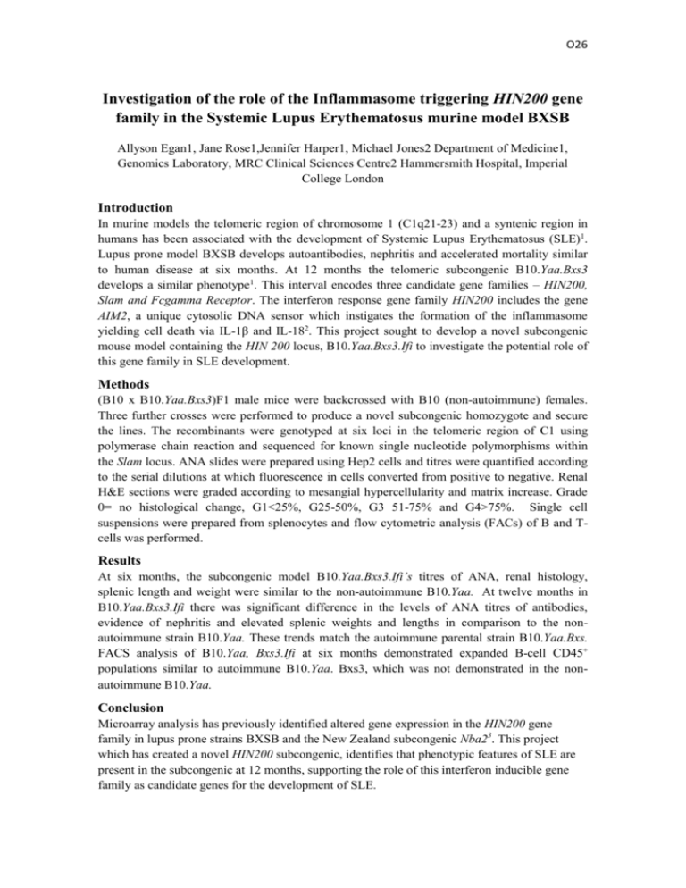
O26 Investigation of the role of the Inflammasome triggering HIN200 gene family in the Systemic Lupus Erythematosus murine model BXSB Allyson Egan1, Jane Rose1,Jennifer Harper1, Michael Jones2 Department of Medicine1, Genomics Laboratory, MRC Clinical Sciences Centre2 Hammersmith Hospital, Imperial College London Introduction In murine models the telomeric region of chromosome 1 (C1q21-23) and a syntenic region in humans has been associated with the development of Systemic Lupus Erythematosus (SLE)1. Lupus prone model BXSB develops autoantibodies, nephritis and accelerated mortality similar to human disease at six months. At 12 months the telomeric subcongenic B10.Yaa.Bxs3 develops a similar phenotype1. This interval encodes three candidate gene families – HIN200, Slam and Fcgamma Receptor. The interferon response gene family HIN200 includes the gene AIM2, a unique cytosolic DNA sensor which instigates the formation of the inflammasome yielding cell death via IL-1β and IL-182. This project sought to develop a novel subcongenic mouse model containing the HIN 200 locus, B10.Yaa.Bxs3.Ifi to investigate the potential role of this gene family in SLE development. Methods (B10 x B10.Yaa.Bxs3)F1 male mice were backcrossed with B10 (non-autoimmune) females. Three further crosses were performed to produce a novel subcongenic homozygote and secure the lines. The recombinants were genotyped at six loci in the telomeric region of C1 using polymerase chain reaction and sequenced for known single nucleotide polymorphisms within the Slam locus. ANA slides were prepared using Hep2 cells and titres were quantified according to the serial dilutions at which fluorescence in cells converted from positive to negative. Renal H&E sections were graded according to mesangial hypercellularity and matrix increase. Grade 0= no histological change, G1<25%, G25-50%, G3 51-75% and G4>75%. Single cell suspensions were prepared from splenocytes and flow cytometric analysis (FACs) of B and Tcells was performed. Results At six months, the subcongenic model B10.Yaa.Bxs3.Ifi’s titres of ANA, renal histology, splenic length and weight were similar to the non-autoimmune B10.Yaa. At twelve months in B10.Yaa.Bxs3.Ifi there was significant difference in the levels of ANA titres of antibodies, evidence of nephritis and elevated splenic weights and lengths in comparison to the nonautoimmune strain B10.Yaa. These trends match the autoimmune parental strain B10.Yaa.Bxs. FACS analysis of B10.Yaa, Bxs3.Ifi at six months demonstrated expanded B-cell CD45+ populations similar to autoimmune B10.Yaa. Bxs3, which was not demonstrated in the nonautoimmune B10.Yaa. Conclusion Microarray analysis has previously identified altered gene expression in the HIN200 gene family in lupus prone strains BXSB and the New Zealand subcongenic Nba23. This project which has created a novel HIN200 subcongenic, identifies that phenotypic features of SLE are present in the subcongenic at 12 months, supporting the role of this interferon inducible gene family as candidate genes for the development of SLE. O26 References 1. Morley et al. JI 2004, 173:4277-4285 2. Burckstummer et al Nature Immunology vol 10, Number 3 March 2009 3. Haywood Morley et al Genes and Immunity (2006) 7, 250-263


![FA801 [4] - University of Kent](http://s3.studylib.net/store/data/006628509_1-5ca1ad65bf344fd7e4f9aba2178de697-300x300.png)