Visualization of human visual processing by MEG and fMRI Sunao
advertisement
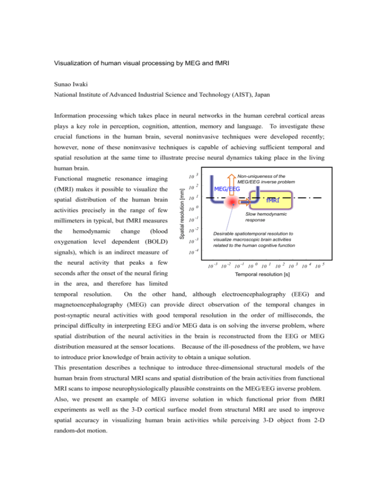
Visualization of human visual processing by MEG and fMRI Sunao Iwaki National Institute of Advanced Industrial Science and Technology (AIST), Japan Information processing which takes place in neural networks in the human cerebral cortical areas plays a key role in perception, cognition, attention, memory and language. To investigate these crucial functions in the human brain, several noninvasive techniques were developed recently; however, none of these noninvasive techniques is capable of achieving sufficient temporal and spatial resolution at the same time to illustrate precise neural dynamics taking place in the living human brain. 3 10 (fMRI) makes it possible to visualize the 10 2 10 1 10 0 spatial distribution of the human brain activities precisely in the range of few millimeters in typical, but fMRI measures the hemodynamic change (blood Spatial resolution [mm] Functional magnetic resonance imaging 10 10 oxygenation level dependent (BOLD) 10 signals), which is an indirect measure of 10 the neural activity that peaks a few seconds after the onset of the neural firing Non-uniqueness of the MEG/EEG inverse problem MEG/EEG fMRI Slow hemodynamic response -1 -2 Desirable spatiotemporal resolution to visualize macroscopic brain activities related to the human cognitive function -3 -4 10 -3 10 -2 10 -1 10 0 10 1 10 2 10 3 10 4 10 5 Temporal resolution [s] in the area, and therefore has limited temporal resolution. On the other hand, although electroencephalography (EEG) and magnetoencephalography (MEG) can provide direct observation of the temporal changes in post-synaptic neural activities with good temporal resolution in the order of milliseconds, the principal difficulty in interpreting EEG and/or MEG data is on solving the inverse problem, where spatial distribution of the neural activities in the brain is reconstructed from the EEG or MEG distribution measured at the sensor locations. Because of the ill-posedness of the problem, we have to introduce prior knowledge of brain activity to obtain a unique solution. This presentation describes a technique to introduce three-dimensional structural models of the human brain from structural MRI scans and spatial distribution of the brain activities from functional MRI scans to impose neurophysiologically plausible constraints on the MEG/EEG inverse problem. Also, we present an example of MEG inverse solution in which functional prior from fMRI experiments as well as the 3-D cortical surface model from structural MRI are used to improve spatial accuracy in visualizing human brain activities while perceiving 3-D object from 2-D random-dot motion.
