Anatomy Of The Heart The Heart Is Located In The Chest, Directly
Anatomy of the Heart
The heart is located in the chest, directly above the diaphragm in the region of the thorax called mediastinum, specifically the middle mediastinum. The normal human heart varies with height and weight .
The tip (apex) of the heart is pointed forward, downward, and toward the left. The (inferior) diaphragmatic surface lies directly on the diaphragm. The heart lies in a double walled fibroserous sac called the pericardial sac,which is divided into (a) fibrous pericardium, and (b) serous pericardium. The fibrous pericardium envelops the heart and attaches onto the great vessels .
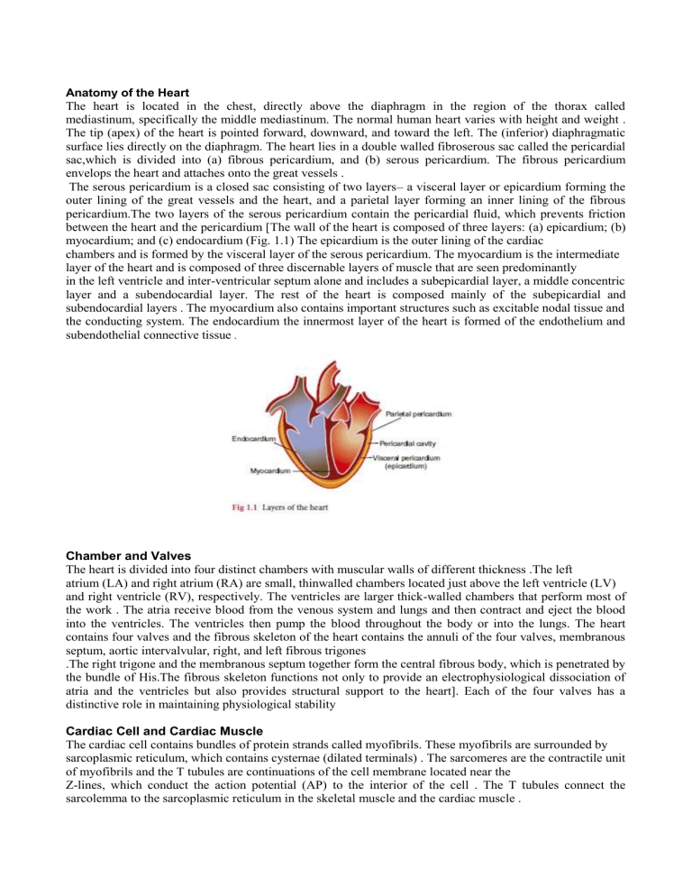
The serous pericardium is a closed sac consisting of two layers– a visceral layer or epicardium forming the outer lining of the great vessels and the heart, and a parietal layer forming an inner lining of the fibrous pericardium.The two layers of the serous pericardium contain the pericardial fluid, which prevents friction between the heart and the pericardium [The wall of the heart is composed of three layers: (a) epicardium; (b) myocardium; and (c) endocardium (Fig. 1.1) The epicardium is the outer lining of the cardiac chambers and is formed by the visceral layer of the serous pericardium. The myocardium is the intermediate layer of the heart and is composed of three discernable layers of muscle that are seen predominantly in the left ventricle and inter-ventricular septum alone and includes a subepicardial layer, a middle concentric layer and a subendocardial layer. The rest of the heart is composed mainly of the subepicardial and subendocardial layers . The myocardium also contains important structures such as excitable nodal tissue and the conducting system. The endocardium the innermost layer of the heart is formed of the endothelium and subendothelial connective tissue .
Chamber and Valves
The heart is divided into four distinct chambers with muscular walls of different thickness .The left atrium (LA) and right atrium (RA) are small, thinwalled chambers located just above the left ventricle (LV) and right ventricle (RV), respectively. The ventricles are larger thick-walled chambers that perform most of the work . The atria receive blood from the venous system and lungs and then contract and eject the blood into the ventricles. The ventricles then pump the blood throughout the body or into the lungs. The heart contains four valves and the fibrous skeleton of the heart contains the annuli of the four valves, membranous septum, aortic intervalvular, right, and left fibrous trigones
.The right trigone and the membranous septum together form the central fibrous body, which is penetrated by the bundle of His.The fibrous skeleton functions not only to provide an electrophysiological dissociation of atria and the ventricles but also provides structural support to the heart]. Each of the four valves has a distinctive role in maintaining physiological stability
Cardiac Cell and Cardiac Muscle
The cardiac cell contains bundles of protein strands called myofibrils. These myofibrils are surrounded by sarcoplasmic reticulum, which contains cysternae (dilated terminals) . The sarcomeres are the contractile unit of myofibrils and the T tubules are continuations of the cell membrane located near the
Z-lines, which conduct the action potential (AP) to the interior of the cell . The T tubules connect the sarcolemma to the sarcoplasmic reticulum in the skeletal muscle and the cardiac muscle .
Cardiac muscle is an involuntary striated muscle,which is mononucleated and has cross-striations formed by alternate segments of thick and thin protein filaments, which are anchored by segments called Z-lines. The cardiac muscle is relatively shorter than skeletal muscle and actin and myosin are the primary structural proteins. When the cardiac muscle is observed by a light microscope, the thinner actin filaments appear as lighter bands, while thicker myosin filaments appear as darker bands . The dark bands are actually the region of overlap between the actin and myosin filaments and the light bands are the region of actin filaments. The thinner actin flaments contain two other proteins called troponin and tropomyosin, which play an important role in contraction .
Cardiac muscle also contains dense bands (specialized cell junctions) called intercalated discs that separate individual cells from one another at their ends and these discs consist of a transverse and a lateral portion.
The transverse portion of the disc acts as a zone of firm adhesion and a route of transmission of contractile force and the lateral portion of the disc acts as a gap junction across which propagation of electrical impulses between the adjacent cardiac cells occurs . This in effect allows the individual cells of the heart to act as a syncytium .
