Additional file 1
advertisement
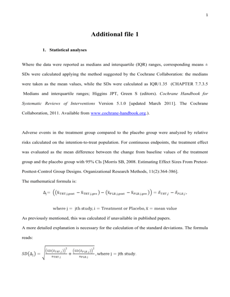
1 Additional file 1 1. Statistical analyses Where the data were reported as medians and interquartile (IQR) ranges, corresponding means ± SDs were calculated applying the method suggested by the Cochrane Collaboration: the medians were taken as the mean values, while the SDs were calculated as IQR/1.35 (CHAPTER 7.7.3.5 Medians and interquartile ranges; Higgins JPT, Green S (editors). Cochrane Handbook for Systematic Reviews of Interventions Version 5.1.0 [updated March 2011]. The Cochrane Collaboration, 2011. Available from www.cochrane-handbook.org.). Adverse events in the treatment group compared to the placebo group were analyzed by relative risks calculated on the intention-to-treat population. For continuous endpoints, the treatment effect was evaluated as the mean difference between the change from baseline values of the treatment group and the placebo group with 95% CIs [Morris SB, 2008. Estimating Effect Sizes From PretestPosttest-Control Group Designs. Organizational Research Methods, 11(2):364-386]. The mathematical formula is: As previously mentioned, this was calculated if unavailable in published papers. A more detailed explanation is necessary for the calculation of the standard deviations. The formula reads: 2 Firstly, the standard deviation of the change from baseline values was calculated using: The variances for pre- and post- treatment/placebo were known, while the degree of correlation was always unknown and had to be guessed. When for a given outcome, δij and the corresponding SD (δij) were available for one or more studies, ρij were calculated rearranging the formula for SD (δij) and the average degree of correlation was used as an ‘educated’ guess for the jth study for which SD (δij)was unknown. On the other hand, when no study reported δij and the corresponding SD (δij) ρij equal to 0.90 was hypothesized. Once SD (δij) for the treatment and the placebo groups had been calculated for each study on each outcome, the SD for the treatment effect SD (Δj) could be calculated using the formula specified above. A meta-analysis was performed on all outcomes and effect sizes were combined to give a pooled estimate of a weighted average of the treatment effects Δj for each study, the weights being the reciprocals of the variance. 2. Heterogeneity As already mentioned in the main body of the article, the studies included in this review were very heterogeneous in the population characteristics. Starting from the main analysis, we explored the pattern of heterogeneity in all subgroup analyses, or sensitivity analyses, performed for all outcomes. Results are herein reported. Cut-offs for the I2 statistic were: low=30%; moderate=3075%; high≥75%. 2.1 Cardiac performance 3 Cardiac Index – All studies reported similar findings for this outcome and an I2 of 0% was calculated from the main analysis (p= 0.66). EF – From the main analysis an I2 of 84% was calculated and the test for heterogeneity resulted highly statistically significant (p<0.001). This remained high in the LVH (I2=79%, p=0.008), while in the non/mild-LVH this reduced to 0% (p=0.19). High heterogeneity was also observed in the age subgroups (<60year-olds: I2=80%, p=0.024; >60year-olds: I2=90%, p<0.001). E/A ratio – The main analysis for this outcome showed very high heterogeneity (I2=93%, p=0.753). p<0.001). No improvement was obtained when the analysis was restricted to left-heart disease (I2=96%, p<0.001) and to <60 years of age (I2=90%, p=0.001). 2.2 Cardiac geometry LVMi – High heterogeneity emerged from the main analysis (I2=96%, p<0.001), and an improvement was found when stratifying the analysis by hypertrophy (LVH: I2=88%, p=0.003; non/mild LVH: I2=80%, p=0.005). Heterogeneity was high when restricting the analysis to age >60 years (I2=95%, p<0.001). EDVi – The main analysis on this outcome showed a high heterogeneity with I2 of 94% (p<0.001), but when restricting the analysis on non/mild LVH subgroup this reduced to 0% (p=0.53). IVS and VTD – The main analysis on these outcomes included only 2 studies largely different from each other (I2=95%, p<0.001 and I2=90%, p<0.001, respectively). 2.3 Cardiac biomarkers NT-proBNP – The high heterogeneity that emerged from the main analysis on this outcome (I2=82%, p<0.001) could be removed in the LVH subgroup (I2=0%, p=0.52), but reached a moderate level in the non/mild LVH group (I2=75%, p=0.017). Restricting the analysis to the left- 4 heart disease and to the group of >60 years of age, the heterogeneity remained moderate-high (I2=84%, p<0.001,and I2=72%, p=0.013, respectively); when stratifying by EF this dropped to 0% in the rEF group (p=0.63), and to a moderate level for the pEF (I2=75%, p=0.017). 2.4 Hemodynamic parameters HR – From the main analysis a moderate heterogeneity emerged for this outcome (I2=56%, p=0.004); this could be removed by stratifying in LVH and non/mild LVH (I2=0.0%, p=0.96 for both groups). The heterogeneity was also removed by restricting the analysis to HF patients with reduced EF (I2=0.0%, p=0.38). In the right-heart disease group the heterogeneity was also 0% (p=0.85). In the left-heart disease this remained high (I2=81%, p<0.001). From the subgroup analysis by compound it emerged that heterogeneity remained moderate in the sildenafil group (I2=64%, p=0.002), while this was removed in the vardenafil and tadalafil groups (respectively, I2=0.0%, p=0.23, and I2=0.0%, p=0.27). Heterogeneity was removed in the <60 years of age group (I2=0.0%, p=0.86), but this increased to 79% (p<0.001) in the >60 years of age group. Lastly, the analysis was restricted to the non-cardiac disease group and an I2=0.0% (p=0.27) was estimated. SBP – Moderate heterogeneity emerged for this outcome (I2=49%, p=0.031), which decreased to 0.0% in the LVH group (p=0.90) and increased to 79% in the non/mild LVH group (p=0.007). Increased heterogeneity in non/mild LVH group could be related to the inclusion of patients with cardiac and non-cardiac diseases. Restricting the analysis to the left-heart disease, no improvement could be observed (I2=66%, p=0.007). In the sildenafil subgroup the heterogeneity remained moderate (I2=60%, p=0.013), and was removed in the tadalafil group (I2=0.0%, p=0.69). In the group of <60 years of age the heterogeneity was absent (I2=0.0%, p=0.43) while this was moderate in the group >60 years of age 5 (I2=72%, p=0.006). Lastly, in the non-cardiac disease group the heterogeneity was absent (I2=0.0%, p=0.50). DBP – Moderate heterogeneity was also present in the main analysis of this outcome (I2=52%, p=0.018). Stratifying the analysis by LVH could not bring any relevant improvement (non/mild LVH: I2=91%, p=0.001, and LVH: I2=66%, p=0.05). The heterogeneity was removed in the rightheart disease group (I2=0.0%, p=0.99), but increased in the left-heart disease group (I2=75%, p=0.001). When considering the treatments, heterogeneity remained moderate in the sildenafil group (I2=63%, p=0.006), but was removed in the tadalafil group (I2=0.0%, p=0.78). From the subgroup analysis by age no heterogeneity emerged in the group of <60 years of age (I2=0.0%, p=0.89), but higher heterogeneity compared to the main analysis in the other group (I2=71%, p=0.007). No heterogeneity was found in the non-cardiac disease group (I2=0.0%, p=0.65). MAP – The main analysis performed on this outcome showed a moderate heterogeneity (I2=71%, p=0.002), which could be removed when restricting the analysis to the LVH group (I2=0.0%, p=0.24). An analysis restricted to the left-heart disease group could not reduce the heterogeneity (I2=75%, p=0.002), nor could an analysis on the sildenafil group only (I2=72%, p=0.003). When stratifying the analysis by age, it emerged that no heterogeneity was present in the group <60 years of age (I2=0.0%, p=0.67), but high heterogeneity was found in the group >60 years of age (I2=83%, p=0.002). SVRi – High heterogeneity was found among all studies reporting findings on this outcome (I2=89%, p<0.001). Subgroup and sensitivity analyses could bring some improvement only in the group <60 years of age the I2 was 0.0% (p=0.014). 6 2.5 Endothelial Function FMD – Very high heterogeneity was present in the main analysis (I2=99%, p<0.001) and no relevant improvement could be obtained by any subgroup or sensitivity analysis. In particular, in the group ≥60 years of age the I2 was 99% (p=0.032), and in the non-cardiac disease group the I2 was 96% (p<0.001). 2.6 Adverse Events No heterogeneity was found among studies, which reported the following adverse events: tinnities, dispnea, skin irritation, and atrial fibrillation. For all other adverse events a high level of heterogeneity was found, but this was statistically significant only for the following: gastric (dyspepsia, pyrexia, gastritis), flushing or rash, musculoskeletal (pain in limb, back pain, myalgia, muscle cramps), headache, and pruritus. 3. Risk of bias All publications reported results from RCTs: however randomization method and allocation concealment were inappropriately described in 54% of studies, so the risk of selection bias was unclear for this group, while for the remaining 46% of trials the risk of selection bias was low. All studies were of low risk for performance and detection bias. Low risk of reporting bias was allocated to 88% of studies: one study 1 did not have enough information, so the risk for this bias was unclear, while 12% were judged to have a medium-high risk of reporting bias due to incomplete reporting of some outcomes of interest (only baseline scores reported, or scores reported in graphs, but not in tables); some outcomes were measured but not reported; some outcomes were reported as indexed scores at baseline but not post-treatment. 7 A medium-high risk for other biases was allocated to some RCTs, in which the same population enrollment is suspected2-5 , or the sample is widely heterogeneous 6, or the study design is a crossover without washout period. 4. Limitations In subgroup analyses we observed specific limitations due to the coupling of studies and the paucity of available data. The analyses of E/A ratio showed no changes, probably due to the paucity of data reported, to the heterogeneity of clinical conditions of the patients enrolled in the trials 3;7;8 and because the baseline value are normal according to age (E/A ratio = 1.880.45; CI: 0.98 to 2.78, from 16 to 20 years; 0.96 0.18; CI: 0.6 to 1.32, over 60 years) 9. For LVMi, subgroup for length of treatment revealed conflicting results (data not shown). In the two studies 10;11 lasting less than 6 months, LVMi increased significantly (+3.011 g/m2; CI -0.506 to 5.515; p=0.018). Andersen et al. included subjects after acute myocardial infarction, in which normal baseline LVMi did not significantly increase after sildenafil (from 9319 g/m2 to 9520 g/m2) whereas in the placebo group LVMi did not decreased significantly (from 9320 g/m2 to 9118 g/m2). And Giannetta et al. performed a RCT on type 2 diabetic patients in which increased LVMi (119.425.7 g/m2) did not significantly decreased after sildenafil and placebo (respectively, -0.675.07 g/m2 and -2.037.64g/m2). Notably, the observed LVMi increase of the subgroup analysis was not clinically significant. In the three studies 6-8 lasting more than 6 months, LVMi decreased significantly and clinically (-8.446 g/m2; 95% CI -15.694 to -1.197; p = 0.022). The two studies by Guazzi M. et al. were performed in patients with LVH due to HF of various etiologies (baseline LVMi = 166.412.1 g/m2 – 8 and 147.230.2 g/m2 7) and were analyzed together with the 8 study of Redfield M. et al 6 that was conducted in non-LVH subjects (baseline LVMi = 65 g/m2; IQR: 54-78). The geometric parameters, IVS and VTD, were assessed in a few selected trials. 9 Supplement Table1. Characteristics of studies: N° patients ITT, N° male vs. female. Study N° Patients (ITT) N° Male vs. Female Aldashev AA, Thorax, 2005 12 9S vs. 8P 5S vs. 8P All male Amin A, Congest Heart Fail, 2013 13 53S vs. 53P Andersen MJ, Circulation, 2013 10 35S vs. 35P Badesch, J Rheumatol, 2007 14 21 S 20 S 21 S 22 P Behling A, J of Cardiac Fail, 2008 15 11S vs. 8P Bharani A, Indian Heart J, 2007 16 8T–P T-P: 4m vs. 4f Bocchio M, Atherosclerosis, 2008 17 18T vs. 18P All male 69 S 68 S 71 S 70 P S: 20m vs. 49f 21m vs. 47f 15m vs. 56f P: 13m vs. 57f Giannetta E, Circulation, 2012 11 29S vs. 25P All male Goldberg DJ, Circulation, 2011 2 28 S - P S-P: 18m vs. 10f Goldberg, Pediatr Cardiol, 2012 3 28 S - P S-P: 18m vs. 10f Groeneweg G, BMC Muscoloskeletan disorders, 2008 1 12T vs.12P T: 3m vs. 9f P: 1m vs. 11f Guazzi M, J Am Coll Cardiol, 2007 19 23S vs. 23P All male Guazzi M, Circulation, 2011 8 22S vs. 22P S: 17m vs. 5f P: 18m vs. 4f Guazzi M, Circ Heart Fail, 2011 7 23S vs. 22P All male Guazzi M, Europ J Heart Failure, 2012 20 16S vs. 16P All male Jing, Z.C, Am J Resp Crit Care Med, 2011 21 43V vs. 16P Lewis GD, Circulation, 20074 17S vs. 17P Lewis GD, Circ Heart Fail, 2008 4;5 15S vs. 15P S&P: 27m vs. 3f Rosano G MC , European Urology, 2005 22 16T vs. 16P All male Redfield MM, JAMA, 2013 6 113S vs. 103P S: 64m vs. 49f P: 48m vs. 55f Sastry BKS, JACC, 2004 23 32 S - P S-P: 20m vs.12f Suntharalingam J, Chest, 2008 24 9S vs. 10P S: 2m vs. 7f P: 7m vs. 3f Van AH, J Sex Med ,2005 25 175V vs. 175P All male Galiè N, NEJM, 2005 18 S: 38m vs. 15f P: 40m vs. 13f S: 32m vs. 3f P: 32m vs. 3f S: 5m vs. 16f 3m vs. 17f 2m vs. 19f P: 4m vs. 18f S: 9m vs. 2f P: 4m vs. 4f V: 8m vs. 35f P: 2m vs. 14f S: 14m vs. 3f P: 15m vs. 2f 10 Supplement Table2. Risk of bias summary per Cochrane metrics. '+' indicates present STUDY Aldashev AA, Thorax, 2005 12 Amin A, Congest Heart Fail, 2013 13 Andersen MJ, Circulation, 2013 10 Badesch, J Rheumatol, 2007 14 Behling A, J of Cardiac Fail, 2008 15 Bharani A, Indian Heart J, 2007 16 Bocchio M, Atherosclerosis, 2008 17 Galiè N, NEJM, 2005 18 Giannetta E, Circulation, 2012 11 Goldberg DJ, Circulation, 2011 2 Goldberg, Pediatr Cardiol, 2012 3 Groeneweg G, BMC Muscoloskeletan disorders, 2008 1 Guazzi M, J Am Coll Cardiol, 2007 19 Guazzi M, Circulation, 2011 8 Guazzi M, Circ Heart Fail, 2011 7 Guazzi M, Europ J Heart Failure, 2012 20 Jing, Z.C, Am J Resp Crit Care Med, 2011 21 Lewis GD, Circulation, 2007 4 Lewis GD, Circ Heart Fail, 2008 5 Rosano G MC , European Urology, 2005 22 Redfield MM, JAMA, 2013 6 Sastry BKS, JACC, 2004 23 Suntharalingam J, Chest, 2008 24 Van Ahlen H, J Sex Med, 2005 25 (la voce giusta è 36 SELECTION BIAS ? ? ? ? ? ? ? ? ? ? ? ? ? PERFORMANCE BIAS - DETECTION BIAS - ATTRITION BIAS - REPORTING BIAS + + + + ? + + ? + OTHER BIAS + + + - 11 Table3. Summary of findings on Adverse Events (AEs) Adverse events Flushing or Rash Headache Gastric (Dyspepsia, Pyrexia, Gastritis) Epistaxis Intestinal (diarrhea, abdominal pain) Nasopharyngitis Musculoskeletal (pain in limb, back pain, myalgia, muscle cramps) Dizziness or tinnitus Visual (Photosensitivity, visual disturbance) Skin irritation Insomnia Pruritus Dyspnea Atrial fibrillation Symptomatic hypotension Death N° Patients (ITT) PDE5i vs. Plb RR 95% CI 1217 3.406 731 vs. 514 ‡ [1.628; 7.126] 1260 2.507 768 vs. 552‡ [1.416; 4.439] 588 4.138 382 vs. 206 [1.564;10.946] 467 4.701 322 vs. 145 [1.314;16.812] 586 1.368 409 vs. 205‡ [0.558; 3.353] 190 1.474 115 vs. 75 [0.271; 8.002] 554 2.622 302 vs. 252‡ [0.822; 8.370] 428 0.910 248 vs. 208‡ [0.516; 1.607] 343 2.303 220 vs. 151‡ [0.650; 8.157] 305 0.929 157 vs. 148 [0.441; 1.959] 527 2.760 366 vs. 161 [0.946; 8.052] 190 1.564 115 vs. 75 [0.068;35.967] 286 1.477 148 vs. 138 [0 .710; 3.069] 78 0.280 39 vs. 39 [0.049; 1.604] 244 2.088 141 vs. 131‡ [0.093;47.072] 301 0.658 215 vs. 118‡ [0.133; 3.252] p value I-square [het. b/w studies] References 0.001 85.83% 2;6;13-15;18-21;25 0.002 80.19% 2;6;10;13-15;18;21;23-25 0.004 92.16% 10;13;14;18;20;24 0.017 89.59% 13;14;18 0.493 88.06% 2;13;14;18;20;21 0.653 98.03% 13;14 0.103 95.07% 2;13;14;18;21 0.746 0% 2;6;13;15;21 0.196 90.60% 0.847 0% 6;10;24 0.063 85.58% 13;14;18;21 0.780 99.48% 13;14 0.296 0% 6;10 0.153 0% 19;20 0.643 99.39% 2;6 0.608 90.03% 2;13;18 ~ 18;21;23 ‡ : inclusion of studies with a cross over design; ~ : Galiè et al. 120 mg/ die and 240 mg/die; ~~Galiè et al. 60 mg/die and 240 mg ~~ 12 Additional File 1. FIGURE LEGEND Additional file_ Figure 1. Effects of PDE5i over Placebo on hemodynamic parameters. HR: main analysis and subgroup analyses in right heart and rEF. DBP, MAP, SVRi: main analysis. Diamond indicates the overall summary estimate for the analysis (width of the diamond represents the 95%CI); boxes, the weight of individual studies in the pooled analysis. 13 Additional File_Figure 1 14 Reference List (1) Groeneweg G, Huygen FJ, Niehof SP et al. Effect of tadalafil on blood flow, pain, and function in chronic cold complex regional pain syndrome: a randomized controlled trial. BMC Musculoskelet Disord 2008;9:143. (2) Goldberg DJ, French B, McBride MG et al. Impact of oral sildenafil on exercise performance in children and young adults after the fontan operation: a randomized, double-blind, placebo-controlled, crossover trial. Circulation 2011;123:1185-1193. (3) Goldberg DJ, French B, Szwast AL et al. Impact of sildenafil on echocardiographic indices of myocardial performance after the Fontan operation. Pediatr Cardiol 2012;33:689-696. (4) Lewis GD, Shah R, Shahzad K et al. Sildenafil improves exercise capacity and quality of life in patients with systolic heart failure and secondary pulmonary hypertension. Circulation 2007;116:1555-1562. (5) Lewis GD, Shah RV, Pappagianopolas PP, Systrom DM, Semigran MJ. Determinants of ventilatory efficiency in heart failure: the role of right ventricular performance and pulmonary vascular tone. Circ Heart Fail 2008;1:227-233. (6) Redfield MM, Chen HH, Borlaug BA et al. Effect of phosphodiesterase-5 inhibition on exercise capacity and clinical status in heart failure with preserved ejection fraction: a randomized clinical trial. JAMA 2013;309:1268-1277. (7) Guazzi M, Vicenzi M, Arena R, Guazzi MD. PDE5 inhibition with sildenafil improves left ventricular diastolic function, cardiac geometry, and clinical status in patients with stable systolic heart failure: results of a 1-year, prospective, randomized, placebo-controlled study. Circ Heart Fail 2011;4:8-17. (8) Guazzi M, Vicenzi M, Arena R, Guazzi MD. Pulmonary hypertension in heart failure with preserved ejection fraction: a target of phosphodiesterase-5 inhibition in a 1-year study. Circulation 2011;124:164-174. (9) Nagueh SF, Appleton CP, Gillebert TC et al. Recommendations for the evaluation of left ventricular diastolic function by echocardiography. Eur J Echocardiogr 2009;10:165193. (10) Andersen MJ, Ersboll M, Axelsson A et al. Sildenafil and diastolic dysfunction after acute myocardial infarction in patients with preserved ejection fraction: the Sildenafil and Diastolic Dysfunction After Acute Myocardial Infarction (SIDAMI) trial. Circulation 2013;127:1200-1208. (11) Giannetta E, Isidori AM, Galea N et al. Chronic Inhibition of cGMP phosphodiesterase 5A improves diabetic cardiomyopathy: a randomized, controlled clinical trial using magnetic resonance imaging with myocardial tagging. Circulation 2012;125:2323-2333. 15 (12) Aldashev AA, Kojonazarov BK, Amatov TA et al. Phosphodiesterase type 5 and high altitude pulmonary hypertension. Thorax 2005;60:683-687. (13) Amin A, Mahmoudi E, Navid H, Chitsazan M. Is chronic sildenafil therapy safe and clinically beneficial in patients with systolic heart failure? Congest Heart Fail 2013;19:99-103. (14) Badesch DB, Hill NS, Burgess G et al. Sildenafil for pulmonary arterial hypertension associated with connective tissue disease. J Rheumatol 2007;34:2417-2422. (15) Behling A, Rohde LE, Colombo FC, Goldraich LA, Stein R, Clausell N. Effects of 5'phosphodiesterase four-week long inhibition with sildenafil in patients with chronic heart failure: a double-blind, placebo-controlled clinical trial. J Card Fail 2008;14:189-197. (16) Bharani A, Patel A, Saraf J, Jain A, Mehrotra S, Lunia B. Efficacy and safety of PDE-5 inhibitor tadalafil in pulmonary arterial hypertension. Indian Heart J 2007;59:323-328. (17) Bocchio M, Pelliccione F, Passaquale G et al. Inhibition of phosphodiesterase type 5 with tadalafil is associated to an improved activity of circulating angiogenic cells in men with cardiovascular risk factors and erectile dysfunction. Atherosclerosis 2008;196:313-319. (18) Galie N, Ghofrani HA, Torbicki A et al. Sildenafil citrate therapy for pulmonary arterial hypertension. N Engl J Med 2005;353:2148-2157. (19) Guazzi M, Samaja M, Arena R, Vicenzi M, Guazzi MD. Long-term use of sildenafil in the therapeutic management of heart failure. J Am Coll Cardiol 2007;50:2136-2144. (20) Guazzi M, Vicenzi M, Arena R. Phosphodiesterase 5 inhibition with sildenafil reverses exercise oscillatory breathing in chronic heart failure: a long-term cardiopulmonary exercise testing placebo-controlled study. Eur J Heart Fail 2012;14:82-90. (21) Jing ZC, Yu ZX, Shen JY et al. Vardenafil in pulmonary arterial hypertension: a randomized, double-blind, placebo-controlled study. Am J Respir Crit Care Med 2011;183:1723-1729. (22) Rosano GM, Aversa A, Vitale C, Fabbri A, Fini M, Spera G. Chronic treatment with tadalafil improves endothelial function in men with increased cardiovascular risk. Eur Urol 2005;47:214-220. (23) Sastry BK, Narasimhan C, Reddy NK, Raju BS. Clinical efficacy of sildenafil in primary pulmonary hypertension: a randomized, placebo-controlled, double-blind, crossover study. J Am Coll Cardiol 2004;43:1149-1153. (24) Suntharalingam J, Treacy CM, Doughty NJ et al. Long-term use of sildenafil in inoperable chronic thromboembolic pulmonary hypertension. Chest 2008;134:229-236. (25) van AH, Wahle K, Kupper W, Yassin A, Reblin T, Neureither M. Safety and efficacy of vardenafil, a selective phosphodiesterase 5 inhibitor, in patients with erectile dysfunction and arterial hypertension treated with multiple antihypertensives. J Sex Med 2005;2:856864.
