3. (StI-HI/EM) HISTOLOGY AND EMBRIOLOGY
advertisement
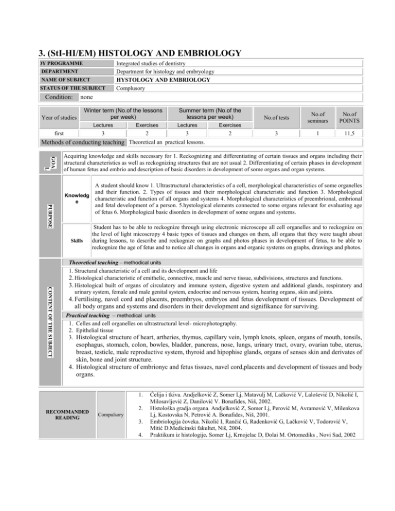
3. (StI-HI/EM) HISTOLOGY AND EMBRIOLOGY STUDY PROGRAMME DEPARTMENT NAME OF SUBJECT STATUS OF THE SUBJECT Condition: Integrated studies of dentistry Department for histology and embryology HYSTOLOGY AND EMBRIOLOGY Complusory none Year of studies Winter term (No.of the lessons per week) first Summer term (No.of the lessons per week) Lectures Exercises Lectures Exercises 3 2 3 2 No.of tests No.of seminars No.of POINTS 3 1 11,5 Methods of conducting teaching Theoretical an practical lessons. GOA L Acquiring knowledge and skills necessary for 1. Reckognizing and differentiating of certain tissues and organs including their structural characteristics as well as reckognizing structures that are not usual 2. Differentiating of certain phases in development of human fetus and embrio and description of basic disorders in development of some organs and organ systems. PURPOSE A student should know 1. Ultrastructural characteristics of a cell, morphological characteristics of some organelles and their function. 2. Types of tissues and their morphological characteristic and function 3. Morphological Knowledg characteristic and function of all organs and systems 4. Morphological characteristics of preembrional, embrional e and fetal development of a person. 5.hystological elements connected to some organs relevant for evaluating age of fetus 6. Morphological basic disorders in development of some organs and systems. Skills Student has to be able to reckognize through using electronic microscope all cell organelles and to reckognize on the level of light micoscropy 4 basic types of tissues and changes on them, all organs that they were taught about during lessons, to describe and reckognize on graphs and photos phases in development of fetus, to be able to reckognize the age of fetus and to notice all changes in organs and organic systems on graphs, drawings and photos. CONTENT OF THE SUBJECT Theoretical teaching – methodical units 1. Structural characteristic of a cell and its development and life 2. Histological characteristic of emithelic, connective, muscle and nerve tissue, subdivisions, structures and functions. 3. Histological built of organs of circulatory and immune system, digestive system and additional glands, respiratory and urinary system, female and male genital system, endocrine and nervous system, hearing organs, skin and joints. 4. Fertilising, navel cord and placents, preembryos, embryos and fetus development of tissues. Development of all body organs and systems and disorders in their development and signifikance for surviving. Practical teaching – methodical units 1. Celles and cell organelles on ultrastructural level- microphotography. 2. Epithelial tissue 3. Histological structure of heart, artheries, thymus, capillary vein, lymph knots, spleen, organs of mouth, tonsils, esophagus, stomach, colon, bowles, bladder, pancreas, nose, lungs, urinary tract, ovary, ovarian tube, uterus, breast, testicle, male reproductive system, thyroid and hipophise glands, organs of senses skin and derivates of skin, bone and joint structure. 4. Histological structure of embrionyc and fetus tissues, navel cord,placents and development of tissues and body organs. 1. RECOMMANDED READING 2. Compulsory 3. 4. Ćelija i tkiva. Andjelković Z, Somer Lj, Matavulj M, Lačković V, Lalošević D, Nikolić I, Milosavljević Z, Danilović V. Bonafides, Niš, 2002. Histološka gradja organa. Andjelković Z, Somer Lj, Perović M, Avramović V, Milenkova Lj, Kostovska N, Petrović A. Bonafides, Niš, 2001. Embriologija čoveka. Nikolić I, Rančić G, Radenković G, Lačković V, Todorović V, Mitić D.Medicinski fakultet, Niš, 2004. Praktikum iz histologije. Somer Lj, Krnojelac D, Đolai M. Ortomediks , Novi Sad, 2002 1. 2. 3. Additional 4. 5. Osnovi histologije, tekst i atlas. Junkvera L, Karneiro J. /Urednici i prevodioci Lačković V, Todorović V. Data Status, Beograd , 2005. Basic histology. Junqueira L, Carneiro J, MC Graw-Hill Companies, 2005. Atlas razvojne morfologije fetalnog perioda.Somer Lj,Đolai M, Lalošević D, Krnojelac D, Mocko-Kaćanski M, Levakov A. Medicinski fakultet Novi Sad-WUS Austrija, Novi Sad 2005. Mikroskopska laboratorijska tehnika u medicini. Lalošević D, Somer Lj, Djolai M, Lalošević V, Mažibrada J, Krnojelac D. Medicinski fakultet Novi Sad-WUS Austrija, Novi Sad, 2005. Hystology, a Text and Atlas, Ross M, Kaye G, Pawlina W. Lippincott Williams&Wilkins, Philadelphia, 2003. Evaluation of students' work – No.of points per individual activity Pre-exam obligations Lectures Exercises Exercises Seminar paper The rest 10 25 15 5 - Final exam Written Oral 20 25 Total 100 List of teachers and assistents Associate 3 1. 2. 3. 4. 5. Assistent Lecturer 2 Prof dr Ljiljana Somer Prof. dr Dušan Lalošević Doc. dr Matilda Đolai Asst. dr Mihaela Mocko-Kaćanski Dr Aleksandra Levakov, Assistent Professor PhD Associate prof. Professor 1 1 1 6. 7. 8. Dr Nenad Šolajić, Associate u nastavi Dr Tamara Lukić, Associate u nastavi Dr Golub Samardžija, Associate u nastavi Chief of the department Prof. dr .Ljiljana Somer Scientist
