Table 4 DTI studies of individuals with prenatal alcohol exposure
advertisement
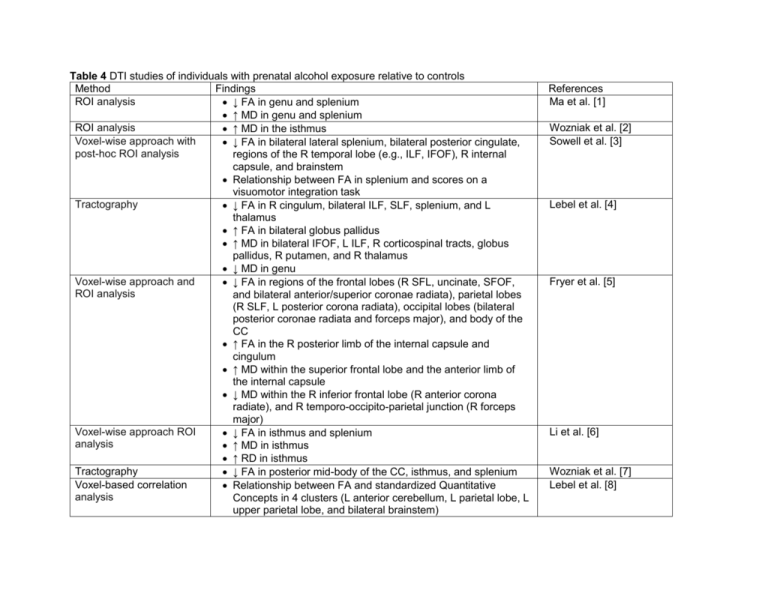
Table 4 DTI studies of individuals with prenatal alcohol exposure relative to controls Method Findings ROI analysis ↓ FA in genu and splenium ↑ MD in genu and splenium ROI analysis ↑ MD in the isthmus Voxel-wise approach with ↓ FA in bilateral lateral splenium, bilateral posterior cingulate, post-hoc ROI analysis regions of the R temporal lobe (e.g., ILF, IFOF), R internal capsule, and brainstem Relationship between FA in splenium and scores on a visuomotor integration task Tractography ↓ FA in R cingulum, bilateral ILF, SLF, splenium, and L thalamus ↑ FA in bilateral globus pallidus ↑ MD in bilateral IFOF, L ILF, R corticospinal tracts, globus pallidus, R putamen, and R thalamus ↓ MD in genu Voxel-wise approach and ↓ FA in regions of the frontal lobes (R SFL, uncinate, SFOF, ROI analysis and bilateral anterior/superior coronae radiata), parietal lobes (R SLF, L posterior corona radiata), occipital lobes (bilateral posterior coronae radiata and forceps major), and body of the CC ↑ FA in the R posterior limb of the internal capsule and cingulum ↑ MD within the superior frontal lobe and the anterior limb of the internal capsule ↓ MD within the R inferior frontal lobe (R anterior corona radiate), and R temporo-occipito-parietal junction (R forceps major) Voxel-wise approach ROI ↓ FA in isthmus and splenium analysis ↑ MD in isthmus ↑ RD in isthmus Tractography ↓ FA in posterior mid-body of the CC, isthmus, and splenium Voxel-based correlation Relationship between FA and standardized Quantitative analysis Concepts in 4 clusters (L anterior cerebellum, L parietal lobe, L upper parietal lobe, and bilateral brainstem) References Ma et al. [1] Wozniak et al. [2] Sowell et al. [3] Lebel et al. [4] Fryer et al. [5] Li et al. [6] Wozniak et al. [7] Lebel et al. [8] Voxel-wise approach ROI Spottiswoode et al. [9] ↓ FA in L middle cerebellar peduncle, positive relationship analysis between trace conditioning performance and FA Along-tract analysis Colby et al. [10] ↓ FA in ILF Tractography Malisza et al. [11] ↓ Total tract volume and number of fibers Voxel-based correlation Green et al. [12] Relationship between FA and saccade reaction time in 4 analysis clusters (R CC, genu, R ILF, and L cerebellum) Tractography longitudinal Treit et al. [13] Altered trajectories of neurodevelopment in SFOF, SLF, and analysis IFOF L left, R right, FA fractional anisotropy, MD mean diffusivity, RD radial diffusivity, CC corpus callosum, IFOF inferior fronto-occipital fasciculus, ILF inferior longitudinal fasciculus, SLF superior longitudinal fasciculus, SFOF superior fronto-occipital fasciculus References 1. Ma X, Coles CD, Lynch ME, LaConte SM, Zurkiya O, Wang D et al. Evaluation of Corpus Callosum Anisotropy in Young Adults With Fetal Alcohol Syndrome According to Diffusion Tensor Imaging. Alcoholism, clinical and experimental research. 2005;29(7):1214-22. 2. Wozniak JR, Mueller BA, Chang PN, Muetzel RL, Caros L, Lim KO. Diffusion tensor imaging in children with fetal alcohol spectrum disorders. Alcoholism, clinical and experimental research. 2006;30(10):1799-806. 3. Sowell ER, Johnson A, Kan E, Lu LH, Van Horn JD, Toga AW et al. Mapping white matter integrity and neurobehavioral correlates in children with fetal alcohol spectrum disorders. The Journal of neuroscience : the official journal of the Society for Neuroscience. 2008;28(6):1313-9. 4. Lebel C, Rasmussen C, Wyper K, Walker L, Andrew G, Yager J et al. Brain diffusion abnormalities in children with fetal alcohol spectrum disorder. Alcoholism, clinical and experimental research. 2008;32(10):1732-40. 5. Fryer SL, Schweinsburg BC, Bjorkquist OA, Frank LR, Mattson SN, Spadoni AD et al. Characterization of white matter microstructure in fetal alcohol spectrum disorders. Alcoholism, clinical and experimental research. 2009;33(3):514-21. 6. Li L, Coles CD, Lynch ME, Hu X. Voxelwise and skeleton-based region of interest analysis of fetal alcohol syndrome and fetal alcohol spectrum disorders in young adults. Human brain mapping. 2009;30(10):3265-74. 7. Wozniak JR, Muetzel RL, Mueller BA, McGee CL, Freerks MA, Ward EE et al. Microstructural corpus callosum anomalies in children with prenatal alcohol exposure: an extension of previous diffusion tensor imaging findings. Alcoholism, clinical and experimental research. 2009;33(10):1825-35. 8. Lebel C, Rasmussen C, Wyper K, Andrew G, Beaulieu C. Brain microstructure is related to math ability in children with fetal alcohol spectrum disorder. Alcoholism, clinical and experimental research. 2010;34(2):354-63. 9. Spottiswoode BS, Meintjes EM, Anderson AW, Molteno CD, Stanton ME, Dodge NC et al. Diffusion tensor imaging of the cerebellum and eyeblink conditioning in fetal alcohol spectrum disorder. Alcoholism, clinical and experimental research. 2011;35(12):2174-83. 10. Colby JB, Soderberg L, Lebel C, Dinov ID, Thompson PM, Sowell ER. Along-tract statistics allow for enhanced tractography analysis. NeuroImage. 2012;59(4):3227-42. 11. Malisza KL, Buss JL, Bolster RB, de Gervai PD, Woods-Frohlich L, Summers R et al. Comparison of spatial working memory in children with prenatal alcohol exposure and those diagnosed with ADHD; A functional magnetic resonance imaging study. Journal of neurodevelopmental disorders. 2012;4(1):12. 12. Green CR, Lebel C, Rasmussen C, Beaulieu C, Reynolds JN. Diffusion tensor imaging correlates of saccadic reaction time in children with fetal alcohol spectrum disorder. Alcoholism, clinical and experimental research. 2013;37(9):1499-507. 13. Treit S, Lebel C, Baugh L, Rasmussen C, Andrew G, Beaulieu C. Longitudinal MRI reveals altered trajectory of brain development during childhood and adolescence in fetal alcohol spectrum disorders. The Journal of neuroscience : the official journal of the Society for Neuroscience. 2013;33(24):10098-109.


