Comparative in-silico analysis of autophagy
advertisement
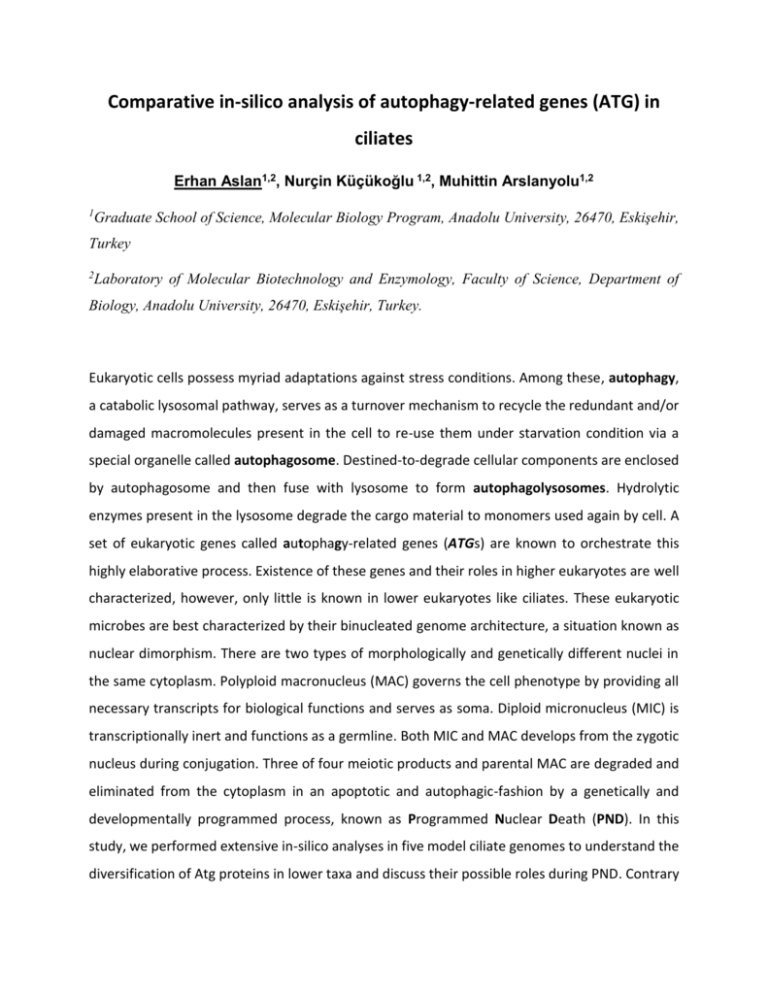
Comparative in-silico analysis of autophagy-related genes (ATG) in ciliates Erhan Aslan1,2, Nurçin Küçükoğlu 1,2, Muhittin Arslanyolu1,2 1 Graduate School of Science, Molecular Biology Program, Anadolu University, 26470, Eskişehir, Turkey 2 Laboratory of Molecular Biotechnology and Enzymology, Faculty of Science, Department of Biology, Anadolu University, 26470, Eskişehir, Turkey. Eukaryotic cells possess myriad adaptations against stress conditions. Among these, autophagy, a catabolic lysosomal pathway, serves as a turnover mechanism to recycle the redundant and/or damaged macromolecules present in the cell to re-use them under starvation condition via a special organelle called autophagosome. Destined-to-degrade cellular components are enclosed by autophagosome and then fuse with lysosome to form autophagolysosomes. Hydrolytic enzymes present in the lysosome degrade the cargo material to monomers used again by cell. A set of eukaryotic genes called autophagy-related genes (ATGs) are known to orchestrate this highly elaborative process. Existence of these genes and their roles in higher eukaryotes are well characterized, however, only little is known in lower eukaryotes like ciliates. These eukaryotic microbes are best characterized by their binucleated genome architecture, a situation known as nuclear dimorphism. There are two types of morphologically and genetically different nuclei in the same cytoplasm. Polyploid macronucleus (MAC) governs the cell phenotype by providing all necessary transcripts for biological functions and serves as soma. Diploid micronucleus (MIC) is transcriptionally inert and functions as a germline. Both MIC and MAC develops from the zygotic nucleus during conjugation. Three of four meiotic products and parental MAC are degraded and eliminated from the cytoplasm in an apoptotic and autophagic-fashion by a genetically and developmentally programmed process, known as Programmed Nuclear Death (PND). In this study, we performed extensive in-silico analyses in five model ciliate genomes to understand the diversification of Atg proteins in lower taxa and discuss their possible roles during PND. Contrary to previous reports, we found that ciliate genomes do not have typical Atg1 proteins since all the reported or newly defined candidate sequences are lack of Atg1-specific C-terminal domain, which is essential for the Atg1 complex formation. We also noticed that several genes in Oxytricha trifallax and Stylonychia lemnae genomes were annotated as ATG16L, a mammalian version of yeast ATG16. However, executed domain analysis showed that these proteins do not contain Atg16 domain. Surprisingly, we failed to find any ATG8 gene, which encodes a ubiquitin-like protein that is critical for autophagosome formation, in parasitic ciliate Ichthyophthirius multifiliis while other four ciliates have varying numbers. On the other hand, phylogenetically ATG8-related gene, ATG12, is only found in O.trifallax. Phylogenetic analysis suggested that Atg8 proteins in ciliates have evolved to function differently. In addition, we also realized that several non-yeast mammalian autophagy genes like UVRAG (in O.trifallax) and VMP1 (in T.thermophila) are found in ciliates. Lastly, we analyzed mRNA expression level of ATGs in T.thermophila and Paramecium tetraurelia transcriptome databases. ATG4 and ATG8 expressions in both species show significant changes during conjugation and autogamy respectively, which may further imply a possible conserved roles during PND. In conclusion, we survey the bioinformatics analyses of autophagy proteins in model ciliates in the perspective of PND. Understanding the role of autophagy proteins in lower eukaryotes like ciliates may help to decipher novel functions of these proteins. Keywords: Ciliate, autophagy-related genes, in silico, autophagy
