placental size and perinatal outcomes
advertisement
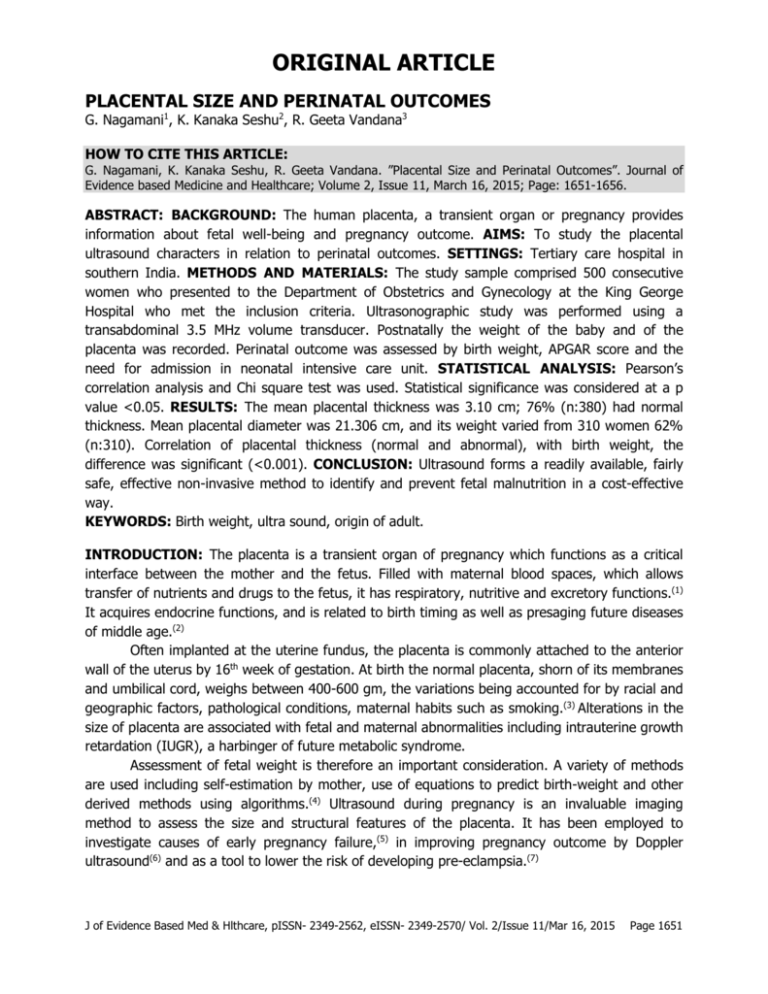
ORIGINAL ARTICLE PLACENTAL SIZE AND PERINATAL OUTCOMES G. Nagamani1, K. Kanaka Seshu2, R. Geeta Vandana3 HOW TO CITE THIS ARTICLE: G. Nagamani, K. Kanaka Seshu, R. Geeta Vandana. ”Placental Size and Perinatal Outcomes”. Journal of Evidence based Medicine and Healthcare; Volume 2, Issue 11, March 16, 2015; Page: 1651-1656. ABSTRACT: BACKGROUND: The human placenta, a transient organ or pregnancy provides information about fetal well-being and pregnancy outcome. AIMS: To study the placental ultrasound characters in relation to perinatal outcomes. SETTINGS: Tertiary care hospital in southern India. METHODS AND MATERIALS: The study sample comprised 500 consecutive women who presented to the Department of Obstetrics and Gynecology at the King George Hospital who met the inclusion criteria. Ultrasonographic study was performed using a transabdominal 3.5 MHz volume transducer. Postnatally the weight of the baby and of the placenta was recorded. Perinatal outcome was assessed by birth weight, APGAR score and the need for admission in neonatal intensive care unit. STATISTICAL ANALYSIS: Pearson’s correlation analysis and Chi square test was used. Statistical significance was considered at a p value <0.05. RESULTS: The mean placental thickness was 3.10 cm; 76% (n:380) had normal thickness. Mean placental diameter was 21.306 cm, and its weight varied from 310 women 62% (n:310). Correlation of placental thickness (normal and abnormal), with birth weight, the difference was significant (<0.001). CONCLUSION: Ultrasound forms a readily available, fairly safe, effective non-invasive method to identify and prevent fetal malnutrition in a cost-effective way. KEYWORDS: Birth weight, ultra sound, origin of adult. INTRODUCTION: The placenta is a transient organ of pregnancy which functions as a critical interface between the mother and the fetus. Filled with maternal blood spaces, which allows transfer of nutrients and drugs to the fetus, it has respiratory, nutritive and excretory functions.(1) It acquires endocrine functions, and is related to birth timing as well as presaging future diseases of middle age.(2) Often implanted at the uterine fundus, the placenta is commonly attached to the anterior wall of the uterus by 16th week of gestation. At birth the normal placenta, shorn of its membranes and umbilical cord, weighs between 400-600 gm, the variations being accounted for by racial and geographic factors, pathological conditions, maternal habits such as smoking.(3) Alterations in the size of placenta are associated with fetal and maternal abnormalities including intrauterine growth retardation (IUGR), a harbinger of future metabolic syndrome. Assessment of fetal weight is therefore an important consideration. A variety of methods are used including self-estimation by mother, use of equations to predict birth-weight and other derived methods using algorithms.(4) Ultrasound during pregnancy is an invaluable imaging method to assess the size and structural features of the placenta. It has been employed to investigate causes of early pregnancy failure,(5) in improving pregnancy outcome by Doppler ultrasound(6) and as a tool to lower the risk of developing pre-eclampsia.(7) J of Evidence Based Med & Hlthcare, pISSN- 2349-2562, eISSN- 2349-2570/ Vol. 2/Issue 11/Mar 16, 2015 Page 1651 ORIGINAL ARTICLE Therefore the present study was undertaken to study the placental ultrasound characters in relation to perinatal outcomes from a large tertiary care hospital in southern India. MATERIAL AND METHODS: The study sample comprised 500 consecutive women who presented to the Department of Obstetrics and Gynecology at the King George Hospital. Inclusion criteria: Singleton pregnancies, gestational age between 37 and 42 weeks. Exclusion criteria: multiple pregnancies, gestation period before 37, history of irregular menstrual cycles, uncertain and unreliable dates. Ultrasonographic study was performed using a transabdominal 3.5 MHz volume transducer. Placental site was determined in two-dimensional real-time mode. The following were studied: fetus, amniotic fluid, placental thickness, diameter, grading and any associated abnormalities. Mode of delivery was noted whether spontaneous or induced, vaginal or cesarean section. The weight of the baby and of the placenta was recorded. Perinatal outcome was assessed by birth weight, APGAR score and the need for admission in neonatal intensive care unit. Congenital anomalies were also looked for. Placental thickness was measured at the level of insertion of umbilical cord in the longitudinal direction, perpendicular to the basal and chorionic plates in the mid placental position. Placental diameter was taken as the maximum length of the chorionic surface in the transverse plane. Volume was calculated as ‘Placental surface area = π x smallest diameter (cm) x largest diameter (cm)/4’. Placental volume was taken as the surface area (expressed in cm2 x thickness (cm)’. After birth, the placenta was examined for weight (gm), diameter (cm), thickness at the insertion of umbilical cord (cm), membrane insertion, colour, fetal surfaces (nodules, masses, thrombosis), maternal surface (masses, calcification, fibrin, hematoma), insertion of cord (cm from placental margin), length and colour of cord, number of vessels in the cord. ETHICS: The Institutional ethical committee gave approval for the conduct of the study STATISTICAL ANALYSIS: Data were analyzed using Microsoft EXCEL spread sheet. Pearson’s correlation analysis and Chi square test were applied to establish the relation among placental parameters (thickness, diameter, volume). Statistical significance was considered at a p value <0.05. RESULTS: The mean placental thickness was 3.10 cm; 380 (76%) had placentae of normal thickness (Table 1). About half subjects had placental diameter between 18 and 22 cm (Table 2); mean placental diameter was 21.306 cm. The placenta weighed between 500 and 600 gm in 310 women (62%) (Table 3). On correlation of placental thickness (normal and abnormal), with birth weight, the difference was significant (x2 value: 273.33; p value <0.001) (Table 4). DISCUSSION: In this study we showed that normal thickness of placenta is associated with normal babies. Placenta, being the first organ to show diseases of pregnancy, can be studied to predict feto-maternal complications. Studies from Mexico,(8) United States of America(9) and J of Evidence Based Med & Hlthcare, pISSN- 2349-2562, eISSN- 2349-2570/ Vol. 2/Issue 11/Mar 16, 2015 Page 1652 ORIGINAL ARTICLE Nigeria(4) have shown that placental ultrasonography is a valid measure of prognosticating pregnancy outcomes. The placenta is the link between the mother and fetus; through this, nutrients and oxygen reach the fetus, which in turn excretes carbon dioxide and metabolic substances. Transplacental transfer can occur either by simple diffusion of gases, water, electrolytes or even some low molecular weight molecules. Selective transfer and facilitated diffusion also occurs in relation to iron. In contrast the maternal hormones traverse slowly, which the placenta secretes chorionic gonadotropins and placental lactogen.(10) Glucose, which forms the main energy substrate for the fetus is delivered by the mother through stores in the liver, muscle and adipose tissue, modulated by insulin. Alterations in the availability and passage of these substances are responsibility for poor growth of fetus with attendant risk of future disease, or of macrosomia as found in gestational diabetes mellitus. Therefore methods are required to assess the adequacy of the placenta and its function as a guide for fetal well-being. Obviously invasive methods are not feasible, and examination of placenta at birth can provide information which can be applied retrospectively. Ultrasonography is a widely available, non-invasive relatively safe method that is extensively used in monitoring during pregnancy. Improvement in the instruments and display methods led to its use for imaging the placental size, position and thickness as a predictor of well-being. Wolf et al studied the placental volume and fetal weight by longitudinal ultrasonographic measurements in a small sample of 18 women between gestational age 16 to 20 weeks.(11) Whereas seven normal subjects, placental and fetal growth showed a nearly linear pattern, among whom four had reduced placental and fetal growth in the last one to three weeks. When evaluated basing on adverse pregnancy outcomes such as fetal death, distress needing surgical intervention or neonatal underweight, it was shown that ultrasound could identify retarded placental growth for at least three weeks before fetal growth retardation. It was one of the early studies when adverse fetal outcome could be presaged by imaging the placenta using ultrasound. Similar results were replicated in Saudi Arabia by Habib in a larger sample of 70 pregnant women.(12) On a longitudinal study, a ‘warning limit’ of placental thickness and diameter was observed. Ultrasound assessment of placenta at 36 weeks was fitted to a logistic regression. The limit of 18 cm placental diameter and 2 cm thickness predicted low birth weight in infants, and thus proved to be useful as a prognostic marker in identifying fetal growth retardation. In a larger population size from Mexico (n: 300 live newborns) Sanin established a model to relate birth weight with placental weight.(8) Placental weight was found to be significantly related to birth weight. For each gram increase of weight of placenta, the birth weight increased by 1.98 gms (p<0.01). The placenta however was shown to have a nonlinear relation to birth weight and could be used as a useful non-invasive predictor of birth weight. The authors concluded that along with age of gestation, the maternal age and size, placental weight could explain 32% of the variability of birth weight and could thus be a “’sentinel’ indicator of nutritional and/or environmental problems.”(8) Thus we have replicated similar relation of normal placental thickness with normal pregnancy outcome in a large series of 500 consecutive pregnancies at a tertiary care hospital from southern India. Evidence is accumulating for adequate maternal and thereby fetal nutrition J of Evidence Based Med & Hlthcare, pISSN- 2349-2562, eISSN- 2349-2570/ Vol. 2/Issue 11/Mar 16, 2015 Page 1653 ORIGINAL ARTICLE for satisfactory outcome. Either under or over nutrition of the fetus and the baby at birth is a well- recognized predictor of future adverse metabolic effects: the ‘thin but fat Indian baby’ syndrome described by Yagnik et al.(13) Babies born of poor maternal nutrition have been shown to be at increased risk of later life metabolic obesity, insulin resistance, type 2 diabetes mellitus, coronary artery disease and premature death. Considering the increasing burden of noncommunicable diseases overlaid upon infections in India, it is important to ensure adequate and proper nutrition in pregnant women so that both under and overweight of newborns are prevented, and thereby adverse vascular risk later in life. Ultrasound forms a readily available, fairly safe, effective non-invasive method to identify and therefore prevent fetal malnutrition in a cost-effective method. The current study replicates the usefulness of placental imaging by ultrasound to satisfactory birth outcomes in a population from southern India. REFERENCES: 1. Donneily L, Campling G. Functions of the placenta. Anaesthes Intens Care Med 2014; 15: 136-9. 2. Kaiser J. Gearing up for a closer look at the human placenta. Science 2014; 344: 1073. 3. Benirschke K. Normal early development. In Creasy RK, Resnik R, Iams JD, Lockwood CJ, Moore TR, Greene MF (eds). Maternal-fetal medicine. Elsevier, Philadelphia 2014; p 37-46. 4. Abu PO, Ohagwu CC, Eze JC, Ochie K. Correlation between placental thickness and estimated fetal weight in Nigerian women. Itnosia J Med Biomed Sci 2009; 1: 80-5. 5. Jauniaux E, Johns J, Burton GJ. The role of ultrasound imaging in diagnosing and investigating early pregnancy failure. Ultrasound Obstet Gynecol 2005; 25: 613-24 6. Stampalija T, Gyte GML, Alfirevic Z. Utero-placental Doppler ultrasound for improving pregnancy outcome. Cochrane Database Syst Rev 2014; (9): CD008363 doi:10.1002/14651858.CD008362.pub2. 7. Smith GCS. Researching new methods of screening for adverse pregnancy outcome: lessons from pre-eclampsia. PLoS Medicine 2012; 9:e1001274. 8. Sanin LH, Lopez SR, Olivares ET, Silva MP, Carrilo ML. Relation between birth weight and placental weight. Biol Neonate 2001; 80: 113-7. 9. Dombrowski MP, Wolfe HM, Saleh A et al. Sonographically thick placenta: a predictor of increased perinatal morbidity and mortality. Ultrasound Obstet Gynecol 1992; 2: 252-5. 10. Embryogenesis and fetal morphological development. In Cunningham FG, Leveno KJ, Bloom SL et al. Williams Obstetrics McGraw Hill N York 2014; p127-53. 11. Wolf H, Oosting H, Treffers PE. A longitudinal study of the relationship between placental and fetal growth as measured by ultrasonography. Am J Obstet Gynecol 1989; 161: 1140-5. 12. Habib FA. Prediction of low birth weight infants from ultrasound measurement of placental diameter and placental thickness. Ann Saudi Med 2002; 22: 312-4. 13. Yajnik CS. Early ife origins of insulin resistance and type 2 diabetes in India and other Asian countries. J Nutr 2004; 134: 205-10. J of Evidence Based Med & Hlthcare, pISSN- 2349-2562, eISSN- 2349-2570/ Vol. 2/Issue 11/Mar 16, 2015 Page 1654 ORIGINAL ARTICLE Placental thickness Number of cases (%) <2.5 cm 40 (8%) 2.5-3.75 cm 380 (76%) >3.75 cm 80 (16%) Table 1: Pattern of placental thickness Placental diameter Number of cases (%) <18 cm 40 (8%) 18-22 cm 290 (58%) >22 cm 170 (34%) Table 2: Pattern of placental diameter Placental weight <500 gm 500-600 gm >600 gm Number of cases (%) 100 (20%) 310 (62%) 90 (18%) Table 3: Pattern of placental weight after parturition Birth weight (Kg) <2.5 kg 2.5-3.5 kg >3.5 kg Placental thickness <2.5 cm 40 0 0 Placental thickness 2.5-3.75 cm 10 360 10 Placental thickness >3.75 cm 40 30 10 Table 4: Relation between placental thickness and birth weight* *When normal and abnormal placental thickness correlated with birth weight, P<0.001. J of Evidence Based Med & Hlthcare, pISSN- 2349-2562, eISSN- 2349-2570/ Vol. 2/Issue 11/Mar 16, 2015 Page 1655 ORIGINAL ARTICLE AUTHORS: 1. G. Nagamani 2. K. Kanaka Seshu 3. R. Geeta Vandana PARTICULARS OF CONTRIBUTORS: 1. Associate Professor, Department of Obstetrics & Gynaecology, Andhra Medical College, Visakhapatnam. 2. Former Post Graduate, Department of Obstetrics & Gynaecology, Andhra Medical College, Visakhapatnam. 3. Assistant Professor, Department of Obstetrics & Gynaecology, Andhra Medical College, Visakhapatnam. NAME ADDRESS EMAIL ID OF THE CORRESPONDING AUTHOR: Dr. G. Nagamani, # 15-12-14, Krishna Nagar, Visakhapatnam-530002. E-mail: nagamanigum@gmail.com Date Date Date Date of of of of Submission: 08/03/2015. Peer Review: 10/03/2015. Acceptance: 11/03/2015. Publishing: 14/03/2015. J of Evidence Based Med & Hlthcare, pISSN- 2349-2562, eISSN- 2349-2570/ Vol. 2/Issue 11/Mar 16, 2015 Page 1656
