Ch. Arning . Department of Neurology, Allgemeines Krankenhaus
advertisement
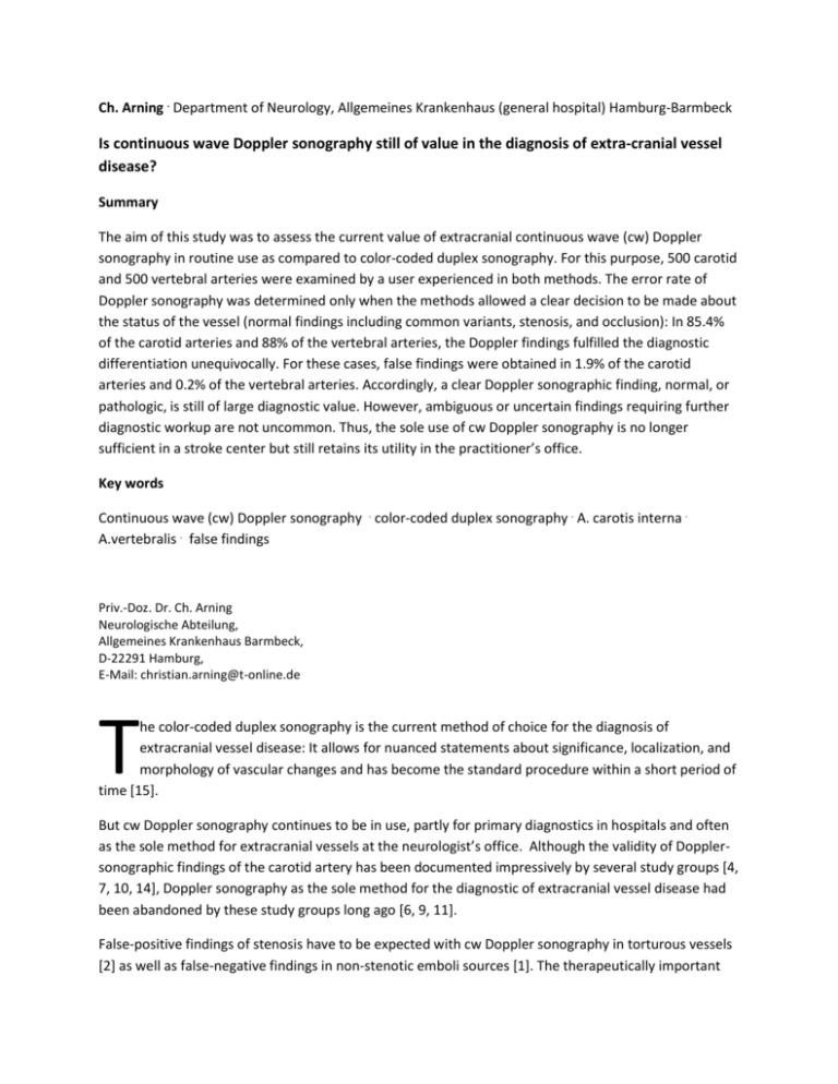
Ch. Arning . Department of Neurology, Allgemeines Krankenhaus (general hospital) Hamburg-Barmbeck Is continuous wave Doppler sonography still of value in the diagnosis of extra-cranial vessel disease? Summary The aim of this study was to assess the current value of extracranial continuous wave (cw) Doppler sonography in routine use as compared to color-coded duplex sonography. For this purpose, 500 carotid and 500 vertebral arteries were examined by a user experienced in both methods. The error rate of Doppler sonography was determined only when the methods allowed a clear decision to be made about the status of the vessel (normal findings including common variants, stenosis, and occlusion): In 85.4% of the carotid arteries and 88% of the vertebral arteries, the Doppler findings fulfilled the diagnostic differentiation unequivocally. For these cases, false findings were obtained in 1.9% of the carotid arteries and 0.2% of the vertebral arteries. Accordingly, a clear Doppler sonographic finding, normal, or pathologic, is still of large diagnostic value. However, ambiguous or uncertain findings requiring further diagnostic workup are not uncommon. Thus, the sole use of cw Doppler sonography is no longer sufficient in a stroke center but still retains its utility in the practitioner’s office. Key words Continuous wave (cw) Doppler sonography . color-coded duplex sonography . A. carotis interna . A.vertebralis . false findings Priv.-Doz. Dr. Ch. Arning Neurologische Abteilung, Allgemeines Krankenhaus Barmbeck, D-22291 Hamburg, E-Mail: christian.arning@t-online.de T he color-coded duplex sonography is the current method of choice for the diagnosis of extracranial vessel disease: It allows for nuanced statements about significance, localization, and morphology of vascular changes and has become the standard procedure within a short period of time [15]. But cw Doppler sonography continues to be in use, partly for primary diagnostics in hospitals and often as the sole method for extracranial vessels at the neurologist’s office. Although the validity of Dopplersonographic findings of the carotid artery has been documented impressively by several study groups [4, 7, 10, 14], Doppler sonography as the sole method for the diagnostic of extracranial vessel disease had been abandoned by these study groups long ago [6, 9, 11]. False-positive findings of stenosis have to be expected with cw Doppler sonography in torturous vessels [2] as well as false-negative findings in non-stenotic emboli sources [1]. The therapeutically important differentiation between filiform stenosis and vessel occlusion remains uncertain [8]. With cw Doppler sonography of the vertebral artery it is not possible to differentiate with certainty between a pathologic situation or a harmless normal variation in cases of missing flow signal or slowed flow [5]. With colorcoded duplex sonography all these findings can be interpreted unambiguously as a rule. Is the use of cw Doppler sonography today still justifiable regardless? How frequent is the occurrence of false results with cw Doppler sonography really? What ranking does the Doppler sonography have in comparison to the color-coded duplex sonography? Which Doppler findings require additional tests? These questions were examined by comparing routine use of both methods at a department of neurology, whereby the value of Doppler sonography as the sole method of the diagnosis of vessel disease was examined. The advantages of Doppler sonography as an addition to duplex sonography, especially in the exam of the periorbital arteries, are generally accepted. The aim of this study is therefore the broader use of both methods in combination (e.g. examination of the periorbital arteries and the subclavian artery with cw- Doppler sonography, evaluation of the carotid and vertebral arteries with duplex sonography). Method Doppler and duplex sonography findings of 500 carotid and 500 vertebral arteries from routine testing at the ultrasound laboratory of the department of neurology were included in the study. The study was done on 250 consecutive patients by a user (DEGUM seminar leader, certified by the German Society for Ultra Sonography in Medicine) very experienced in both methods. A complete cw Doppler sonography (DWL Multi-Dop P, probes 4 and 8 MHz) with frequency spectrum analysis was performed at first in all cases. The following vessels were compared side by side with the usual technique [13, 15]: A. supratrochlearis, A. carotis communis, A. carotis interna, A. carotis externa, A. vertebralis at the atlas loop and if possible at the origin as well as A. subclavia. For each Doppler sonography finding it was determined if it allowed for a clear determination of the vessel condition. The differentiation between normal finding including common variants, stenosis, and vessel occlusion had to be unambiguous. All vessel findings with localized narrowing leading to hemodynamic changes (from a degree of about 50-60% stenosis on) and principally detectable with Doppler sonography were considered a stenosis. In cases where the assignment of the Doppler sonography finding to one of the 3 categories was not possible without ambiguity, the finding was rated as ambiguous/uncertain; a presumable diagnosis was not sufficient. Hence a missing visualization of the internal carotid artery at its origin was labeled as an ambiguous result even if direct signs of stenosis were missing (exclusion of a middle-grade stenosis is with this constellation of findings not possible). Likewise, the typical findings for an occluded internal carotid artery were always rated as ambiguous since the possibility for a differential diagnosis of filiform stenosis exists. A unilateral slowing of the vertebral artery flow was always, even with a normal pulse curve, rates as an inconclusive finding since a common variant can only be determined with certainty if the lumen diameter is known and, alternatively, a pathologic situation could exist. Table 1 shows all findings that were classified as ambiguous for the carotid and vertebral arteries. Table 1 Findings with cw Doppler sonography classified as ambiguousa A. carotis A. carotis interna not visualized at its origin A.carotis communis not visualized Questionable stenosis finding of the A. carotis interna (DDx torturous vessel) Questionable finding of a pathologic increase of flow (AV-fistula?) A. vertebralis A. vertebralis not visualized A. vertebralis not unequivocally identifiable A. vertebralis slowed unilaterally Questionable stenosis finding at the origin (finding ambiguous) ___________________________________________________________________________________________ a Differentiation between normal finding (including common variant), stenosis, and vessel occlusion not possible Doppler sonography was followed by color duplex sonography (Acuson 128 XP 10, standard probe L5, nominal frequency of 5 MHz). The following vessels were examined with the usual technique [3, 13, 15]: A. carotis communis, A. carotis interna, and A. carotis externa bilaterally each as well as the A. vertebralis in the 1st and 2nd segment bilaterally. The criteria published by us in 1999 [3] were used for the duplex-sonographic diagnosis of stenoses, occlusions, and normal variants. The results of Doppler and duplex sonography were compared. If there were deviations between the methods and an unambiguous result of the cw Doppler sonography, the Doppler result was regarded as a false finding. The reference method duplex sonography delivered an unambiguous result in all cases. It was always attempted to detect the cause of the false Doppler findings. In cases classified as ambiguous after the Doppler sonography, the duplex findings were consulted to determine the frequency of important pathologic results compared to harmless common variants. Results Table 2 shows the frequency of unambiguous and ambiguous/uncertain diagnoses with Doppler sonography of the carotid and vertebral artery: An unambiguous finding for the vessel (normal finding including common variants, stenosis, and occlusion) could be determined for the carotid artery in more than 85% of the cases and in more than 88% of the cases for the vertebral artery. Table 2 Frequency of unambiguous and uncertain/ambiguous diagnoses with Doppler sonography of the A. carotis and A. vertebralis (n=500) Diagnosis unambiguousa ___________________ [n] [%] Diagnosis uncertain/ambiguous __________________________ [n] [%] _______________________________________________________________________________ A. carotis 427 85.4 73 14.6 A. vertebralis 440 88.0 60 12.0 _________________________________________________________________________________________ a Reliable differentiation possible between normal finding (including common variant), stenosis, and occlusion The rate of false results with the cw Doppler sonography is very low, with a total of 1.9% for the carotid artery and 0.2% for the vertebral artery if only the findings determined to be unambiguous by the examiner are considered (table 3). Causes for the false-negative findings for the carotid artery were unrecognized low- to mid-grade stenoses, for the vertebral artery collateralized vessel occlusions. One false-positive case for the carotid artery occurred because of collateral flow increased with a contralateral occlusion. Table 3 False findings with cw Doppler sonography of the A. carotis and A. vertebralisa Findings with False findings False findings clear diagnosis [n] [n] [%] ______________________________________________________________________________________ A. carotis Normal finding 397 7 1.8 Pathologic finding 30 1 3.3 Findings total 429 8 1.9 A. vertebralis Normal finding 438 1 0.2 Pathologic finding 2 0 0 Findings total 440 1 0.2 _______________________________________________________________________________________ a Without consideration of ambiguous/uncertain findings All the findings for the carotid and vertebral artery classified as ambiguous/uncertain with Doppler sonography were able to be clarified with duplex sonography (table 4). Table 4 Causes of uncertain/ambiguous findings with cw Doppler sonography (n+500) after resolution with color-coded duplex sonography Doppler sonography [%] Color-coded duplex sonography [%] __________________________________________________________________________________________ ACI origins segment not visualized 10.8 Dorso-medial origins variant 4.6 Vessel occlusion 4.4 Filiform stenosis (proximal) 0.8 Slowing of flow in occlusion/distal stenosis 1 ACC not visualized 0.8 Occlusion Pathway anomaly (loop formation) 0.6 0.2 ACI: questionable stenosis (DDx torturous vessel) 2.8 Stenosis Torturous vessel Increased flow 1.2 1.2 0.2 ACI: questionable pathologic increase in flow (AV-fistula?) 0.2 AV-fistula 0.2 AVT origins not visualized 4.6 Hypoplasia Vessel occlusion Slowing of flow in proximal disturbance Slowing of flow in distal disturbance Unremarkable (poor test conditions) 1.8 2.0 0.2 0.2 0.4 AVT not distinctly identifiable 1.8 AVT unilaterally slowed, normal pulsatility 1.8 Hypoplasia Unremarkable (poor test conditions) Hypoplasia Proximal occlusion 0.6 1.2 1.6 0.2 Hypoplasia Distal disturbance 1.4 1.8 Proximal stenosis Proximal occlusion 0.2 0.2 AVT unilaterally slowed, increased pulsatility AVT unilaterally slowed, reduced pulsatility 1.4 AVT: questionable stenosis finding at origin 0.2 Stenosis at origin 0.2 __________________________________________________________________________________________ ACI A.carotis interna, ACC A. carotis communis, AVT A. vertebralis Discussion The study shows that cw Doppler sonography has a very low error rate as long as it delivers an unequivocal and certain finding, either normal or pathologic, from the hand of an experienced user. A routine duplex sonography is not necessary in such cases. In the evaluation of the determined error rate it is to be considered that the definition of “ambiguous or uncertain finding” was kept very broad here. All constellations that would allow any different, also rather improbable, diagnoses were included. For example, even the typical constellation of findings for internal carotid artery occlusion was considered uncertain because of the possibility of the filiform stenosis as differential diagnosis. If the Doppler sonography finding would have been evaluated as a definite occlusion, which is possible with a predictive value of 90% [13], four significant false results would have occurred in our collective in this manner. The same is valid for other constellations of findings in the carotid or vertebral artery. With a missing flow signal or slowed flow in the vertebral artery, it cannot be clearly differentiated with cw Doppler sonography if it is a pathologic situation or a common variant [5]. However, this differentiation is of certain relevance; therefore, all similar findings must be rated as “uncertain”. The rate of false findings in the carotid artery and especially in the vertebral artery would be significantly higher if all – even uncertain – Doppler sonography findings would be part of the equation. The experienced examiner would be able to prove “most” clinically relevant findings if cw Doppler sonography would be solely available, but this is not enough for today’s demand of a sophisticated vascular diagnostic. The relatively high rate of ambiguous and uncertain findings in our study, 14.6% with the carotid artery and 12% with the vertebral artery, can be explained by about 50% with variations in pathway and caliber of the vessels as well as pathologic vascular changes. In a collective with a greater share of normal findings, possibly at a neurologist’s office, one should expect less ambiguous cases than in our study population. A department of neurology with a “stroke focus” on the other hand, has to expect a comparatively rate of ambiguous cases, requiring further clarification with duplex sonography. In such an environment the color-coded duplex sonography is certainly the method of choice in primary diagnostics. Outside of facilities serving the acute diagnostics of stokes, the use of cw Doppler sonography appears justified to this day as long as there is an option to clarify uncertain findings with duplex sonography. Clinical relevance certainly needs to be considered because further investigation is not required in all cases of ambiguous or uncertain Doppler sonography findings. If, for example, hemodynamically relevant stenoses must be excluded prior to a cardio-surgical intervention, the detailed search for an emboli source is not necessary. The numbers determined here regarding the error rate of cw Doppler sonography are valid for the experienced examiner; the dependence of the results on the qualifications of the examiner and the difficulties learning the methods are well-known [12]. Conclusions Continuous wave Doppler sonography of the carotid and vertebral artery is a valuable method for the experienced examiner. Such findings always have a great validity when they are clear and unambiguous, either normal or pathologic. But ambiguous and uncertain findings occur in a significant share of cases, demanding further diagnostics. The application appears therefore only to make sense if there is a possibility to resolve uncertain findings with duplex sonography. Half of the uncertain findings are caused by anatomical variants and by pathologic changes. Complementary diagnostic will therefore be necessary more frequently; the sooner relevant pathologic findings can be expected. For this reason, cw Doppler sonography alone is not sufficient for a stroke center. In the practitioner’s office, on the other hand, it still retains its utility. Literature 1. Arning C, Hermann HD (1988) Floating thrombus in the internal carotid artery disclosed by B-module ultrasonography. J Neurol 235: 425-427 2. Arning C (1994) False diagnosis carotid stenosis in conventional Doppler sonography. Causes of false diagnoses with use in clinic and practice. Ultraschall Klin. Prax 9: 144-148 3. Arning C (1999) Color-coded duplex sonography of the arteries supplying the brain. A text-picture atlas of methodical foundations, normal and pathologic findings, 2. Aufl. Thieme, Stuttgart 4. Daffershofer M, Hennerici M (1990) Spectrum analysis of Doppler signals. Ultraschall Med 11: 219-26 5. Delcker A. Diener HC (1992) The different ultrasound methods for examining A. vertebralis – a comparing evaluation. Ultraschall Med 13: 213-220 6. Delcker A, Diener HC, Timmann D, Faustmann P (1993) The role of vertebral and internal carotid artery disease in the pathogenesis of vertebrobasilar transient ischemic attacks. Eur Arch Psychiatry Clin Neurosci 242: 179-183 7. Diener HC, Dichgans J, Voigt K (1981) Functional anatomy of extracranial arteries in occlusive vascular disease by direct continues wave Doppler sonography. Cadiovasc Intervent Radiol 4: 193-201 8. Görtler M, Niethammer R, Widder B (1994) Differentiating subtotal carotid artery stenoses from occlusion by colour-coded duplex sonography. J Neurol 241: 301-305 9. Hennerici MG, Meairs SP (1999) Cerebrovascular ultrasound. Curr Opin Neurol 12: 57-63 10. Neuerburg-Heusler D (1984) Doppler sonographic diagnostic in extracranial occlusive disease. Validity of direct, indirect, and combined methods. Vasa (Suppl 12): 59-70 11. Neuerburg-Heusler D, Hennerici MG (1999) Vascular diagnostic with ultrasound, 3. Aufl. Thieme, Stuttgart 12. Reutern GM von (1982) Thoughts for the education in ultrasound diagnostic in brain-supplying arteries. Ultraschall 3: 58-60 13. Reutern GM von, Kaps M, Büdingen HJ von (2000) Ultrasound diagnostic of brain-supplying arteries. 3. Aufl. Thieme, Stuttgart 14. Trockel U, Hennerici M, Aulich A, Sandmann W (1984) The superiority of combined continuous wave Doppler examination over periorbital Doppler for the detection of extracranial carotid disease. J Neurol Neurosurg Psychiatry 47: 43-50 15. Widder B (1999) Doppler and duplex sonography of brain-supplying arteries, 5. Aufl. Springer, Berlin Heidelberg


