MR brain with block - Radiology Associates of the Fox Valley, SC
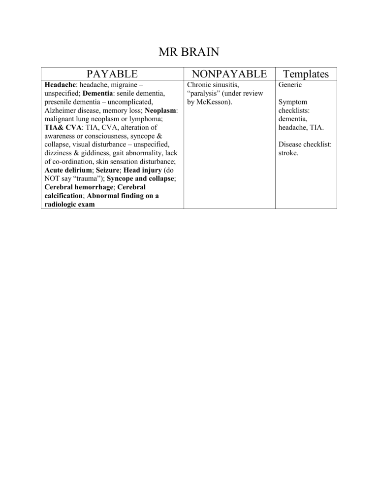
MR BRAIN
PAYABLE
Headache : headache, migraine – unspecified; Dementia : senile dementia, presenile dementia – uncomplicated,
Alzheimer disease, memory loss; Neoplasm : malignant lung neoplasm or lymphoma;
TIA& CVA : TIA, CVA, alteration of awareness or consciousness, syncope & collapse, visual disturbance – unspecified, dizziness & giddiness, gait abnormality, lack of co-ordination, skin sensation disturbance;
Acute delirium ; Seizure ; Head injury (do
NOT say “trauma”);
Syncope and collapse ;
Cerebral hemorrhage ; Cerebral calcification ; Abnormal finding on a radiologic exam
NONPAYABLE Templates
Chronic sinusitis,
“paralysis” (under review by McKesson).
Generic
Symptom checklists: dementia, headache, TIA.
Disease checklist: stroke.
File name: “MR brain”
UNENHANCED [<AND CONTRAST ENHANCED>] HEAD MR
INDICATION:
[+Payable: Headache : headache, migraine – unspecified; Dementia : senile dementia, presenile dementia – uncomplicated, Alzheimer disease, memory loss; Neoplasm : malignant lung neoplasm or lymphoma; TIA& CVA : TIA, CVA, alteration of awareness or consciousness, syncope & collapse, visual disturbance – unspecified, dizziness & giddiness, gait abnormality, lack of co-ordination, skin sensation disturbance; Acute delirium ; Seizure ; Head injury (do
NOT say “trauma”);
Syncope and collapse ; Cerebral hemorrhage ; Cerebral calcification ;
Abnormal finding on a radiologic exam+]
[+Nonpayable: chronic sinusitis.+]
COMPARISON: []
TECHNIQUE: Unenhanced head MR was performed. [<An enhanced head MR was performed following intravenous injection of>] [+(volume/type of IV contrast).+] [+The patient was scanned within 24 hours of admission to the hospital.+]
INTERPRETATION:
Brain and CSF spaces: [<Normal.>]
Pituitary gland and pineal: [<Normal.>]
Vasculature: [<Normal.>]
Paranasal sinuses: [<Normal.>]
Nasal cavity and nasopharynx: [<Normal.>]
Otomastoid findings: [<Normal.>]
Bones and joints: [<Normal.>]
Orbits: [<Normal.>]
IMPRESSION:
[<Normal brain MR examination.>]
[+Signature/location+]
File name: “MR brain for dementia”
UNENHANCED [<AND CONTRAST ENHANCED>] HEAD MR
INDICATION: [<Dementia.>] [+Additional clinical features.+]
COMPARISON: []
TECHNIQUE: Unenhanced head MR was performed. [<An enhanced head MR was performed following intravenous injection of>] [+(volume/type of IV contrast).+] [+The patient was scanned within 24 hours of admission to the hospital.+]
INTERPRETATION:
Brain and CSF spaces: [+Atrophy: frontal = Pick, temporal = Alzheimer, cerebellar = alcohol, mass, swelling, prior white or gray matter infarcts, demyelinating disease. Subdural hematoma or mass. Hydrocephalus; ventricle distortion, hemorrhage, mass.+]
Pituitary gland and pineal: [<Normal.>]
Vasculature: [+Anerysm, stenosis.+]
Paranasal sinuses: [+Sinusitis, mass+]
Nasal cavity and nasopharynx: [+Mass, lymphadenopathy, abscess, inflammation+]
Otomastoid findings: [+Skull fracture, fluid level in sinus, ear, mastoid air cells.+]
Bones and joints: [<Normal.>]
Orbits: [<Normal.>]
IMPRESSION:
[<Normal brain MR examination.>]
[+Signature/location+]
File name: “MR brain for headache”
UNENHANCED [+AND CONTRAST ENHANCED+] HEAD MR
INDICATION: [<Headache.>] [+Additional clinical features.+]
COMPARISON: []
TECHNIQUE: Unenhanced head MR was performed. [<An enhanced head MR was performed following intravenous injection of>] [+(volume/type of IV contrast).+] [+The patient was scanned within 24 hours of admission to the hospital.+]
INTERPRETATION:
Brain and CSF spaces: [+Brain mass (primary or metastatic tumor, abscess, AVM, intraparenchymal hemorrhage, sarcoidosis), swelling (infiltrating tumor, encephalitis, severe contusion/concussion, pseudotumor cerebri, stroke),, abnormal enhancement (tumor, sarcoidosis, encephalitis, stroke, AVM, aneurysm). Extra-axial crescent (subdural hematoma, subdural effusion from intracranial hypotension), lens (epidural hematoma), mass or masses (menigioma, other primary tumors, metastatic disease), or enhancing meninges (meningitis both infectious and malignant, intracranial hypotension, sarcoidosis). Subarachnoid hemorrhage. Ventricular distension (hydrocephalus), fluid-level (hemorrhage), mass, or compression (pseudotumor cerebri. Cerebellar tonsil position (Arnold Chiari malformation).+]
Pituitary gland and pineal: [+Mass, hemorrhage, abscess, cyst+]
Vasculature: [+Focally enlarged intracranial artery (aneurysm, AVM). Absent filling or significant stenosis (stroke). Venous filling defect (venous sinus thrombus). Abnormal thickened or enhancing temporal arteries (temporal arteritis).+]
Paranasal sinuses: [+Sinus opacification, membrane thickening, or fluid level (sinusitis).+]
Nasal cavity and nasopharynx: [+Mass, lymphadenopathy, abscess, polyps.+]
Otomastoid findings: [Mastoid air cell opacification, membrane thickening, or fluid level
(mastoiditis).]
Bones and joints: [+Skull metastatic deposit. TMJ arthritis. Cervico-basilar and upper cervical disc and facet disease.+]
Orbits: [+Mass, abscess, inflammation, ophthalmopathy.+]
IMPRESSION:
[<Normal brain MR examination.>]
[+Signature/location+]
File name: “MR brain for TIA”
MRI BRAIN UNENHANCED [<AND ENHANCED>] [<DONE FOR TIA>]
INDICATION: Suspect [+TIA+] [+on the basis of what sign or symptom. Payables include: alteration of awareness or consciousness, syncope & collapse, visual disturbance – unspecified, dizziness & giddiness, gait abnormality, lack of co-ordination, skin sensation disturbance; acute delirium; seizure, syncope and collapse.+]
COMPARISON: []
TECHNIQUE: Unenhanced head MR was performed. [<An enhanced head MR was performed following intravenous injection of>] [+(volume/type of IV contrast).+] [+The patient was scanned within 24 hours of admission to the hospital.+]
INTERPRETATION:
Brain and CSF spaces: [+Brain mass (primary or metastatic tumor, abscess, AVM, intraparenchymal hemorrhage, sarcoidosis), swelling (infiltrating tumor, encephalitis, severe contusion/concussion, pseudotumor cerebri, stroke), abnormal enhancement (tumor, sarcoidosis, encephalitis, stroke, AVM, aneurysm). Extra-axial crescent (subdural hematoma, subdural effusion from intracranial hypotension), lens (epidural hematoma), mass or masses (menigioma, other primary tumors, metastatic disease), or enhancing meninges (meningitis both infectious and malignant, intracranial hypotension, sarcoidosis). Ventricular distension (hydrocephalus), fluid-level (hemorrhage), mass, or compression (pseudotumor cerebri+.]
Pituitary gland and pineal: [+Mass, hemorrhage, abscess, cyst+]
Vasculature: [+Focally enlarged intracranial artery (aneurysm, AVM). Absent filling or significant stenosis (stroke). Venous filling defect (venous sinus thrombus). Abnormal thickened or enhancing temporal arteries (temporal arteritis).+]
Paranasal sinuses: [<Normal.>]
Nasal cavity and nasopharynx: [<Normal.>]
Otomastoid findings: [<Normal.>]
Bones and joints: [<Normal.>]
Orbits: [<Normal.>]
IMPRESSION:
[+Mention indication(s).+]
[+Signature/location+]
File name: “MR brain for Stroke”
MRI BRAIN UNENHANCED [<AND ENHANCED>] [<SHOWING STROKE>]
INDICATION: [+Describe neurologic deficit.+]
COMPARISON: []
TECHNIQUE: Unenhanced head MR was performed. [+An enhanced head MR was performed following intravenous injection of (volume/type of IV contrast).+] [+The patient was scanned within 24 hours of admission to the hospital.+]
INTERPRETATION:
Brain and CSF spaces: [+Describe restricted diffusion, T1 and T2 prolongation, and contrast enhancement of abnormality. Describe size, location, vascular distribution, and any associated hemorrhage. No mass (primary or metastatic tumor, intraparenchymal hemorrhage), swelling
(infiltrating tumor, stroke), or abnormal enhancement (tumor, stroke). No subdural hematoma identified.+]
Pituitary gland and pineal: [<Normal.>]
Vasculature: [<Normal.>]
Paranasal sinuses: [<Normal.>]
Nasal cavity and nasopharynx: [<Normal.>]
Otomastoid findings: [<Normal.>]
Bones and joints: [<Normal.>]
Orbits: [<Normal.>]
IMPRESSION:
MR features of an [+acute, subacute, chronic+] stroke, measuring [] mm in maximum dimension, located in the [+stroke location+], compatible with the [] arterial distribution. [<No associated hemorrhage.>] [<No intracranial mass.>].
[+Signature/location+]
