Sports Medicine II Shoulder Special Tests Name SC Glide Test
advertisement
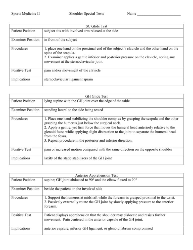
Sports Medicine II Shoulder Special Tests Name _______________________ Patient Position SC Glide Test subject sits with involved arm relaxed at the side Examiner Position in front of the subject Procedures 1. place one hand on the proximal end of the subject’s clavicle and the other hand on the spine of the scapula. 2. Examiner applies a gentle inferior and posterior pressure on the clavicle, noting any movement at the sternoclavicular joint. Positive Test pain and/or movement of the clavicle Implications sternoclavicular ligament sprain Patient Position GH Glide Test lying supine with the GH joint over the edge of the table Examiner Position standing lateral to the side being tested Procedures 1. Place one hand stabilizing the shoulder complex by grasping the scapula and the other grasping the humerus just below the surgical neck. 2. Apply a gentle, yet firm force that moves the humeral head anteriorly relative to the glenoid fossa while applying slight distraction to the joint to separate the humeral head from the fossa. 3. Repeat procedure in the posterior and inferior direction. Positive Test pain or increased motion compared with the same direction on the opposite shoulder Implications laxity of the static stabilizers of the GH joint Patient Position Anterior Apprehension Test supine; GH joint abducted to 90° and the elbow flexed to 90° Examiner Position beside the patient on the involved side Procedures 1. Support the humerus at midshaft while the forearm is grasped proximal to the wrist. 2. Passively externally rotate the GH joint by slowly applying pressure to the anterior forearm. Positive Test Patient displays apprehension that the shoulder may dislocate and resists further movement. Pain centered in the anterior capsule of the GH joint. Implications anterior capsule, inferior GH ligament, or glenoid labrum compromised Patient Position AC Compression Test sitting or standing with the arm hanging naturally at the side Examiner Position standing on the involved side Procedures 1. Cup hands over the anterior and posterior joint structures. 2. Squeeze hands together, compressing the AC joint. Positive Test pain at the AC joint or excursion of the clavicle over the acromion process Implications damage to the AC ligament and possibly the coracoclavicular ligament Patient Position AC Traction Test sitting or standing; arm hanging naturally from the side Examiner Position standing lateral to the involved side Procedures 1. grasp patient’s humerus proximal to the elbow and palpate the AC joint with the opposite hand. 2. Apply a downward traction on the humerus Positive Test humerus and scapula move inferior to the clavicle, causing a step deformity, pain, or both Implications sprain of the AC or coracoclavicular ligaments Patient Position Posterior Apprehension Test Supine; shoulder flexed to 90° and the elbow flexed to 90°; GH joint being tested off the side of the table Examiner Position standing on the involved side Procedures 1. Grasp the forearm with one arm and stabilize the posterior scapula with the opposite hand. 2. Apply a longitudinal force to the humeral shaft, encouraging the humeral head to move posteriorly on the glenoid fossa. 3. Examiner may choose to alter the amount of flexion and rotation of the humerus Positive Test patient displays apprehension and produces muscle guarding to prevent the shoulder from subluxating posteriorly. Implications laxity in the poster GH capsule, torn posterior labrum Patient Position Sulcus Sign sitting, arm hanging at the side Examiner Position standing lateral to the involved side Procedures 1. Grip patient’s arm distal to the elbow. 2. Apply a downward traction force to the humerus. Positive Test Indentation appears beneath the acromion process. Implications humeral head slides inferiorly on the glenoid fossa, indicating laxity in the superior GH ligament Patient Position Yergason’s Test sitting or standing; GH joint in the anatomical position; elbow flexed to 90°; forearm in neutral position Examiner Position positioned lateral to the patient on the involved side Procedures 1. Stabilize the olecranon inferiorly and maintain close to the thorax; stabilize forearm proximal to the wrist. 2. patient provides resistance while the examiner concurrently moves the GH joint into external rotation and the proximal radioulnar joint into supination. Positive Test pain or snapping (or both) in the bicipital groove Implications Primary: snapping or popping in the bicipital groove indicates a tear or laxity of the transverse humeral ligament. Secondary: pain with no associated popping in the bicipital groove may indicate bicipital tendinitis Patient Position Speed’s Test sitting or standing; elbow extended; GH joint in neutral position or slightly extended to stretch biceps brachii Examiner Position Standing lateral to and in front of the involved limb Procedures 1. Position fingers of one hand over the bicipital groove while stabilizing the shoulder. Stabilize the forearm proximal to the wrist. 2. Resist flexion of the GH joint and elbow while palpating for tenderness over the bicipital groove. Positive Test pain along the long head of the biceps brachii tendon, especially in the bicipital groove Implications inflammation of the long head of the biceps tendon as it passes through the bicipital groove Patient Position Drop Arm Test sitting or standing; humerus fully abducted and externally rotated and the forearm supinated Examiner Position standing lateral to, or behind, the involved extremity Procedures 1. patient slowly lowers the arm to the side Positive Test arm falls uncontrollably from a position of approximately 90° abduction to the side Implications lesions to the rotator cuff, especially the supraspinatus Patient Position Neer Impingement Test standing or sitting; shoulder, elbow, and wrist in the anatomical position Examiner Position standing lateral or forward of the involved side Procedures 1. Stabilize the patient’s shoulder on the poster aspect. Grip patient’s forearm distal to the elbow joint. 2. With elbow extended, humerus is placed in internal rotation and the forearm is pronated. 3. GH joint is forcefully moved through forward flexion as the scapula is stabilized. Positive Test pain with motion, especially near the end of ROM Implications pathology in rotator cuff or the long head of the biceps brachii tendon Patient Position Hawkin’s Shoulder Impingement Test sitting or standing; shoulder, elbow, and wrist in the anatomical position Examiner Position standing lateral or forward of the involved side Procedures 1. Grip the patient’s arm at the elbow joint and wrist. 2. With elbow flexed, GH joint elevated to 90° in the scapular plane. 3. Passively internally rotate humerus. Positive Test pain with motion, especially near the end of ROM Implications pathology is present in rotator cuff or the long head of the biceps brachii tendon Patient Position Empty Can Test sitting or standing; GH abducted to 90° in the scapular plane, elbow extended, and the humerus internally rotated and the forearm pronated so that the thumb points downward Examiner Position standing facing the patient Procedures 1. Place one hand on the superior portion of the midforearm to resist the motion of abduction in the scapular plane. 2. Resist abduction (apply a downward pressure) Positive Test weakness or pain accompanying the movement Implications supraspinatus tendon is: 1-being impinged between the humeral head and the coracoacromial arch 2-is inflamed or 3-contains a lesion Patient Position Adson’s Test sitting; shoulder abducted to 30°, elbow extended with the thumb pointing upward, humerus externally rotated Examiner Position standing behind the patient Procedures 1. Palpate the radial pulse. 2. Externally rotate and extend the patient’s shoulder while the face is rotated toward the involved side and extends the neck. 3. Patient is instructed to inhale deeply and hold the breath. Positive Test radial pulse disappears or markedly diminishes Implications subclavian artery is being occluded between the anterior and middle scalene muscles and the pectoralis minor Patient Position Allen’s Test sitting; the head facing forward Examiner Position standing behind the patient Procedures 1. Palpate the radial pulse. 2. Elbow is flexed to 90° while the clinician abducts the shoulder to 90°. 3. Passively horizontally abduct and externally rotate the shoulder. 4. Patient rotates head towards the opposite shoulder. Positive Test radial pulse disappears Implications pectoralis minor muscle is compressing the neurovascular bundle
