Exploring Post-Translational Modification With DFT
advertisement
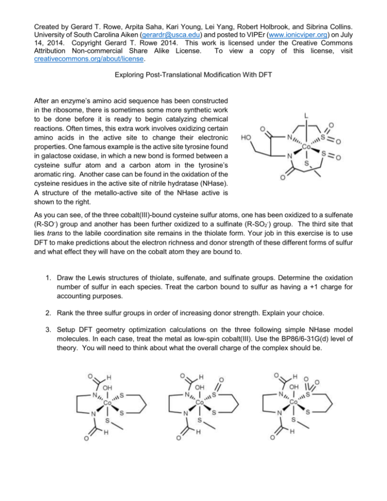
Created by Gerard T. Rowe, Arpita Saha, Kari Young, Lei Yang, Robert Holbrook, and Sibrina Collins. University of South Carolina Aiken (gerardr@usca.edu) and posted to VIPEr (www.ionicviper.org) on July 14, 2014. Copyright Gerard T. Rowe 2014. This work is licensed under the Creative Commons Attribution Non-commercial Share Alike License. To view a copy of this license, visit creativecommons.org/about/license. Exploring Post-Translational Modification With DFT After an enzyme’s amino acid sequence has been constructed in the ribosome, there is sometimes some more synthetic work to be done before it is ready to begin catalyzing chemical reactions. Often times, this extra work involves oxidizing certain amino acids in the active site to change their electronic properties. One famous example is the active site tyrosine found in galactose oxidase, in which a new bond is formed between a cysteine sulfur atom and a carbon atom in the tyrosine’s aromatic ring. Another case can be found in the oxidation of the cysteine residues in the active site of nitrile hydratase (NHase). A structure of the metallo-active site of the NHase active is shown to the right. As you can see, of the three cobalt(III)-bound cysteine sulfur atoms, one has been oxidized to a sulfenate (R-SO-) group and another has been further oxidized to a sulfinate (R-SO2-) group. The third site that lies trans to the labile coordination site remains in the thiolate form. Your job in this exercise is to use DFT to make predictions about the electron richness and donor strength of these different forms of sulfur and what effect they will have on the cobalt atom they are bound to. 1. Draw the Lewis structures of thiolate, sulfenate, and sulfinate groups. Determine the oxidation number of sulfur in each species. Treat the carbon bound to sulfur as having a +1 charge for accounting purposes. 2. Rank the three sulfur groups in order of increasing donor strength. Explain your choice. 3. Setup DFT geometry optimization calculations on the three following simple NHase model molecules. In each case, treat the metal as low-spin cobalt(III). Use the BP86/6-31G(d) level of theory. You will need to think about what the overall charge of the complex should be. Created by Gerard T. Rowe, Arpita Saha, Kari Young, Lei Yang, Robert Holbrook, and Sibrina Collins. University of South Carolina Aiken (gerardr@usca.edu) and posted to VIPEr (www.ionicviper.org) on July 14, 2014. Copyright Gerard T. Rowe 2014. This work is licensed under the Creative Commons Attribution Non-commercial Share Alike License. To view a copy of this license, visit creativecommons.org/about/license. 4. After your geometry optimizations are finished, open up the output files in a viewer like WebMO or GaussView. Bring up the population analysis charge display (Results->Charges in GaussView) and write down the charges of the cobalt and sulfur atoms in each molecule. 5. Measure the Co-S bond lengths in each of the three model compounds. Don’t freak out if you don’t see a bond between the metal and all the ligands. That’s just a consequence of how the program decides when to draw a bond between two atoms, and it doesn’t usually work for metalligand interactions. 6. Do your computational results match your predictions based on oxidation number and donor strength? Explain.
