Li-ion Batteries by Soft X-ray Absorption and Emission Spectroscopy
advertisement

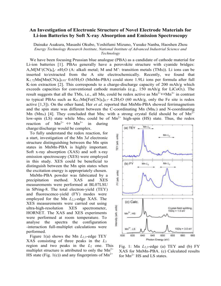
An Investigation of Electronic Structure of Novel Electrode Materials for Li-ion Batteries by Soft X-ray Absorption and Emission Spectroscopy Daisuke Asakura, Masashi Okubo, Yoshifumi Mizuno, Yusuke Nanba, Haoshen Zhou Energy Technology Research Institute, National Institute of Advanced Industrial Science and Technology We have been focusing Prussian blue analogue (PBA) as a candidate of cathode material for Li-ion batteries [1]. PBAs generally have a perovskite structure with cyanide bridges: AxM[M’(CN)6]y·nH2O (A: alkali metal; M and M’: transition metals (TMs)). Li ions can be inserted to/extracted from the A site electrochemically. Recently, we found that K1.72Mn[Mn(CN)6]0.93·0.65H2O (MnMn-PBA) could store 1.9Li ions per formula after full K-ion extraction [2]. This corresponds to a charge-discharge capacity of 200 mAh/g which exceeds capacities for conventional cathode materials (e.g., 150 mAh/g for LiCoO2). The result suggests that all the TMs, i.e., all Mn, could be redox active as Mn2+⇔Mn3+ in contrast to typical PBAs such as K0.1Mn[Fe(CN)6]0.7·4.2H2O (60 mAh/g, only the Fe site is redox active [1,3]). On the other hand, Her et al. reported that MnMn-PBA showed ferrimagnetism and the spin state was different between the C-coordinating Mn (MnC) and N-coordinating Mn (MnN) [4]. They concluded that MnC with a strong crystal field should be of Mn2+ low-spin (LS) state while MnN could be of Mn2+ high-spin (HS) state. Thus, the redox reaction of Mn2+ ⇔ Mn3+ in during charge/discharge would be complex. To fully understand the redox reaction, for a start, investigation of the Mn 3d electronic structure distinguishing between the Mn spin states in MnMn-PBA is highly important. Soft x-ray absorption (XAS) and soft x-ray emission spectroscopy (XES) were employed in this study. XES could be beneficial to distinguish between the Mn spin states when the excitation energy is appropriately chosen. MnMn-PBA powder was fabricated by a precipitation method. XAS and XES measurements were performed at BL07LSU in SPring-8. The total electron-yield (TEY) and fluorescence-yield (FY) modes were employed for the Mn L2,3-edge XAS. The XES measurements were carried out using ultra-high-resolution XES spectrometer, HORNET. The XAS and XES experiments were performed at room temperature. To analyse the spectra the configuration -interaction full-multiplet calculations were performed. Figure 1(a) shows the Mn L2,3-edge TEY XAS consisting of three peaks in the L3 region and two peaks in the L2 one. This Fig. 1: Mn L2,3-edge (a) TEY and (b) FY multiplet structure is attributed to only the Mn2+ XAS for MnMn-PBA. (c) Calculated results HS state (Fig. 1(c)) and any fingerprints of Mn2+ for Mn2+ HS and LS states. LS state could not be observed. In general, the probing depth of TEY mode is less than 5 nm. Thus, the Mn2+ LS state should not exist in the surface region. As Fig. 1(b) shows, the Mn L2,3-edge FY XAS is quite different from the TEY XAS while the peak positions are almost similar. The enhancements of the peaks at 644, 652.5 eV should be intrinsic although the saturation effect in the FY mode distorts the spectrum. The calculated result for Mn2+ LS state indicates the peak at 652.4 eV and the shoulder at 655 eV could be of the Mn2+ LS state. On the other hand, the peak at 644 eV in the experimental FY spectrum was not reproduced. In general, the distortion due to the saturation effect increases in going to higher energy region. Most likely, the calculated Mn2+-LS main peak at 642 eV in the L3 region would be hidden in the broadened base line around 642-645 eV. The shoulders at 639 eV in the FY result could be of the unoccupied t2g orbital for the Mn2+ LS state. In the calculated spectrum the energy separation between that peak and the main peak is 3 eV, which is equal to the Fig. 2: (a) Mn 2p RXES and (b) the crystal-field splitting 10Dq. Thus, the peaks calculated results with polarization at 639 eV and 642 eV can be attributed to t2g dependence. In (b), the solid and dotted lines and eg orbitals, respectively. The similar mean the in-plane and out-of-plane situation was observed in Fe L3-edge XAS geometries, respectively. for Fe3+ LS state [3], i.e., 3d5 LS state with a hole in the t2g orbital. To further clarify the Mn2+ LS state, high energy resolution Mn 2p resonant XES (RXES) was carried out. The excitation energies of 640 and 643 eV were chosen, because the Mn2+ HS character could be dominant at 640 eV and the Mn2+ LS one would be included around 643 eV as discussed above. Figure 2(a) displays the RXES. As for the 640-eV spectrum, the d-d excitation from -2 to -5 eV is similar to that for MnO [5], suggesting the Mn2+ HS state is dominant at 640 eV. In fact, the calculation for Mn2+ HS well reproduced the experimental spectrum (Fig. 2(b)). For the 643-eV spectrum, the d-d excitation, particularly the enhanced peak at -4 eV is attributed to the Mn2+ LS state. This peak could not be observed in MnO [5]. In summary, we succeeded in distinguishing between the spin states in MnMn-PBA by combined use of TEY XAS, FY XAS and XES. In future, the redox reaction of MnMn-PBA during Li-ion insertion/extraction will be investigated. References [1] M Okubo et al., J. Phys. Chem. Lett. 1, 2063 (2010). [2] D. Asakura et al., J. Phys. Chem. C 116, 8364 (2012). [3] D. Asakura et al., Phys. Rev. B 84, 045117 (2011). [4] J.-H. Her et al., Inorg. Chem. 49, 1524 (2010). [5] G. Ghiringhelli et al., Phys. Rev. B 73, 035111 (2006).


