ND_Semi_Ann - SCIPP - University of California, Santa Cruz
advertisement

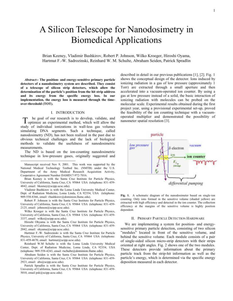
1 A Silicon Telescope for Nanodosimetry in Biomedical Applications Brian Keeney, Vladimir Bashkirov, Robert P. Johnson, Wilko Kroeger, Hiroshi Oyama, Hartmut F.-W. Sadrozinski, Reinhard W. M. Schulte, Abraham Seiden, Patrick Spradlin Abstract-- The position- and energy-sensitive primary particle detectors of a nanodosimetry system are described. They consist of a telescope of silicon strip detectors, which allow the determination of the particle’s position from the hit strip address and its energy from the specific energy loss. In our implementation, the energy loss is measured through the timeover-threshold (TOT). I. INTRODUCTION he goal of our research is to develop, validate, and Toptimize an experimental method, which will allow the described in detail in our previous publications [1], [2]. Fig. 1 shows the conceptual design of the detector. Ions induced by ionizing radiation in a gas of low pressure (approximately 1 Torr) are extracted through a small aperture and then accelerated into a vacuum-operated ion counter. By using a gas at low pressure instead of a solid, the basic interaction of ionizing radiation with molecules can be probed on the molecular scale. Experimental results obtained during the first project year, using a provisional experimental set-up, proved the feasibility of the ion counting technique with a vacuumoperated multiplier and demonstrated the possibility of nanometer spatial resolution [3]. study of individual ionizations in wall-less gas volumes simulating DNA segments. Such a technique, called nanodosimetry (ND), has not been realized in the past due to obvious technical challenges and the lack of biological methods to validate the usefulness of nanodosimetric measurements. The ND is based on the ion-counting nanodosimetric technique in low-pressure gases, originally suggested and Manuscript received Nov 9, 2001. This work was supported by the National Medical Technology Testbed Inc. (NMTB) under the U.S. Department of the Army Medical Research Acquisition Activity, Cooperative Agreement Number DAMD17-972-7016. Brian Keeney is with the Santa Cruz Institute for Particle Physics, University of California, Santa Cruz, CA 95064 USA (telephone: 831-4594842, email: bkeeney@scipp.ucsc.edu). Vladimir Bashkirov is with the Loma Linda University Medical Center, Dept. of Radiation Medicine, Loma Linda, CA 92354, USA (telephone: 909-558-8366, email: vbashkirov@dominion.llumc.edu). Robert P. Johnson is with the Santa Cruz Institute for Particle Physics, University of California, Santa Cruz, CA 95064 USA (telephone: 831-4592125, email: johnson@scipp.ucsc.edu). Wilko Kroeger is with the Santa Cruz Institute for Particle Physics, University of California, Santa Cruz, CA 95064 USA (telephone: 831-4593337, email: wilko@scipp.ucsc.edu). Hiroshi Ohyama is with the Santa Cruz Institute for Particle Physics, University of California, Santa Cruz, CA 95064 USA (telephone: 831-4592042, email: ohyama@scipp.ucsc.edu). Hartmut F.-W. Sadrozinski is with the Santa Cruz Institute for Particle Physics, University of California, Santa Cruz, CA 95064 USA (telephone: 831-459-4670, email: hartmut@scipp.ucsc.edu). Reinhard W.M Schulte is with the Loma Linda University Medical Center, Dept. of Radiation Medicine, Loma Linda, CA 92354, USA (telephone: 909-558-4243, email: rschulte@dominion.llumc.edu). Abraham Seiden is with the Santa Cruz Institute for Particle Physics, University of California, Santa Cruz, CA 95064 USA (telephone: 831-4592923, email: abs@scipp.ucsc.edu). Patrick Spradlin is with the Santa Cruz Institute for Particle Physics, University of California, Santa Cruz, CA 95064 USA (telephone: 831-4595010, email psk@scipp.ucsc.edu). Fig. 1. A schematic diagram of the nanodosimeter based on single-ion counting. Only ions formed in the sensitive volume (shaded yellow) are extracted with high efficiency and detected in the ion counter. The collection efficiency at the margins of the sensitive volume is highly position dependent. II. PRIMARY PARTICLE DETECTION HARDWARE We are implementing a system for position- and energysensitive primary particle detection, consisting of two silicon ”modules” located in front of the sensitive volume, and behind the sensitive volume. Each module consists of a pair of single-sided silicon micro-strip detectors with their strips oriented at right angles. Fig. 2 shows one of the two modules. These detectors provide information about the primary particle track from the strip-hit information as well as the particle’s energy, which is determined via the specific energy deposition measured in each detector. 2 Fig. 3. The Time-over-threshold (TOT) calibration curve. The curve is linear below 100 fC input charge and saturates at 100s (not shown). Fig. 2. One of the two silicon modules. The 6.4cm x 6.4 cm SSD and the MCM with 5 GTFE and 1 GTRC ASIC's are visible. Alignment of the SSD's on the two sides to each other is accomplished by the Delrin posts and the precision cutting of the SSD to 20 m. At relatively low proton energies (< 100 MeV) the energy loss is a rapidly changing function of the proton energy [4], [5]. This fact allows quantifying the ND spectra as a function of the primary particle energy. Locating the proton track with high precision permits an accurate ion collection efficiency map of the sensitive volume to be obtained and a measurement of the ND spectra as a function of distance from the primary particle track. For the readout of the fast silicon detector signals, we are using a low-noise, low-power front-end ASIC Global Trigger Front End (GTFE) developed for the GLAST mission [6]. All GTFEs on one silicon detector are read out via a digital controller ASIC Global Trigger Readout Controller (GTRC) into a computer. The GTFE is a binary chip, with a threshold able to be set for every chip, and has a fast output of the time over threshold (TOT). It allows self-triggering through an OR of the TOT of all channels on a detector. The GTRC also allows digitization of the TOT, yielding a measurement of the input charge through the pulse width, i.e. the TOT signal, over a large dynamic range. The measured electronic calibration of the chip TOT vs. input charge is shown in Fig. 3, which exhibits a linear dependence of the TOT on the charge input up to a duration of 100s. At that point, saturation sets in very quickly. Thus we expect valuable energy measurement up to an input charge of 100fc, which corresponds to the charge deposited by 20 MeV protons or about 20 minimum ionizing particles (MIPs) in 400m Si (see Table 1 below). One feature of the selected ASIC is that only one TOT value is returned per detector, even if more then one strip is hit. The TOT value is the OR of all pulse widths. Thus to achieve good energy resolution, events with only one hit have to be selected, such that there is no appreciable charge sharing between strips. The TOT values for hits next to any non-functional strips were also eliminated because the extent of charge sharing is unknown. Fig. 4. Time-over-threshold (TOT) spectrum for 40MeV protons. The effect of the fiducial and hit cut are shown. Open circles: TOT in all events, open squares: TOT in events with single hits and with applied fiducial cuts. Fig. 4 shows the TOT spectrum for 40 MeV protons for all events and for single-hit events and other fiducial cuts as mentioned. 3 TABLE I ENERGY AND TOT Fig. 5 shows the TOT spectrum from the SLAC beam test for all events, for those with one hit, two adjacent hits and three and more adjacent hits, respectively. The ASIC voltages Vanalog and threshold settings Vthr were slightly different for the two set-ups, resulting in slightly different TOT calibration curves, which were taken into consideration in the data analysis. (LLUMC data: Vanalog = 1.92V, Vthr = 5fC; SLAC data Vanalog = 2.3V, Vthr = 1.4fC). III. SILICON DETECTOR PERFORMANCE In order to determine the energy measurement capabilities of the silicon strip detector-TOT system, we exposed silicon detectors to proton beams of various energies and recorded the TOT spectra. Table 1 shows the proton beam energies selected. The TOT spectra for single hits allow finding the mean and the FWHM of the deposited charge for different incident proton energies. We used a prototype module to take TOT spectra in the proton beam of the proton synchrotron at Loma Linda University Medical Center (LLUMC). Monoenergetic beam energies of 250 MeV and 40 MeV were selected. Beams of lower proton energies were produced by degrading the proton beam with polystyrene absorbers. In addition, we have analyzed the data taken in a 13.5GeV proton beam during the GLAST beam test in January 2000 [7], using identical Si detectors and front-end ASIC's. Fig. 6. Time-over-threshold TOT spectrum for two mono-energetic proton beams (250 MeV and 40 MeV) and two degraded proton beams (24 MeV and 17 MeV) with fiducial and hit cuts applied. The TOT spectra for single hit tracks are shown in Fig.6 for selected energies of the LLUMC runs. The energies, mean and RMS of all TOT spectra are shown in Table 1. In addition, the calculated deposited charge in 400 m silicon and the expected TOT mean value are shown in Table 1. Fig. 7. Measured and predicted mean time-over-threshold (TOT) signal as a function of the proton energy. The proton energy can be measured uniquely at energies above 20 MeV (and below 3 MeV, where the signal of stopping protons is not saturating the TOT). The error bars shown are the RMS width of the TOT spectra in Fig. 6. Fig. 5. Time-over-threshold (TOT) spectrum for 13.5GeV protons. The effect of the hit cut is shown. The mean measured and calculated TOT values vs. the energy of the primary protons are shown in Fig. 7. The RMS of the TOT spectra derived from the core of the distributions are shown as error bars. The agreement between measured and predicted TOT values is excellent over the entire energy range from 20 MeV to 13.5 GeV. It is clear from Fig. 7 that there is less energy discrimination for proton energies above 100 MeV due to the smaller dependence of the energy loss on 4 primary proton energy. We can derive the energy resolution of the system E from the observed TOT RMS TOT and the derivative of the TOT vs. energy curve (Fig. 7): E TOT TOT E VI. REFERENCES [1] [2] [3] [4] [5] [6] [7] Fig. 8. Relative width of the measured TOT spectra TOT/TOT, and derived energy resolution E/E. The TOT resolution is of the order 10-20% and represents the LET resolution of a single plane. The energy resolution is about 15% below 100MeV. At energies above 100 MeV the dE/dx curve is relatively flat, which leads to a relatively low energy resolution. Fig. 8 shows the relative width of the measured TOT spectra TOT TOT and the energy resolution, E E . Below 40 MeV, the energy resolution of the detector is on the order of 15%, and increases to 25% at 250 MeV. IV. CONCLUSION We have demonstrated that the primary energy of the protons can be derived from their specific energy deposition using the TOT signal of a single silicon strip detector for proton energies above 20 MeV. In addition, there is an applicable region below about 3 MeV, where the particle stops in the detector. In the energy range of 3-20 MeV, where we observe TOT saturation, protons deposit a significant fraction of their energy in the silicon detector, so that the TOT signal of at least one of the four detectors used will be within the measurable range, and thus will provide sufficient information to reconstruct the energy of the particle passing the sensitive gas volume. At high energies, i.e., above 100 MeV, the TOT signal is a relatively shallow function of incident proton energy, which intrinsically yields poor resolution, but for these energies all four detectors will provide a measurement, thereby reducing the measurement uncertainty. V. ACKNOWLEDGMENT This work was supported by the National Medical Technology Testbed Inc. (NMTB) under the U.S. Department of the Army Medical Research Acquisition Activity, Cooperative Agreement Number DAMD17-972-7016. The views and conclusions contained in this presentation are those of the authors and do not necessarily reflect the position or the policy of the U.S. Army or NMTB. We thank Bill Rowe and Alec Webster for valuable technical assistance. Shchemelinin S., Breskin A., Chechik R., Pansky A., Colautti P., Conte V., De Nardo L., and G. Tornielli. “Ionization measurements in small gas samples by single ion counting”. Nucl. Instr. & Meth. A368, 895-861, 1996. Shchemelinin S., Breskin A., Chechik R., Pansky A., and P. Colautti. A nanodosimeter based on single ion counting. In: “Microdosimetry – An interdisciplinary approach.” Goodhead D., O’Neel P. & Menzel H. (Eds.), The Royal Society of Chemistry (Cambridge), pp 375-378, 1997. Shchemelinin S., Breskin A., Chechik R., Colautti P., and R. Schulte. “First ionization cluster measurements on the DNA scale in a wall-less sensitive volume”. Radiat. Prot. Dosim. 82, 43-50, 1999. Particle Data Group, D.E. Groom ed., ”Review of Particle Physics”, Eur. Phys. J, vol. C15, pp.1-878, 2000. Sadrozinski, H. F.-W., ”Applications of Silicon Detectors”, IEEE Nucl. Science Symposium 2000, Lyon, France, Oct 2000. R. P. Johnson, P. Poplevin, H. F.-W. Sadrozinski, and E. Spencer, “An amplifier-discriminator chip for the GLAST silicon-strip tracker,” IEEE Trans. Nucl. Sci., vol. 45, pp. 927-932, 1998. E. do Couto e Silva, G. Godfrey, P. Anthony, R. Arnold, H. Arrighi, et al, “ Results from the Beam Test of the Engineering Model of the GLAST LAT”, Nucl. Inst. Meth. A (2001) Vol 474/1, 19-37, 2001.


