LAB #11: AXIAL SKELETON
advertisement
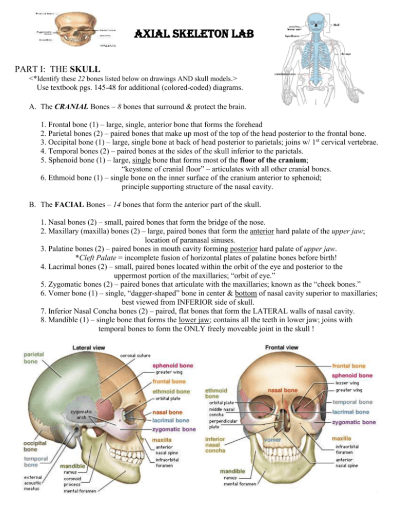
AXIAL SKELETON Lab PART I: THE SKULL <*Identify these 22 bones listed below on drawings AND skull models.> Use textbook pgs. 145-48 for additional (colored-coded) diagrams. A. The CRANIAL Bones – 8 bones that surround & protect the brain. 1. Frontal bone (1) – large, single, anterior bone that forms the forehead 2. Parietal bones (2) – paired bones that make up most of the top of the head posterior to the frontal bone. 3. Occipital bone (1) – large, single bone at back of head posterior to parietals; joins w/ 1st cervical vertebrae. 4. Temporal bones (2) – paired bones at the sides of the skull inferior to the parietals. 5. Sphenoid bone (1) – large, single bone that forms most of the floor of the cranium; “keystone of cranial floor” – articulates with all other cranial bones. 6. Ethmoid bone (1) – single bone on the inner surface of the cranium anterior to sphenoid; principle supporting structure of the nasal cavity. B. The FACIAL Bones – 14 bones that form the anterior part of the skull. 1. Nasal bones (2) – small, paired bones that form the bridge of the nose. 2. Maxillary (maxilla) bones (2) – large, paired bones that form the anterior hard palate of the upper jaw; location of paranasal sinuses. 3. Palatine bones (2) – paired bones in mouth cavity forming posterior hard palate of upper jaw. *Cleft Palate = incomplete fusion of horizontal plates of palatine bones before birth! 4. Lacrimal bones (2) – small, paired bones located within the orbit of the eye and posterior to the uppermost portion of the maxillaries; “orbit of eye.” 5. Zygomatic bones (2) – paired bones that articulate with the maxillaries; known as the “cheek bones.” 6. Vomer bone (1) – single, “dagger-shaped” bone in center & bottom of nasal cavity superior to maxillaries; best viewed from INFERIOR side of skull. 7. Inferior Nasal Concha bones (2) – paired, flat bones that form the LATERAL walls of nasal cavity. 8. Mandible (1) – single bone that forms the lower jaw; contains all the teeth in lower jaw; joins with temporal bones to form the ONLY freely moveable joint in the skull ! Practice Questions: Part I -- The Skull 1. The total number of cranial bones is ________. 2. The dagger-shaped bone in nasal cavity is the _________________. 3. Bone that forms the forehead is the ____________________. 4. The cheek bones are the: ____________________. 5. The total number of facial bones is _________. 6. Bone that forms the bridge of the nose is the _____________. 7. Bone that forms back of skull: _______________. 8. Bone that makes up the posterior hard palate: _____________. 9. Bone that makes up the anterior hard palate: _______________. 10. Bone that makes up the lower jaw: ___________________. PART II: Vertebral Column, Sternum, & Ribs <*Identify these bones listed below on drawings AND skeleton models.> Use textbook pgs. 150-158 for additional (colored-coded) diagrams. A. Vertebral Column – bones that surround and protect the spinal cord. 1. Parts of a vertebrae: a. Spinous process – posterior projection that serves as a point of attachment for muscles; most posterior part of vertebrae. b. Vertebral foramen – central canal through which spinal cord passes. c. Body (centrum) – large circular area anterior to vertebral foramen that forms the joints at the intervertebral discs. d. Transverse process – lateral projections; serve as muscle attachments. e. Articular process – smooth surfaces that join with other vertebrae. 2. Types of Vertebrae a. Cervical – 7 vertebrae in the neck region. The first two are highly specialized to form the type of joint that enables the head to turn/pivot: the first cervical vertebrae (C1) is called the atlas and the second cervical vertebrae (C2) is called the axis. b. Thoracic – 12 vertebrae in the upper back region. They are characterized by long spinous process. Each vertebrae joins with a pair of ribs. c. Lumbar – 5 vertebrae in the lower back. They are generally larger & heavier than other vertebrae to bear the large amount of the body’s weight. d. Sacral – single, large vertebrae; for added strength for a triangle-shaped bone. e. Coccyx – one vestigial (nonfunctional) bone of vertebral column which represents the tailbone. B. Sternum & Ribs 1. Sternum – 3 bones composing the breastbone. a. Manubrium – superior portion b. Body (gladiolus) – middle/largest portion c. Xiphoid Process – inferior/smallest portion 2. Ribs – 12 pairs that join with vertebral column. a. “True” Ribs – 7 pairs that join with sternum by their OWN costal cartilage. b. “False” Ribs – 3 pairs that connect to sternum by attaching to the cartilage of rib #7. c. “Floating” Ribs – 2 pairs that do NOT connect to sternum; only attach to thoracic vertebrae. Axial Skeleton Lab Anatomy/Physiology Name __________________________ Date ________________ Period _____ <Use Textbook pages 145-158 and the skull coloring sheet to answer the following questions.> 1. Name the 3 major components of the axial skeleton: a. b. c. 2. Define a suture: 3. What is the only freely moveable bone of the skull? _____________________ 4. List 2 functions of sinuses: a. b. 5. Why is the sphenoid bone called the “keystone of the cranial floor?” 6. Name the SKULL bone that is described below: a. b. c. d. e. f. g. h. bone forming anterior cranium bone; “forehead” : ___________________ “cheekbone”: ___________________ anterior bone of upper jaw: _________________ bony skeleton of nose: ______________ site of ear canal (auditory meatus); “sides of skull”: ________________ facial bone with sinuses: _________________ posterior cranial bone that joins with atlas: _________________ *not really a skull bone (no articulation with other bones): ________________ 7. Label the specific regions of the sternum & ribs in the thorax diagram below: a. “ “ ribs b. __________________ (sternum region) 8. Name the part of the thoracic vertebrae that forms the point of articulation with a rib: _________________ 9. What is the name given to the first cervical vertebrae (C1)? ________________ 10. What is the cavity in the vertebra through which the spinal cord passes? ________________________ 11. What is the function of the transverse process? ________________________________ 12. Which part of a vertebra is the most posterior? __________________ 13. What is a herniated (“slipped”) disc? 14. What is the structural difference between an individual thoracic vertebrae and a lumbar vertebrae? 15. Name the sections of the vertebral column AND designate the # of vertebrae in each: = ____ = ____ = ____ = ____ = ____


