Serie - Zielinski Family Home Page
advertisement
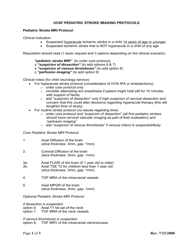
UCSF PEDIATRIC STROKE IMAGING PROTOCOLS Pediatric Stroke MRI Protocol Clinical indication: Suspected hyperacute ischemic stroke in a child 14 years of age or younger Suspected ischemic stroke that is NOT hyperacute in a child of any age Requisition should read (1 basic request and 3 options depending on the clinical scenario): “pediatric stroke MRI” (to order core protocol) ± “suspicion of dissection” (to add options 6 & 7) ± “suspicion of venous thrombosis” (to add option 8) ± “perfusion imaging” (to add option 9) Clinical notes (for child neurology service): For hyperacute stroke protocol (consideration of IV/IA tPA or embolectomy): o order core protocol only o consider attempting w/o anesthesia if patient might hold still for 10 minutes with support of family o add “suspicion of dissection” only if high suspicion of cervical dissection and concern that this could alter decisions regarding hyperacute therapy (this will lengthen time of study) For routine stroke protocol (no issues regarding time) o order core protocol and “suspicion of dissection” (all first pediatric strokes should have cervical vascular imaging as part of their evaluation) and “perfusion imaging” o add “suspicion of venous thrombosis” if venous infarct is suspected/likely Core Pediatric Stroke MRI Protocol 1. Axial Diffusion of the brain (slice thickness: 3mm, gap: 1mm) 2. Coronal Diffusion of the brain (slice thickness: 3mm, gap: 1mm) 3a. 3b. Axial FLAIR of the brain (if 1 year old or older) Axial TSE T2 for children less than 1 year old (slice thickness: 3mm, gap: 1mm) 4. TOF MRA of the intracranial vessels 5. Axial MPGR of the brain (slice thickness: 3mm, gap: 1mm) Optional Pediatric Stroke MRI Protocol if dissection is suspected: option 6. Axial T1 fat sat of the neck option 7. TOF MRA of the neck vessels if venous thrombosis is suspected: option 8. TOF MRV of the intracranial veins/sinuses Page 1 of 3 Rev. 7/23/2008 perfusion MR imaging: option 9. axial perfusion imaging of the brain (requires the injection of contrast, 0.1 mmol/kg chased by 0.1 cc/kg of saline, injection rate: 2 cc/sec between 1-10 years and 4 cc/sec between 10-18 years) Pediatric Stroke CT Protocol Clinical indication: Suspected hyperacute ischemic stroke in a child 15 years of age or older Suspected ischemic stroke in a child who cannot receive MRI (e.g., pacemaker, clinical instability) and risks of radiation are acceptable given clinical scenario Requisition should read (1 basic request and 3 options depending on the clinical scenario): “pediatric stroke CT” (to order core protocol) ± “perfusion imaging” (to add option 2) ± “intracranial CTA” (to add option 3) ± “cervical and intracranial CTA” (to add option 4) Clinical notes (for child neurology service): For hyperacute stroke protocol (consideration of IV/IA tPA or embolectomy): o order core protocol, perfusion imaging, and intracranial CTA o add “cervical CTA” only if high suspicion of cervical dissection and concern that this could alter decisions regarding hyperacute therapy (this exposes the thyroid to radiation) For routine stroke protocol (no issues regarding time) o order MRI unless contraindicated o if contraindications to MRI, and radiation risks are acceptable, order core protocol and all options Core Pediatric Stroke CT Protocol 1. Noncontrast CT of the brain (slice thickness: 2.5mm, reconstruction interval: 2.5mm, pitch as close to 1 as possible, rotation time: 1 sec, noise index 4, min-max mA: 50-300, 120 kVp) Optional Pediatric Stroke MRI Protocol option 2. 1 single Perfusion-CT series according to dedicated pediatric protocol Perfuson-CT protocols 1-10 years 10-18 years acquisition parameters 80 kVp 80 mAs 80 kVp 100 mAs duration of the CT acquisition 35 sec 40 sec every 1 sec every 1 sec CT image sampling rate Page 2 of 3 Rev. 7/23/2008 total number of images per level 35 images 40 images 1 cc/kg 1 cc/kg saline chase 0.1 cc/kg 0.1 cc/kg injection rate 2 cc/sec 4 cc/sec 5 sec 5 sec amount of contrast material for the bolus delay between beginning of intravenous administration of contrast material and data acquisition ° option 3. CTA of the intracranial vessels (slice thickness: 1.25mm, reconstruction interval: 1mm, pitch as close to 1 as possible, rotation time: 0.4 sec, noise index 4, min-max mA: 100-300, 100 kVp, 1.5 cc/kg of contrast chased by 0.1 cc/kg of saline, same injection rate as for PCT) option 4. extension of the CTA acquisition to the level of the clavicles (NOT to include the aortic arch or the heart) 5. post-contrast CT of the brain ALWAYS TO BE OBTAINED IF CONTRAST MATERIAL IS INJECTED (slice thickness: 2.5mm, reconstruction interval: 2.5mm, pitch as close to 1 as possible, rotation time: 1 sec, noise index 4, min-max mA: 50-300) Page 3 of 3 Rev. 7/23/2008

