Mostafa alsaid saleh _9 Outcome after Hepatectomy VS RFA Final
advertisement
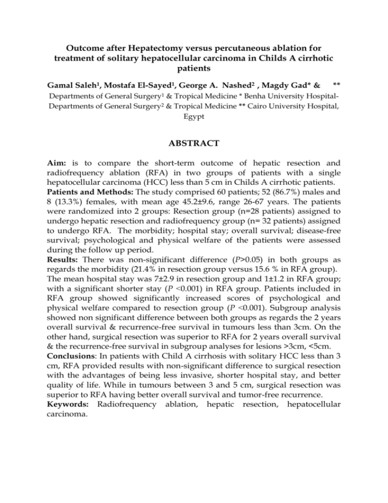
Outcome after Hepatectomy versus percutaneous ablation for treatment of solitary hepatocellular carcinoma in Childs A cirrhotic patients Gamal Saleh1, Mostafa El-Sayed1, George A. Nashed2 , Magdy Gad* & ** Departments of General Surgery1 & Tropical Medicine * Benha University HospitalDepartments of General Surgery2 & Tropical Medicine ** Cairo University Hospital, Egypt ABSTRACT Aim: is to compare the short-term outcome of hepatic resection and radiofrequency ablation (RFA) in two groups of patients with a single hepatocellular carcinoma (HCC) less than 5 cm in Childs A cirrhotic patients. Patients and Methods: The study comprised 60 patients; 52 (86.7%) males and 8 (13.3%) females, with mean age 45.2±9.6, range 26-67 years. The patients were randomized into 2 groups: Resection group (n=28 patients) assigned to undergo hepatic resection and radiofrequency group (n= 32 patients) assigned to undergo RFA. The morbidity; hospital stay; overall survival; disease-free survival; psychological and physical welfare of the patients were assessed during the follow up period. Results: There was non-significant difference (P>0.05) in both groups as regards the morbidity (21.4% in resection group versus 15.6 % in RFA group). The mean hospital stay was 7±2.9 in resection group and 1±1.2 in RFA group; with a significant shorter stay (P ˂0.001) in RFA group. Patients included in RFA group showed significantly increased scores of psychological and physical welfare compared to resection group (P ˂0.001). Subgroup analysis showed non significant difference between both groups as regards the 2 years overall survival & recurrence-free survival in tumours less than 3cm. On the other hand, surgical resection was superior to RFA for 2 years overall survival & the recurrence-free survival in subgroup analyses for lesions >3cm, <5cm. Conclusions: In patients with Child A cirrhosis with solitary HCC less than 3 cm, RFA provided results with non-significant difference to surgical resection with the advantages of being less invasive, shorter hospital stay, and better quality of life. While in tumours between 3 and 5 cm, surgical resection was superior to RFA having better overall survival and tumor-free recurrence. Keywords: Radiofrequency ablation, hepatic resection, hepatocellular carcinoma. INTRODUCTION Hepatocellular carcinoma (HCC) is the fifth most common cancers in the world, with an estimated 500,000 deaths per year[1]. Advances in diagnostic imaging and widespread application of screening programs in high-risk populations have allowed detection of small HCC, which can be curable by partial hepatic resection (HR), liver transplantation, or local ablation therapies. Out of these, liver transplantation, which offers the potential to both resect the entire potentially tumor-bearing liver and eliminate the cirrhosis, achieves the best results but can be offered only to a minority of patients because of the shortage of donors and high cost[2]. Therefore, HR has generally been accepted as the first treatment of choice for HCC in many centres. Nevertheless, the associated cirrhosis carries a high risk of intraoperative hemorrhage and limits the extent of surgery thus increases the risk of postoperative liver failure. So, many nonsurgical ablative methods have been developed for patients with small HCC not eligible for surgery, such as cryoablation , percutaneous ethanol injection (PEI) , acetic acid injection , radiofrequency ablation (RFA) , microwave coagulation and transcatheter arterial chemoembolization [3-8]. Among these therapies, RFA is a promising and recently developed ablation technique. It induces deep thermal injury in hepatic tissue while sparing the normal parenchyma. Its basic principle includes generation of high-frequency alternating current which causes ionic agitation and conversion to heat, with subsequent evaporation of intracellular water which leads to irreversible cellular changes, including intracellular protein denaturation, melting of membrane lipid bilayers, and coagulative necrosis of [9] individual tumor cells . Cohort studies have shown RFA to give encouraging results in terms of tumor control, with complete tumor ablation rates of 90% to 95%, and low local recurrence rate of 5% to 10%. The treatment has also been shown to be safe, with a 3-year survival rate of 62% to 68%[10-14]. More recently, RFA has been also successfully offered in patients eligible for liver resection or transplantation [15,16]. Few studies in literature evaluate the outcomes of percutaneous treatments in comparison with surgical treatment [17–19]. The aim of this study is to compare the short-term outcome of hepatic resection and radiofrequency ablation in two groups of patients with a single HCC less than 5 cm in Childs A cirrhotic patients. PATIENTS AND METHODS This prospective study was conducted in General Surgery & Tropical Medicine Departments, Benha and Cairo University Hospitals from January 2008 to Mayo 2011 on 60 patients. Patients were diagnosed to have HCC based on histopathological examination (via percutaneous ultrasound guided 18 gauge needle biopsy) with typical radiological features of HCC by two imaging techniques (US and spiral contrast-enhanced CT) plus or minus alpha fetoprotein level higher than 400ng/ml. Inclusion criteria: Solitary HCC smaller than 5 cm in diameter. No extra-hepatic metastasis. No radiological evidence of invasion into the major portal/ hepatic vein branches. Good liver function with Child Class A, with no history of encephalopathy, ascites refractory to diuretics, or variceal bleeding. Platelet count of >50,000/mm3 and prothrombin activity higher than 60%. No previous treatment of HCC. Patient should be suitable to be treated by either surgical resection or local ablation therapies. The patients were randomized into 2 groups: Resection group (n=28 patients) assigned to undergo hepatic resection. Radiofrequency group (n= 32 patients) assigned to undergo radiofrequency thermal ablation. Written consent was obtained before surgery from all patients after explanation & discussion of the procedure and its possible complications. All patients were subjected to: History taking Thorough clinical examination including hepatomegaly, splenomegaly or ascites. Laboratory investigations including: hepatitis viral markers, complete blood count, liver function tests and serum alpha fetoprotein. Imaging studies including - Abdominal ultrasound for assessment of hepatic focal lesion: site, size, number, echopattern, and detection of splenomegaly or ascites. - Colour-Doppler detection of intralesional arterial signal (before, during and after treatment). - Spiral contrastenhanced CT to detect hepatic lesion with contrast uptake in early arterial phase and rapid wash-out in late venous (portal) phase Liver biopsy under ultrasound guidance was done using a true cut needle 18 gauge. Resection Group: During the study period, 28 patients were submitted to surgical resection of HCC. Resection aiming at a free resection margin of at least 1 cm over the tumour by visual estimation was performed intraoperatively. All surgical resections had negative resection margins confirmed with histopathology. Surgical specimen examination confirmed the presence of liver cirrhosis in all patients. Nonanatomic resections were performed in 17 cases, in the other 11 cases anatomic resections were performed [4 left lateral lobectomy (segments 2,3); 5 bisegmentectomies (segments 5,6);and 2 segmentectomies ( segment 5)]. Anatomic resection was defined as resection of the lesion together with the portal vein related to the lesion and the corresponding hepatic territory. Nonanatomic resection was defined as resection of a lesion without regard to segmental, sectional, or lobar anatomy[20]. Surgery was carried out under general anaesthesia using a bilateral subcostal incision. Formal abdominal exploration was done to exclude other intraabdominal pathology. After mobilization of the lobe with the lesion, attention was then turned to the porta hepatis which was dissected, followed by extrahepatic pedicle occlusion of the respective portal and hepatic artery branch in anatomic resections. In nonanatomic resections hilar dissection was omitted and direct parenchymal transaction along an estimated plane 1 cm over the tumor using scalpel or cutting current diathermy after application of bipolar radiofrequency device[21] (which consists of 2x2 array of needles arranged in a rectangle, introduced perpendicular into the liver along the intended transection line producing coagulative necrosis of liver parenchyma and sealing biliary radicles and blood vessels) to assure haemostasis on the raw liver surface with ligation of the Intra-parenchymatous pedicles. Suction drains were left after hepatic resection, Fig 1-6. RFA Group: During the study period, 32 patients were submitted to RFA with a percutaneous approach under ultrasound guidance in an operative room setting under conscious sedation or general anaesthesia. Both subcostal and intercostal approaches were used while the patient was in an antiTrendelenburg position. Either 3 or 5cm expandable electrode needles, according to tumour size, with multiple retractable lateral-exit J-hooks on the tip were introduced into the center of the tumour enabling a substantial and reproducible enlargement of the volume of thermal necrosis produced with single needle insertion and offer the potential of large volume coagulation necrosis. RF thermal ablation was performed with a gradual increase in power until either the power roll off was achieved or 15 minutes of treatment time had elapsed. Thermal coagulation of the track was performed during needle withdrawal. Immediately after the procedure, sterile dressings were applied on the site of puncture; the patients were asked to lie down on the site of puncture for at least 2 hours with observation of vital signs every half an hour. Assessment of patients after treatment and follow up: HCC treatment was ended when the entire tumour appeared echogenic with ultrasound and disappearance of intralesional arterial Colour-Doppler signals with the next strategy of follow up: Abdominal ultrasound to detect the change of echopattern of hepatic focal lesion, one week after treatment, 1 and 3 months later then every 6 months. Needle biopsy one week after the end of treatment, response was considered complete when specimens revealed necrotic tissue with no viable cells. Spiral CT one month after treatment, response was considered complete if there is no contrast enhancement of hepatic lesions in arterial phase and partial if there were areas of enhancement within the original lesion. Serum alpha-fetoprotein 3 months later and every 6 months. The morbidity, hospital stay, overall survival, and disease-free survival for both groups were accounted. Psychological and physical welfare of the patients were assessed on a 4-point (4: normal, 3: partially disturbed, 2: disturbed and 1: distressing) questionnaire of three subscales including: physical well-being; relational life and psychological well-being; and total psychological and physical welfare. Statistical analysis: The collected data were tabulated and analyzed using t-test and z test (test of proportion). Statistical analysis was conducted using the SPSS (Version 16) for Windows statistical package. Values of P <0.05 were considered significant. Fig1: HCC in right lobe of the liver. Fig 2: Radiofrequency device used, 1cm away from the margin of the tumour, for haemostasis. Fig 3: Cutting through the liver parenchyma using current diathermy. Fig 4: The dissected tumor with 1cm safety margin before complete excision. Fig 5: Cut surface of the liver after resection. Fig 6: Resected specimen. RESULTS The study comprised 60 patients; 52 (86.7%) males and 8 (13.3%) females, with mean age 45.2±9.6, range 26-67 years. There was a non-significant difference (P>0.05) between patients enrolled in both groups as regards the age and sex presentation, with a significant (P<0.001) male predominance in either group. The characteristics of the patients submitted to the study are reported in Table 1. In the early post-operative period; transient liver failure was reported in 4 patients, 2 (7.1%) in resection group and 2 (6.2%) in RFA group, they were responded to conservative treatment; mild pleural effusion in 3 patients, 1 (3.6%) in resection group and 2 (6.2%) in RFA group; bile leak in 1 (3.6%) patient in resection group; hepatic abscess in 1(3.1%) case in RFA group; and wound infection in 2 (7.1%) patients in resection group. There was nonsignificant difference (P>0.05) in both groups as regards the morbidity (21.4% in resection group versus 15.6 % in RFA group), Table 2. The mean hospital stay was 7±2.9 in resection group and 1±1.2 in RFA group; with a significant shorter stay (P ˂0.001) in RFA group, Table 3. Patients included in RFA group showed significantly increased scores of psychological and physical welfare compared to resection group (P ˂0.001), Table 4. The 2 years overall survival rates were 82.1% (85% in patients with HCC < 3cm & 75% in patients with HCC>3, <5cm) in resection group; and 68.7% (77.7% & 57.1% in those had HCC of < 3cm & >3, <5cm respectively) in RFA group; with a significant longer survival (P ˂0.05) in resection group. Also the recurrence-free survival rates at the end of follow up period were 71.4% (75% & 62.5% in cases with HCC< 3cm >3, <5cm respectively) in resection group; and 59.3% (72.2% & 42.8% in those had HCC of < 3cm & >3, <5cm respectively) in RFA group; with a significant higher recurrence (P ˂0.05) in RFA group. Subgroup analysis showed non significant difference (P >0.05) between both groups as regard the 2 years overall survival & recurrence-free survival in tumours less than 3cm. On the other hand, surgical resection was significant superior (P ˂0.05) to RFA for 2 years overall survival & the recurrence-free survival in subgroup analyses for lesions >3cm, <5cm, Table 5&6. Table 1.Patients’ demographic characteristics Resection RFA group group N=28 % N=32 Age (years) * 44±2.4 46±1.9 Range (26-65) (31-67) Sex , 24 85.7 28 Males Females Cause of liver cirrhosis: Hepatitis C virus Hepatitis B virus Hepatitis B & C virus Tumor size: < 3cm >3cm, <5cm AFP: <4oong/ml >4oong/ml Mean (X-) ±SD * % P value 87.5 >0.05 4 14.29 4 12.5 >0.05 18 64.29 20 62.5 >0.05 4 14.29 8 25 >0.05 6 21.43 4 12.5 >0.05 20 8 71.43 28.57 18 14 56.25 43.75 >0.05 >0.05 12 16 42.86 57.14 20 12 62.5 37.5 >0.05 >0.05 Table 2. Complications for each method Resection group RFA group N=28 N=32 % No. % No. Wound 2 7.14 0 0 infection Liver failure 2 7.14 2 6.25 Hepatic 0 0 1 3013 abscess Biliary leak 1 3.57 0 0 Pleural 1 3.57 2 6.25 effusion Cutaneous 0 0 0 0 P value >0.05 >0.05 >0.05 >0.05 >0.05 metastasis Renal failure Intraabdominal bleeding Total 0 0 0 0 0 0 0 0 6 21.43 5 15.63 Table 3.Early outcome Resection group N=28 No. % Hospital 0 0 mortality 6 21.43 Complications hospital stay 7±2.9 (days) * Mean (X-) ±SD * P value RFA group N=32 % No. 0 0 5 15.63 1±1.2 >0.05 >0.05 <0.001 Table 4. Psychological and physical welfare scores among studied groups RFA P value Resection group group N=28 N=32 Physical well-being 2.78±0.67 Psychological well3.1±0.78 being Psychological and 2.78±0.75 physical welfare Data are shown as mean ±SD 3.86±0.38 <0.001 3.71±0.49 <0.001 3.76±0.44 <0.001 Table 5. Overall patients’ survival rates during the follow up period. 6 12 Variable 18 months 24 months months months No % No % No % No % Overall: Resection 28 100 26 92.8 25 89.3 23 82.1 (N=28) 75 22 68.7 RFA (N=32) 31 96.8 27 84.4 24 HCC <3cm: Resection 20 100 19 95 18 90 17 85 (N=20) RFA ( 18 100 16 88.8 15 83.3 14 77.7 N=18) HCC >3cm, 5cm: Resection ( 8 100 7 87.5 7 87.5 6 75 N=8) 9 64.3 8 57.1 RFA (N=14) 13 92.8 11 78.6 Table 6. Recurrence-free survival rates during the follow up period. 6 12 Variable 18 months 24 months months months N % N % N % N % o o o o Overall: Resection (N=28) RFA (N=32) HCC <3cm: Resection (N=20) RFA (N=18) HCC >3cm, 5cm: Resection (N=8) RFA (N=14) 27 96.4 22 78.6 21 75 20 71.4 28 87.5 22 68.7 21 65.6 19 59.4 20 100 16 75 15 75 17 94.4 14 77.7 14 77.7 13 72.2 7 87.5 6 75 6 75 5 62.5 11 78.6 8 57.1 7 50 6 42.8 80 15 DISCUSSION The management of hepatocellular carcinoma on cirrhosis involves nowadays many treatment options in relation to the tumor stage and the severity of underlying chronic liver disease[22,23]. Among these, liver transplantation has the best results in terms of overall survival and disease-free survival, but only few patients can be submitted to this treatment because of organ shortage[24,25]. Currently, liver resection is the gold standard treatment for resectable liver tumours whenever functional hepatic reserve allows it; however it is not possible or appropriate in up to 80% of cases due to a low predicted hepatic reserve in cirrhosis, significant comorbidity or technical issues related to the location, number or size of the lesions, subsequently other modalities must be fully examined [26-28]. RFA presents a valid alternative to hepatic resection on many levels, especially by improving the overall survival compared to standard chemotherapy or palliative treatments. Despite this, overall survivals at 5 years still do not match those of hepatic resection and these outcome differences have been attributed to the fact that hepatic resection patients had resectable lesions while those treated by RFA were unresectable. It is this explanation, which has been taken by some authors to imply that in matched patients, results with hepatic resection and RFA would be similar, that has resulted in some units advocating a randomized prospective trial for resectable lesions. If proven, the advantage of a minimal invasive technique, with the greater preservation of liver, reduced complications and shorter hospital stays would expand the indications considerably[27,29,30]. In the current study, only patients submitted to surgical or ablative treatment with curative intent were included because the strong prognostic value of complete response of treatment both in surgical therapies and in RFA has been clearly [31,32] demonstrated . Both treatments in this series were confirmed to be safe, with no death occurring in either group. The current study showed a lower incidence of complications in the RFA group. In addition the length of hospital stay was significantly shorter in the RFA group. These results were likely explained by the less invasive nature of RFA compared with surgical resection. Similar figures were reported by other authors[33-36]. In the current study, no patient had postoperative haemorrhage in resection group and the rate of biliary leak was also low (3.5%), this could be attributed to effective biliary control as well as blood vessel occlusion with the bipolar radiofrequency device. Also, all our resections were performed without applying Pringle’s manoeuvre and therefore the rate of postoperative liver failure in our series was low; since avoidance of hepatic pedicle clamping prevents ischemia-reperfusion injury to the liver, which is known to predispose to postoperative liver failure[37-41]. Apart from achieving a low rate of resection specific complications, the rate of overall postoperative complications in this series was 21.4% and is consistent with that reported in other series ranging from 16 to 45%[42-47]. In this study, RFA provided better quality of life–adjusted survival than that observed with resection group. On the other hand Molinari and Helton[48] reported that hepatic resection had better quality of life– adjusted survival as ablation therapy. There was superior survival benefit for patients undergoing surgical resection as compared with RFA. However, in subgroup analysis of lesions less than 3 cm, there was no significant difference in recurrence-free survival between RFA and surgical resection. This corresponds with the findings of other studies[18,19,34]. Viral hepatitis could contribute to the HCC recurrences and it could influence the overall outcome. According to the results of this study, recurrence was the main reason of death which directly affected the overall survival analyses (68.7% in RFA group and 82.1% in surgical resection group). The difference of local tumor clearance between the two modalities might be the essential factor that affected recurrence. HCC mainly disseminates through portal and hepatic veins. The tumor embolus could shed in the neighbouring branches of vessels and form the [49-52] microsatellite . Partial hepatectomy especially anatomic resection removed at least 1 cm rim of normal liver parenchyma together with the original lesion macroscopically, and thus theoretically eliminated both the primary tumor and possible venous tumor thrombi[53,54]. This was impossible to be achieved by any local ablation modalities. Furthermore, in the RFA procedure, repeated insertion and overlapping the ablation areas were necessary when encountering tumours larger than one single session ablative area. Via the guidance of twodimensional ultrasound, a viable seam could be possibly left undetected in the actual lesion area which existed in a threedimensional formation during the process of overlaying the ablation sessions. This hypothesis had actually been proved by Toyosaka et al.,[ 53]. In cases of solitary HCC less than 3 cm, overlaying ablation was usually not necessary because the necrosis area produced by one session of a single-needle electrode was closed to a sphere with a diameter of 3 cm[55]. The viable tumor nest was consequently hard to survival due to homogeneously heat effect. This might at least in part explain why no significant difference in recurrence-free survival between RFA and surgical resection for HCC less than 3 cm was found. The results in this study were comparable with other series, Chagnon[56] study on solitary HCC measuring less than 5 cm observed similar overall survival with HR and RFA . In the retrospective study of Hasegawa et al.,[57] although for HCC higher recurrence rates were found for RFA, overall survival was similar to HR for tumours less than 3 cm. Vivarelli et al.,[17] showed that percutaneous radiofrequency had a higher recurrence rate than liver resection and a high number of recurrences, 31.6%, developed at the site of the treated tumor. It could be concluded that in patients with Child A cirrhosis with solitary HCC less than 3 cm, RFA provided results with nonsignificant difference to surgical resection with the advantages of being less invasive, shorter hospital stay, and better quality of life. While in tumours between 3 and 5 cm, surgical resection was superior to RFA having better overall survival and tumor-free recurrence rate. REFERRENCES 1. Liovet JM, Burroughs A, Bruix J: Hepatocellular carcinoma. Lancet 2003, 362:1907-17. 2. Befeler AS, Hayashi PH, Di Bisceglie AM: Liver transplantation for hepatocellular carcinoma. Gastroenterology 2005; 128:1752-64. 3. Zhou XD, Tang ZY: Cryotherapy for primary liver cancer. Semin Surg Oncol 1998; 14:171-4. 4. Danila M, Sporea I, Sirli R, Popescu A: Percutaneous ethanol injection therapy in the treatment of hepatocarcinomaresults obtained from a series of 88 cases. J Gastrointestin Liver Dis. 2009; 18:317-22. 5. Huo TI, Huang YH, Huang HC, Wu JC, Lee PC, Chang FY, Lee SD: Fever and infectious complications after percutaneous acetic acid injection therapy for hepatocellular carcinoma: incidence and risk factor analysis. J Clin Gastroenterol. 2006; 40:63942. 6. Lau WY, Lai EC: The current role of radiofrequency ablation in the management of hepatocellular carcinoma: a systematic review. Ann Surg. 2009; 249:20-5. 7. Kawamoto C, Ido K, Isoda N, Hozumi M, Nagamine N, Ono K, Sato Y, Kobayashi Y, Nagae G, Sugano K: Longterm outcomes for patients with solitary hepatocellular carcinoma treated by laparoscopic microwave coagulation. Cancer 2005; 103:985-93. 8. Pelletier G, Roche A, Ink O, Anciaux ML, Derhy S, Rougier P, Lenoir C, Attali P, Etienne JP: A randomized trial of hepatic arterial chemoembolization in patients with unresectable hepatocellular carcinoma. J Hepatol. 1990; 11:181-4. 9. Bruix J, Sherman M: Management of hepatocellular carcinoma. Hepatology 2005; 42:1208-36. 10.Curley SA, Izzo F, Ellis LM. Radiofrequency ablation of hepatocellular carcinoma in 110 patients with cirrhosis. Ann Surg. 2000; 232: 381–91. 11.Rossi S, Buscarini E, Garbagnati F, et al. Percutaneous treatment of small hepatic tumors by an expandable RF needle electrode. AJR Am J Roentgenol. 1998; 170:1015– 22. 12.Wood TF, Rose DM, Chung M. Radiofrequency ablation of 231 unresectable hepatic tumors: indications, limitations and complications. Ann Surg Oncol. 2000; 7:593– 600. 13.Nicoli N, Casari A, Marchiori L. Intraoperative and percutaneous radiofrequency thermal ablation in the treatment of hepatocellular carcinoma. Chir Ital. 2000; 52:29–40. 14.Buscarini L, Buscarini E, Di Stasi M. Percutaneous radiofrequency ablation of small hepatocellular carcinoma: long-term results. Eur Radiol. 2001; 11:914 –21. 15.Ng KKC, Lam CM, Poon RT. Thermal ablative therapy for malignant liver tumors: a critical appraisal. J Gastroenterol Hepatol 2003; 18:616–29. 16.Curley S. Radiofrequency ablation of malignant liver tumors. Ann Surg Oncol 2003; 10:338–47. 17.Vivarelli M, Guglielmi A, Ruzzenente A, Cucchetti A, Bellusci R, Cordiano C, Cavallari A. Surgical resection versus percutaneous radiofrequency ablation in the treatment of hepatocellular carcinoma on cirrhotic liver. Ann Surg. 2004; 240(1):102–7. 18.Hong SN, Lee SY, Choi MS, Lee JH, Koh KC, Paik SW, Yoo BC, Rhee JC, Choi D, Lim HK, Lee KW, Joh JW. Comparing the outcomes of radiofrequency ablation and surgery in patients with a single small hepatocellular carcinoma and wellpreserved hepatic function. J ClinGastroenterol 2005; 39(3):247–252. 19.Wakai T, Shirai Y, Suda T, Yokoyama N, Sakata J, Cruz P, Kawai H, Matsuda Y, Watanabe M, Aoyagi Y, Hatakeyama K. Long-term outcomes of hepatectomyvs percutaneous ablation for treatment of hepatocellular carcinoma < or =4 cm. World J Gastroenterol 2006; 12(4):546–52. 20.Belghiti J, Clavien PA, Gadzijev E, Garden JO, Lau WY, Makuuchi M, Strong RW. The Brisbane 2000 terminology of liver anatomy and resection. Terminology Committee of the International HepatoPancreato-Biliary Association. HPB. 2000; 2:333–9. 21.Pai M, Jiao LR, Khorsandi S, Canelo R, Spalding DRC, Habib NA. Liver resection with bipolar radiofrequency device: HabibTM 4X. HPB. 2008; 10: 256-60. 22.Taura K, Ikai I, Hatano E, Fujii H, Uyama N, Shimahara Y. Implication of frequent local ablation therapy for intrahepatic recurrence in prolonged survival of patients with hepatocellular carcinoma undergoing hepatic resection: an analysis of 610 patients over 16 years old. Ann Surg 2006;244(2):265– 273. 23.El-Serag HB, Mallat DB, Rabeneck L. Management of the single liver nodule in a cirrhotic patient: a decision analysis model. J Gastroenterol 2005;39(2):152– 9. 24.Mazzaferro V, Battiston C, Perrone S, Pulvirenti A, Regalia E, Romito R, Sarli D, Schiavo M, Garbagnati F, Marchiano A, Spreafico C, Camerini T, Mariani L, Miceli R, Andreola S. Radiofrequency ablation of small hepatocellular carcinoma in cirrhotic patients awaiting liver transplantation: a prospective study. Ann Surg 2004; 240(5):900–9. 25.Hashikura Y, Kawasaki S, Terada M, Ikegami T, Nakazawa Y, Urata K, Chisuwa H, Mita A, Ohno Y, Miyagawa S. Long-term Results of Living-Related Donor Liver Graft Transplantation: a Single Center Analysis of 110 Patients. Transplantation. 2001;72:95–99. 26.Johson PJ. Hepatocellular carcinoma: is currently therapy really altering outcome? Gut. 2002; 51:459– 62. 27.Mulier, S., Ni, Y., Jamart, J., Michel, L., Marchal, G., and Ruers, T. Radiofrequency ablation versus resection for resectable colorectal liver metastases: time for a randomized trial? Annals of Surgical Oncology. 2008; 15: 144–57. 28.Bruix J, Llovet JM. Prognostic prediction and treatment strategies in hepatocellular carcinoma. Hepatology. 2002; 35: 519–24. 29.Birth, M., Hildebrand, P., Dahmen, G., Ziegler, A., Broring, D. C., Hillert, C., and Bruch, H. P. Present state of radio frequency ablation of liver tumors in Germany. Der Chirurg; Zeitschrift fur alle Gebiete der operativen Medizen. 2004; 75: 417–23. 30.Adams, R. B., Haller, D. G., and Roh, M. S. Improving resectability of hepatic colorectal metastases: expert consensus statement by Abdalla et al. Annals of Surgical Oncology.2006; 13: 1281–3. 31.Guglielmi A, Ruzzenente A, Sandri M, Pachera S, Pedrazzani C, Tasselli S, Iacono C. Radiofrequency ablation for HCC in cirrhotic patients: prognostic factors for survival. J Gastrointest Surg 2007; 11(2):143–9. 32.Sala M, Llovet JM, Vilana R, et al. Initial response to percutaneous ablation predicts survival in patients with hepatocellular carcinoma. Hepatology 2004; 40(6):1352–60. 33.Livraghi T, Solbiati L, Meloni MF, Gazelle GS, Halpern EF, Goldberg SN. Treatment of focal liver tumors with percutaneous radiofrequency ablation: complications encountered in a multicentric study. Radiology. 2003; 226: 441-51. 34.Chen MS, Li QJ, Zheng Y, Guo RP, Liang HH, Zhang YQ,. A prospective randomized trial comparing percutaneous local ablative therapy and partial hepatectomy for small hepatocellular carcinoma. Ann Surg. 2006; 243: 321-8. 35.Giorgio A, Tarantino L, de Stefano G, Coppola C, Ferraioli G. Complications after percutaneous salineenhanced radiofrequency ablation of liver tumors: 3year experience with 336 patients at a single centre. Am J Roentgenol. 2005; 184: 207-11. 36.Shimada K, Sano T, Sakamoto Y, Kosuge T. A long-term follow-up and management study of hepatocellular carcinoma patients surviving for 10 years or longer after curative hepatectomy. Cancer 2005; 104: 1939-47. 37.Huguet C, Nordlinger B, Galopin JJ, Bloch P, Gallot D. Normothermic hepatic vascular exclusion for extensive hepatectomy. Surg Gynecol Obstet. 1978; 147: 689-93. 38.Huguet C, Gavelli A, Bona S. Hepatic resection with ischemia of the liver exceeding one hour. J Am Coll Surg 1994; 178: 454-8. 39.Bismuth H, Castaing D, Garden OJ. Major hepatic resection under total vascular exclusion. Ann Surg. 1989; 210:13-9. 40.Elias D, Lasser P, Debaene B, Doidy L, Billard V, Spencer A, Leclercq B. Intermittent vascular exclusion of the liver (without vena cava clamping) during major hepatectomy. Br J Surg 1995; 82: 1535-9. 41.Delva E, Camus Y, Nordlinger B, Hannoun L, Parc R, Deriaz H, et al. Vascular occlusions for liver resections. Operative management and tolerance to hepatic ischemia: 142 cases. Ann Surg. 1989; 209: 211-8. 42.Romano F, Franciosi C, Caprotti R, Uggeri F. Hepatic surgery using the Ligasure vessel sealing system. World J Surg. 2005;29:110-2. 43.Weber JC, Bachellier P, Oussoultzoglou E, Jaeck D. Simultaneous resection of colorectal primary tumour and synchronous liver metastases. Br J Surg. 2003; 90: 956-62. 44.Makuuchi M, Takayama T, Gunven P, Kosuge T, Yamazaki S, Hasegawa H. Restrictive versus liberal blood transfusion policy for hepatectomies in cirrhotic patients. World J Surg. 1989; 13: 644-8. 45.Gozzetti G, Mazziotti A, Grazi GL, Jovine E, Gallucci A, Gruttadauria S. Liver resection without blood transfusion. Br J Surg. 1995; 82: 1105-10. 46.Adam R, Pascal G, Azoulay D, Tanaka K, Castaing D, Bismuth H. Liver resection for colorectal metastases: the third hepatectomy. Ann Surg. 2003; 238:871-84. 47.Scatton O, Massault PP, Dousset B, Houssin D, Bernard D, Terris B, Soubrane O. Major liver resection without clamping: a prospective reappraisal in the era of modern surgical tools. J Am Coll Surg. 2004; 199:702-8. 48.Molinari M, Helton S. Hepatic resection versus radiofrequency ablation for hepatocellular carcinoma in cirrhotic individuals not candidates for liver transplantation: A Markov model decision analysis. The American Journal of Surgery. 2009; 198, 396–406. 49.Hasegawa K, Kokudo N, Imamura H, Matsuyama Y, Aoki T, Minagawa M, Sano K, Sugawara Y, Takayama T, Makuuchi M. Prognostic Impact of Anatomic Resection for Hepatocellular Carcinoma. Ann Surg. 2005; 242:252–9. 50.Shi M, Zhang CQ, Zhang YQ, Liang XM, Li JQ. Micrometastasis of Solitary Hepatocellular Carcinoma and Appropriate Resection Margin. World J Surg. 2004; 28:376–81. 51.Sasaki A, Kai S, Iwashita Y, Hirano S, Ohta M, Kitano S. Microsatellite Distribution and Indication for Locoregional Therapy in Small Hepatocellular Carcinoma. Cancer. 2005; 103:299–306. 52.Regimbeau JM, Kianmanesh R, Farges O, Dondero F, Sauvanet A, Belghiti J. Extent of Liver Resection Influences the Outcome in Patient with Cirrhosis and Small Hepatocellular Carcinoma. Surgery. 2002; 131:311–7. 53.Toyosaka A, Okamoto E, Mitsunobu M, Oriyama T, Nakao N, Miura K. Intrahepatic Metastases in Hepatocellular Carcinoma: Evidence for Spread via the Portal Vein as Efferent Vessel. Am J Gastroenterol. 1996; 91(8):1610–15. 54.Shi M, Guo RP, Lin XJ, Zhang YQ, Chen MS, Zhang CQ, Lau WY, Li JQ. Partial Hepatectomy with Wide Versus Narrow Resection Margin for Solitary Hepatocellular Carcinoma. Ann Surg.2007; 245:36–43. 55.Takuma Teratani, Haruhiko Yoshida, Shuichiro Shiina, Shuntaro Obi, Shinpei Sato, Ryosuke Tateishi, Norio Mine, Yuji Kondo, Takao Kawabe, Masao Omata. Radiofrequency Ablation for Hepatocellular Carcinoma in So-called High-Risk Locations. Hepatology2006; 45(3),1101–8. 56.Chagnon, S. Prospective randomized trial of radiofrequency ablation versus surgical resection for small hepatocellular carcinomas. Journal de Radiologie. 2007; 88: 1842–4. 57.Hasegawa, K., Makuuchi, M., Takayama, T., Kokudo, N., Arii, S., Okazaki, M., Okita, K., Omata, M., Kudo, M., Kojiro, M., Nakanuma, Y., Takayasu, K., Monden, M., Matsuyama, Y., and Ikai, I. Surgical resection vs. percutaneous ablation for hepatocellular carcinoma: a preliminary report of the Japanese nationwide survey. Journal of Hepatology. 2008; 49: 589–94.
