Hemiarthroplasty Operation
advertisement
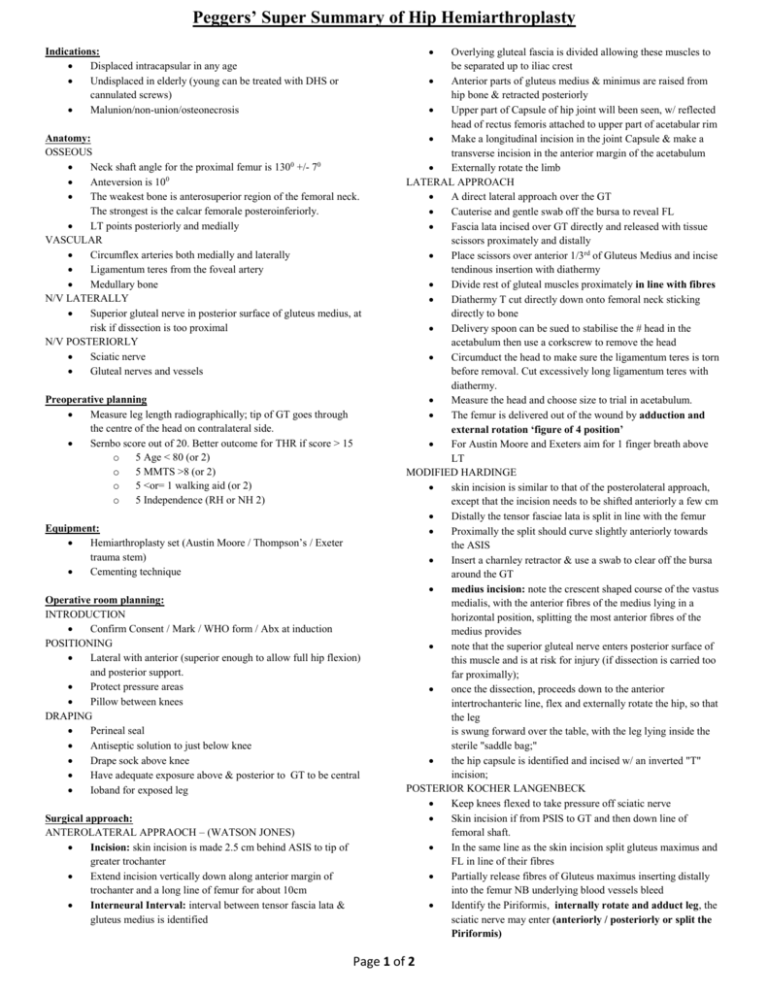
Peggers’ Super Summary of Hip Hemiarthroplasty Indications: Displaced intracapsular in any age Undisplaced in elderly (young can be treated with DHS or cannulated screws) Malunion/non-union/osteonecrosis Anatomy: OSSEOUS Neck shaft angle for the proximal femur is 1300 +/- 70 Anteversion is 100 The weakest bone is anterosuperior region of the femoral neck. The strongest is the calcar femorale posteroinferiorly. LT points posteriorly and medially VASCULAR Circumflex arteries both medially and laterally Ligamentum teres from the foveal artery Medullary bone N/V LATERALLY Superior gluteal nerve in posterior surface of gluteus medius, at risk if dissection is too proximal N/V POSTERIORLY Sciatic nerve Gluteal nerves and vessels Preoperative planning Measure leg length radiographically; tip of GT goes through the centre of the head on contralateral side. Sernbo score out of 20. Better outcome for THR if score > 15 o 5 Age < 80 (or 2) o 5 MMTS >8 (or 2) o 5 <or= 1 walking aid (or 2) o 5 Independence (RH or NH 2) Equipment: Hemiarthroplasty set (Austin Moore / Thompson’s / Exeter trauma stem) Cementing technique Operative room planning: INTRODUCTION Confirm Consent / Mark / WHO form / Abx at induction POSITIONING Lateral with anterior (superior enough to allow full hip flexion) and posterior support. Protect pressure areas Pillow between knees DRAPING Perineal seal Antiseptic solution to just below knee Drape sock above knee Have adequate exposure above & posterior to GT to be central Ioband for exposed leg Surgical approach: ANTEROLATERAL APPRAOCH – (WATSON JONES) Incision: skin incision is made 2.5 cm behind ASIS to tip of greater trochanter Extend incision vertically down along anterior margin of trochanter and a long line of femur for about 10cm Interneural Interval: interval between tensor fascia lata & gluteus medius is identified Overlying gluteal fascia is divided allowing these muscles to be separated up to iliac crest Anterior parts of gluteus medius & minimus are raised from hip bone & retracted posteriorly Upper part of Capsule of hip joint will been seen, w/ reflected head of rectus femoris attached to upper part of acetabular rim Make a longitudinal incision in the joint Capsule & make a transverse incision in the anterior margin of the acetabulum Externally rotate the limb LATERAL APPROACH A direct lateral approach over the GT Cauterise and gentle swab off the bursa to reveal FL Fascia lata incised over GT directly and released with tissue scissors proximately and distally Place scissors over anterior 1/3rd of Gluteus Medius and incise tendinous insertion with diathermy Divide rest of gluteal muscles proximately in line with fibres Diathermy T cut directly down onto femoral neck sticking directly to bone Delivery spoon can be sued to stabilise the # head in the acetabulum then use a corkscrew to remove the head Circumduct the head to make sure the ligamentum teres is torn before removal. Cut excessively long ligamentum teres with diathermy. Measure the head and choose size to trial in acetabulum. The femur is delivered out of the wound by adduction and external rotation ‘figure of 4 position’ For Austin Moore and Exeters aim for 1 finger breath above LT MODIFIED HARDINGE skin incision is similar to that of the posterolateral approach, except that the incision needs to be shifted anteriorly a few cm Distally the tensor fasciae lata is split in line with the femur Proximally the split should curve slightly anteriorly towards the ASIS Insert a charnley retractor & use a swab to clear off the bursa around the GT medius incision: note the crescent shaped course of the vastus medialis, with the anterior fibres of the medius lying in a horizontal position, splitting the most anterior fibres of the medius provides note that the superior gluteal nerve enters posterior surface of this muscle and is at risk for injury (if dissection is carried too far proximally); once the dissection, proceeds down to the anterior intertrochanteric line, flex and externally rotate the hip, so that the leg is swung forward over the table, with the leg lying inside the sterile "saddle bag;" the hip capsule is identified and incised w/ an inverted "T" incision; POSTERIOR KOCHER LANGENBECK Keep knees flexed to take pressure off sciatic nerve Skin incision if from PSIS to GT and then down line of femoral shaft. In the same line as the skin incision split gluteus maximus and FL in line of their fibres Partially release fibres of Gluteus maximus inserting distally into the femur NB underlying blood vessels bleed Identify the Piriformis, internally rotate and adduct leg, the sciatic nerve may enter (anteriorly / posteriorly or split the Piriformis) Page 1 of 2 Peggers’ Super Summary of Hip Hemiarthroplasty Identify the short external rotators and quadrates femoris Divide the tendinous insertion of the Piriformis and external rotators, place a stay suture and reflect to protect the sciatic nerve Obturator internis elevated to reveal posterior column down to ischium and hamstring insertion RETRACTORS Self retainers Hohmann Bennetts Langerbeck REAMING Aim posterior & laterally knock out the cancellous cavity with a box osteotome Circular reamers aiming down femoral cavity scrape posteriorly and laterally Specific set Reamers or Broach with 100 anteversion until appropriate fit and length looking at measuring points Trail head and reduce to check for o Stability o flexion to 900 o Extension to 00 o kick back o leg length o IR/ER CANAL PREPARATION Irrigate Measure and place plug 1-2cm distal to tip Hydrogen peroxide swab with a knot in situ down canal Mix cement and introduce when doughy via pressurized technique sucking any blood proximately Remove excess cement and place implant in being careful to place implant in anteversion and up to the measured point. Hold in situ as cement expands during setting process Anterior 1/3rd of the Gluteus medius elevated with the capsule to expose the # Implant positioning: LT needs to be cut at appropriate level for implant Ream to middle hole of implant on neck or tip of neck to GT 100 anteversion Subcut LMWH injections Right lateral, spinal, sedation, IV Cefuroxime 1.5g, sterile prep and drape, WHO checks Findings : Procedure : Femoral stem prepared Hardinge plug inserted Washout given Standard stem Thompson's prosthesis inserted with cement Acetabulum washed out Reduction achieved & stable in ROM Washout given Closure : Wound closed in layers, #1 vicryl deep and to fascia, 2/0 vicryl to fat and subcut caprosyn 3/0 to skin Opsite, pressure dressings Post Op Instructions : Monitor CSM Keep legs ABDucted with a wedge between the legs Analgesics Routine blood check – Hb and electrolytes Check Xray – Left Hip lateral and AP pelvis to check leg length Operative note: Preparation and Position: Anterolateral approach to the hip joint. All tissues divided in the line of incision Head extracted measuring 53mm Routine post hemiarthroplasty mobilisation Closure Irrigate Haemostasis No1 vicryl to Piriformis and external rotators via an intraosseous canal / FL in centre then continuous / No1 vicryl to deep fat & dermal / 2/0 vicryl to subcutaneous tissues / 3/0 caprisyn to subcuticular closure. Incision and Approach : Femoral neck osteotomised IV fluids for 24hrs Evidence: Posterior approach reduces dislocation. Pellicci PM et al. Clin Orthop Relat Research 1998. THR better than bipolar. Keating JF et al. JBJS (Am) 2006 THR better than Hemi or bipolar in independent mobile patients. Bannister GC et al. JBJS (Am) 2006 also supported by Sernbo score Complications: Intraoperative Instability Perforation of femoral canal or # Early Infection if wound not dry by 10th day return to theatre Failure of fixation Dislocation Late Septic or aseptic failure Thrombosis Mortality 10% at 1 month, 30% at 1 year Page 2 of 2
