Extremity Thrust Manipulation Techniques
advertisement
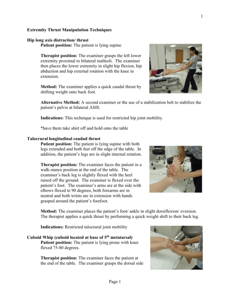
1 Extremity Thrust Manipulation Techniques Hip long axis distraction/ thrust Patient position: The patient is lying supine. Therapist position: The examiner grasps the left lower extremity proximal to bilateral malleoli. The examiner then places the lower extremity in slight hip flexion, hip abduction and hip external rotation with the knee in extension. Method: The examiner applies a quick caudal thrust by shifting weight onto back foot. Alternative Method: A second examiner or the use of a stabilization belt to stabilize the patient’s pelvis at bilateral ASIS. Indications: This technique is used for restricted hip joint mobility. *have them take shirt off and hold onto the table Talocrural longitudinal caudad thrust Patient position: The patient is lying supine with both legs extended and both feet off the edge of the table. In addition, the patient’s legs are in slight internal rotation. Therapist position: The examiner faces the patient in a walk-stance position at the end of the table. The examiner’s back leg is slightly flexed with the heel raised off the ground. The examiner is flexed over the patient’s foot. The examiner’s arms are at the side with elbows flexed to 90 degrees, both forearms are in neutral and both wrists are in extension with hands grasped around the patient’s forefoot. Method: The examiner places the patient’s foot/ ankle in slight dorsiflexion/ eversion. The therapist applies a quick thrust by performing a quick weight shift to their back leg. Indications: Restricted talocrural joint mobility Cuboid Whip (cuboid located at base of 5th metatarsal) Patient position: The patient is lying prone with knee flexed 75-80 degrees. Therapist position: The examiner faces the patient at the end of the table. The examiner grasps the dorsal side Page 1 2 of the patient’s forefoot and places both thumbs (overlapped) on the medial plantar surface of the cuboid. Method: The examiner places the patient’s foot/ ankle into slight dorsiflexion. The therapist applies a quick thrust into slight plantarflexion while taking the knee into extension. Indications: This technique is used for plantar flexed cuboid, decreased mobility of the calcaneocuboid joint. *If have pain with walking and swelling over cuboid = indicative of needing manip PA (volar glide) thrust of capitate on lunate Patient position: The patient is seated. Therapist position: The examiner faces the patient in a walk-stance position. The examiner holds the patients left hand using both hands. The examiner’s fingers are placed on the palmar aspect of the patient’s hand (index fingers supporting the patient’s hand over the palmar aspect of the proximal carpal row, and the remaining fingers supporting the palm of the hand). The pads of both thumbs are placed directly over the capitate on the dorsal side of the patient’s hand. Method: The examiner applies a slight traction to the wrist by gently leaning back. The examiner places the wrist and hand into slight wrist flexion. The therapist applies a quick thrust into neutral while maintaining posterior to anterior pressure on the capitate. Alternative Method: Technique can be modified for other carpal bones. Indications: Restricted wrist mobility. *Go from flexion and thrust to neutral. Thumbs are on top of one another. MCP thrust Patient position: The patient is lying supine or sitting with upper arm supported on table or supported on the ipsilateral thigh. Therapist position: The therapist stabilizes the distal metacarpal with thumb and index finger with one hand and grasps the proximal phalanx with the other thumb and index finger. Page 2 3 Method: The therapist bunches up the skin at the joint, then distracts the joint, and then applies a thrust in a volar direction. Indications: Restricted MCP joint mobility Page 3

