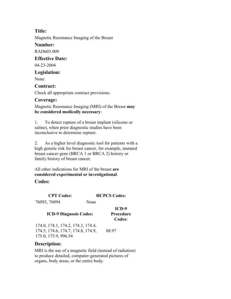
Title:
Magnetic Resonance Imaging of the Breast
Number:
RAD603.009
Effective Date:
04-23-2004
Legislation:
None
Contract:
Check all appropriate contract provisions.
Coverage:
Magnetic Resonance Imaging (MRI) of the Breast may
be considered medically necessary:
1. To detect rupture of a breast implant (silicone or
saline), when prior diagnostic studies have been
inconclusive to determine rupture.
2. As a higher level diagnostic tool for patients with a
high genetic risk for breast cancer, for example, mutated
breast cancer gene (BRCA 1 or BRCA 2) history or
family history of breast cancer.
All other indications for MRI of the breast are
considered experimental or investigational.
Codes:
CPT Codes:
76093, 76094
HCPCS Codes:
None
ICD-9 Diagnosis Codes:
174.0, 174.1, 174.2, 174.3, 174.4,
174.5, 174.6, 174.7, 174.8, 174.9,
175.0, 175.9, 996.54
ICD-9
Procedure
Codes:
88.97
Description:
MRI is the use of a magnetic field (instead of radiation)
to produce detailed, computer-generated pictures of
organs, body areas, or the entire body.
MRI of the breast can be performed using magnetic
resonance (MR) scanners and intravenous MR contrast
agents. Specialized breast coils are available to enhance
the test outcome.
Rationale:
In June of 2003, the American Society of Clinical
Oncology (ASCO) presented three studies on MRI
showing that MRI has significant implications for
women at high risk for breast cancer. Mammography
remains the gold standard in detecting and diagnosing
breast cancer and, it should be noted, MRI should not be
used as a screening method for breast cancer. MRI is
intended for patients who have a BRCA-1 or BRCA-2
gene mutation or who have a strong history of breast
cancer in their families. In October of 2003, Blue Cross
Blue Shield Association Technology Evaluation Center
released an assessment supporting the use of MRI of the
breast in patients considered to be at high genetic risk of
breast cancer. MRI has also been demonstrated to be an
excellent diagnostic tool in women with dense breast
tissue and for the augmented breast.
Pricing:
None
References:
MRI: Best for Breast Screening of High-Risk Women.
Medinews.com (30 July 2003)
http://www.medinews.com/
Iijima, K, Origuchi, J, Yoshida, M, et al. Efficiency of
coronal breast MRI for breast conserving therapy
American Society of Clinical Oncology (2003) Abstract
Number 239.
Kuhl, CK. MRI of breast tumors European Radiology
(2000) 10(1): 46-58.
Liberman, L, Morris, EA, Dershaw, DD, et al. MR
imaging of the ipsilateral breast in women with
percutaneously proven breast cancer. American Journal
of Roentgenology (2003 April) 180(4): 901-10.
Liberman, L, Morris, EA, Kim, CM, et al. MR imaging
findings in the contralateral breast of women with
recently diagnosed breast cancer. American Journal of
Roentgenology (2003 February) 180(2): 333-41.
Hlawatsch, A, Teifke, A, Schmidt, M. Preoperative
assessment of breast cancer: sonography versus MR
imaging. American Journal of Roentgenology (2002
December) 179(6): 1493-501.
Partridge, SC, Gibbs, JE, Lu, Y, Esserman, LJ.
Accuracy of MR imaging for revealing residual breast
cancer in patients who have undergone neoadjuvant
chemotherapy. American Journal of Roentgenology
(2002 November) 179(5): 1193-9.
Rieber, A, Schirrmeister, H, Gabelmann, A. Preoperative staging of invasive breast cancer with MR
mammography and/or PET: boon or bunk. British
Journal of Radiology (2002 October) 75(898): 789-98.
Munot, K, Dall, B, Achnuthan, R, Parkin, G. Role of
magnetic resonance imaging in the diagnosis and singlestage surgical resection of invasive lobular carcinoma of
the breast. British Journal of Surgery (2002 October)
89(10): (1296-301).
Bedrosian, I, Mick, R, Orel, SG, Schnall, M, et al.
Changes in the surgical management of patients with
breast carcinoma based on preoperative magnetic
imaging. Cancer (2003 August 1) 98(3): 468-73.
Lieberman, L, Morris, EA, Benton, CL, et al. Probably
benign lesions at breast magnetic resonance imaging:
preliminary experience in high-risk women. Cancer
(2003 July 15) 98(2): 377-88.
Lee, SG, Orel, SG, Woo, IJ, et al. MR imaging
screening of the contralateral breast in patients with
newly diagnosed breast cancer: preliminary results.
Radiology (2003 March) 226(3): 733-8.
MRI of the Breast in Screening Women considered to be
at high genetic risk of breast cancer. BCBSA TEC
Assessments in Press (October 2003)
http://www.bcbs.com/tec/tecinpress/03.html
CPT® only copyright 2003 American Medical
Association. All Rights Reserved.
Blue Cross and Blue Shield of Illinois, a Division of
Health Care Service Corporation, a Mutual Legal
Reserve Company, an Independent Licensee of the Blue
Cross and Blue Shield Association.
© Copyright 2004. Health Care Service Corporation. All
Rights Reserved.
Legal Disclaimer | Privacy Statement | Code Of
Conduct
Breast Cancer Screening with
MRI — What Are the Data for
Patients at High Risk?
Laura Liberman, M.D.
Return to Search
Result
PDF
PDA Full Text
Add to Personal
Archive
Add to Citation
Manager
Notify a Friend
E-mail When Cited
E-mail When
Letters Appear
Related Article
by Kriege, M.
Find Similar
Articles
PubMed Citation
More than 275,000 women in the United States will receive a diagnosis of breast cancer this
year, and 40,110 women will die of the disease.1 Randomized trials have shown that the use of
screening mammography in the general population reduces mortality associated with breast
cancer by at least 24 percent.2 Cancer is detected in 5 to 7 of every 1000 women on the first
screening mammogram and in 2 or 3 of every 1000 women who undergo regular screening
mammography. Although the average lifetime risk of breast cancer in an American woman is
one in seven,1 the risk increases in women who have a history of breast cancer, atypia or lobular
carcinoma in situ, mantle irradiation for Hodgkin's disease, or a strong family history of breast
cancer. Women with inherited mutations of the BRCA1 or BRCA2 gene have the highest risk of
breast cancer. They make up 5 to 10 percent of women with breast cancer and are also at
increased risk for ovarian cancer. The cumulative risk of breast cancer in women with BRCA1
mutations is 3.2 percent by the age of 30 years, 19.1 percent by the age of 40, 50.8 percent by the
age of 50, 54.2 percent by the age of 60, and 85.0 percent by the age of 70; the cumulative
lifetime risk for carriers of BRCA1 or BRCA2 mutations is 50 to 85 percent.3 Breast cancers in
mutation carriers often occur at a young age, have "pushing margins" and a high nuclear grade,
and lack estrogen receptors.4
How can we prevent breast cancer or make an early diagnosis of the disease in women with
BRCA mutations? The strategies include bilateral prophylactic mastectomy, chemoprevention,
and close surveillance, including yearly mammograms beginning at 25 to 35 years of age.2,3,5
However, screening mammography detects less than half of the breast cancers in mutation
carriers, perhaps owing to young age, dense breasts, or pathological features of the tumor.5,6,7,8
Cancers in mutation carriers grow rapidly; half of them appear in the interval between annual
mammograms. The median size of such "interval cancers" is 1.7 cm, and half have spread to
axillary lymph nodes by the time they are detected.5,6,7,8 It has been suggested that
supplementing mammography with other imaging techniques, shorter screening intervals, or
both may be valuable in mutation carriers.2,5,6,7,8
Magnetic resonance imaging (MRI) of the breast provides information about tissue vascularity
that is not available from mammography. In many breast cancers there is neovascularity, which
causes enhancement of the tumor after the injection of intravenous contrast material
(gadolinium). The pattern (morphology) and time course (kinetics) of enhancement can
determine the likelihood of malignancy.9 Breast MRI is highly sensitive; its disadvantages
include cost, variations in technique and interpretation, imperfect specificity, variation in
parenchymal enhancement during the menstrual cycle (the midcycle is optimal), exclusion
criteria (e.g., the presence of pacemakers or aneurysm clips or a patient's claustrophobia), and an
unproved survival benefit.10 Studies that have cumulatively evaluated breast MRI in more than
1000 high-risk patients found that the technique identified cancer that was not seen on
mammography in 4 percent of cases (Table 1). 10,11,12,13,14,15
View this
table:
[in this
window]
Table 1. Results of Prior Nonrandomized Studies of the Screening of HighRisk Women with Breast MRI.
[in a new
window]
In this issue of the Journal, Kriege et al.16 report a prospective, nonrandomized study of clinical
breast examination, mammography, and MRI in 1909 women who had a genetic or familial
predisposition to breast cancer (lifetime risk,
15 percent) in the Netherlands. Of these
women, 358 (19 percent) had BRCA mutations. This work makes important contributions.
Kriege et al. provide data on almost twice as many patients and twice as many mutation carriers
as were included in all previously published evaluations of MRI in high-risk patients combined.
Those who interpreted the MRIs and mammograms were unaware of the results of the other
technique. The investigators analyzed their data in subgroups according to quantified levels of
risk. Their study confirms the high sensitivity of MRI in identifying invasive breast cancer in
high-risk patients.
Kriege et al. found that the breast-cancer detection rate was 9.5 per 1000 woman-years of
follow-up overall: 7.8 per 1000 for women with a 15 to 29 percent lifetime risk, 5.4 per 1000 for
those with a 30 to 49 percent lifetime risk, and 26.5 per 1000 for carriers of BRCA1 or BRCA2
mutations. Among 45 cancers, 22 (49 percent) were identified by MRI but not mammography,
10 (22 percent) were identified by both MRI and mammography, and 8 (18 percent) were
identified by mammography but not MRI. Of these 45 tumors, 4 were interval cancers, and 1
was identified by clinical examination only. Certain features appeared in more than half of
cancers in mutation carriers: they were diagnosed in women between the ages of 30 and 39
years; they were invasive cancers; and the tumors were of high nuclear grade, estrogen receptor–
negative, and node-negative. Only 17 percent of cancers in mutation carriers were interval
cancers. In their analyses, MRI, as compared with mammography, had higher sensitivity (71
percent vs. 40 percent) but lower specificity (90 percent vs. 95 percent).
Kriege et al. report that short-term follow-up MRI was recommended in 7 percent of
examinations, as compared with 10 to 25 percent in prior reports.11,17 MRI had limited
sensitivity (17 percent) in detecting ductal carcinoma in situ; in prior studies, the sensitivity of
MRI for this type of lesion ranged from 0 percent13 to 100 percent.11,14 Kriege et al. also report
that MRI had lower specificity than mammography, but Kuhl et al.11 found that MRI had higher
sensitivity and specificity than mammography. Refinement and standardization of MRI
technique and interpretation may improve specificity while retaining high sensitivity. Not
addressed by Kriege et al. is the potential role of ultrasonography in screening high-risk women.
In studies that supplemented mammography with both MRI and ultrasonography, MRI had
higher sensitivity and specificity than ultrasonography and was superior in detecting ductal
carcinoma in situ (Table 2).11,13,14
View this
table:
[in this
window]
Table 2. Sensitivity and Specificity of Mammography, MRI, and
Ultrasonography for Detecting Tumors in High-Risk Women.
[in a new
window]
The report by Kriege et al. highlights an important issue: How do we evaluate the efficacy of a
screening test, and what is the desirable balance between sensitivity and specificity? Any method
of breast-cancer screening has the potential for benefit (lifesaving cancer detection) and for
harm (cost, anxiety, follow-up imaging, or benign biopsy). The prognosis is better for small,
early cancers, but detecting small cancers at an early stage does not guarantee improved survival
rates; detecting nonlethal cancers or cancers that have already metastasized will not decrease
mortality. Only a randomized, controlled trial with death as the end point can definitively prove
that any screening intervention improves survival.18
Without information provided by randomized, controlled trials, the management of breast
cancer may be guided by other reports, such as observational studies, data extrapolation, and
expert opinion.19 Whereas breast cancer develops in only a minority of women in the general
population, the disease develops in most women who are BRCA mutation carriers (50 to 85
percent). For mutation carriers, the benefit of high sensitivity may outweigh the effects of
imperfect specificity. The Blue Cross–Blue Shield Association's Technology Evaluation Center
has adopted criteria for technology assessment, including that the technology improve the net
health outcome; a recent report concluded that using MRI to screen women at high genetic risk
for breast cancer meets this criterion.20 The new data reported by Kriege et al. provide further
evidence of a benefit.
MRI can detect otherwise occult breast cancer in high-risk patients and is probably most
beneficial to those at highest risk. Data are accumulating in support of supplementing
mammography with MRI to detect cancer in carriers of BRCA mutations. MRI may also be
valuable in screening women with an increased risk due to nongenetic factors (e.g., prior breast
cancer), but more work is needed to substantiate this possibility, including analysis of the
contribution of MRI in subgroups with defined risk factors and quantified levels of risk. No data
support the use of MRI in screening women at normal risk. Ideally, breast MRI should be
performed at facilities that follow technical and interpretive guidelines9 and that can perform
biopsies of lesions detected by MRI alone.21 Whether the excellent results reported in the
literature can be achieved in practice remains to be determined. Further outcomes research is
essential to develop evidence-based recommendations for methods of breast-cancer screening
that are tailored to the specific needs of women at various levels of risk.
Source Information
From the Memorial Sloan-Kettering Cancer Center, New York.
References
1. Jemal A, Tiwari RC, Murray T, et al. Cancer statistics, 2004. CA Cancer J Clin
2004;54:8-29.[Abstract/Full Text]
2. Smith RA, Saslow D, Sawyer KA, et al. American Cancer Society guidelines for breast
cancer screening: update 2003. CA Cancer J Clin 2003;53:141-169.[Abstract/Full Text]
3. Burke W, Daly M, Garber J, et al. Recommendations for follow-up care of individuals
with an inherited predisposition to cancer. II. BCRA1 and BRCA2. JAMA
1997;277:997-1003.[Abstract]
4. Lakhani SR, Van De Vijver MJ, Jacquemier J, et al. The pathology of familial breast
cancer: predictive value of immunohistochemical markers estrogen receptor,
progesterone receptor, HER-2, and p53 in patients with mutations in BRCA1 and
BRCA2. J Clin Oncol 2002;20:2310-2318.[Abstract/Full Text]
5. Meijers-Heijboer H, van Geel B, van Putten WL, et al. Breast cancer after prophylactic
mastectomy in women with a BRCA1 or BRCA2 mutation. N Engl J Med 2001;345:159164.[Abstract/Full Text]
6. Brekelmans CTM, Seynaeve C, Bartels CCMM, et al. Effectiveness of breast cancer
surveillance in BRCA1/2 gene mutation carriers and women with high familial risk. J
Clin Oncol 2001;19:924-930.[Abstract/Full Text]
7. Scheuer L, Kauff N, Robson M, et al. Outcome of preventive surgery and screening for
breast and ovarian cancer in BRCA mutation carriers. J Clin Oncol 2002;20:12601268.[Abstract/Full Text]
8. Komenaka IK, Ditkoff BA, Joseph KA, et al. The development of interval breast
malignancies in patients with BRCA mutations. Cancer 2004;100:20792083.[CrossRef][ISI][Medline]
9. ACR breast imaging reporting and data system atlas. Reston, Va.: American College of
Radiology, 2003.
10. Morris EA, Liberman L, Ballon DJ, et al. MRI of occult breast carcinoma in a high-risk
population. AJR Am J Roentgenol 2003;181:619-626.[Abstract/Full Text]
11. Kuhl CK, Schmutzler RK, Leutner CC, et al. Breast MR imaging screening in 192
women proved or suspected to be carriers of a breast cancer susceptibility gene:
preliminary results. Radiology 2000;215:267-279.[Abstract/Full Text]
12. Tilanus-Linthorst MMA, Obdeijn IMM, Bartels KCM, de Koning HJ, Oudkerk M. First
experiences in screening women at high risk for breast cancer with MR imaging. Breast
Cancer Res Treat 2000;63:53-60.[CrossRef][ISI][Medline]
13. Warner E, Plewes DB, Shumak RS, et al. Comparison of breast magnetic resonance
imaging, mammography, and ultrasound for surveillance of women at high risk for
hereditary breast cancer. J Clin Oncol 2001;19:3524-3531.[Abstract/Full Text]
14. Podo F, Sardanelli F, Canese R, et al. The Italian multi-centre project on evaluation of
MRI and other imaging modalities in early detection of breast cancer in subjects at high
genetic risk. J Exp Clin Cancer Res 2002;21:Suppl:115-124.
15. Stoutjesdijk MJ, Boetes C, Jager GJ, et al. Magnetic resonance imaging and
mammography in women with a hereditary risk of breast cancer. J Natl Cancer Inst
2001;93:1095-1102.[Abstract/Full Text]
16. Kriege M, Brekelmans CTM, Boetes C, et al. Efficacy of MRI and mammography for
breast-cancer screening in women with a familial or genetic predisposition. N Engl J
Med 2004;351:427-437.[Abstract/Full Text]
17. Liberman L, Morris EA, Benton CL, Abramson AF, Dershaw DD. Probably benign
lesions at breast magnetic resonance imaging: preliminary experience in high-risk
women. Cancer 2003;98:377-388.[CrossRef][ISI][Medline]
18. Kopans DB, Monsees B, Feig SA. Screening for cancer: when is it valid? Lessons from
the mammography experience. Radiology 2003;229:319-327.[Abstract/Full Text]
19. Harris RP, Helfand M, Woolf SH, et al. Current methods of the US Preventive Services
Task Force: a review of the process, 2004. (Accessed July 9, 2004, at
http://www.ahrq.gov/clinic/ajpmsuppl/harris3.htm.)
20. Blue Cross Blue Shield. Magnetic resonance imaging of the breast in screening women
considered to be at high genetic risk of breast cancer, December 2003. (Accessed July 9,
2004, at http://www.bluecares.com/tec/vol18/18_15.html.)
21. Liberman L, Morris EA, Dershaw DD, Thornton CM, Van Zee KJ, Tan LK. Fast MRIguided vacuum-assisted breast biopsy: initial experience. AJR Am J Roentgenol
2003;181:1283-1293.[Abstract/Full Text]
Return to Search
Result
PDF
PDA Full Text
Add to Personal
Archive
Add to Citation
Manager
Notify a Friend
E-mail When Cited
E-mail When
Letters Appear
Related Article
by Kriege, M.
Find Similar
Articles
PubMed Citation
This article has been cited by other articles:
Robson, M. E., Offit, K. (2004). Breast MRI for Women With Hereditary Cancer Risk.
JAMA 292: 1368-1370 [Full Text]
(2004). Breast Cancer Screening in High-Risk Women: Does MRI Add Value?. Journal
Watch Women's Health 2004: 1-1 [Full Text]
(2004). Breast Cancer Screening by MRI in High-Risk Women. Journal Watch
Gastroenterology 2004: 9-9 [Full Text]
(2004). Breast Cancer Screening by MRI in High-Risk Women. Journal Watch (General)
2004: 4-4 [Full Text]
Article
Options
Extract
PDF
Send to a Friend
Related articles in
this issue
• Similar articles in
this journal
•
•
•
•
Literature
Track
• Add to File Drawer
• Download to
Citation Manager
• PubMed citation
Breast MRI for Women With Hereditary
Cancer Risk
• Articles in PubMed
by
•Robson ME
•Offit K
• Contact me when
this article is cited
Mark E. Robson, MD; Kenneth Offit, MD, MPH
JAMA. 2004;292:1368-1370.
Topic
Collections
• Women's Health,
Other
• Oncology
• Breast Cancer
• Magnetic
Resonance Imaging
• Collection E-mail
Alerts
Approximately a decade ago, germline mutations in BRCA1 and
BRCA2 were identified as the most common detectable causes
of a hereditary predisposition to breast (and ovarian) cancer.1-2
A recent meta-analysis of 22 studies indicated that the average
risk of breast cancer by 70 years is 65% for women with
BRCA1 mutations and 45% for BRCA2 mutations,3 although the
risk may be substantially higher in some families. Women with
BRCA1 mutations in their fourth and fifth decade of life have on average
approximately a 30-fold higher risk of breast cancer than women without mutations,
and BRCA2 mutation carriers are at 10-fold to 16-fold higher risk.3
Confronted by breast cancer risks of this magnitude, it is not surprising that a
significant fraction of mutation carriers elect to undergo prophylactic mastectomy, a
procedure that has been shown to reduce breast cancer risk by 90% or more.4-6
However, for many women, the physical and psychological morbidity of risk-reducing
surgery is unacceptable. Although adjuvant therapy with tamoxifen appears to
reduce contralateral breast cancer risk in affected mutation carriers,7-8 its value as
primary prevention in unaffected women remains uncertain.9 While our group and
other researchers have described a significant reduction in breast cancer risk among
women with mutations who enter premature menopause as the result of a riskreducing oophorectomy,10-11 protection is clearly incomplete.
Women at hereditary risk who choose not to undergo preventive mastectomy have
been advised to undergo breast self-examination, clinical breast examination (CBE),
and annual mammography beginning at an early age (25-30 years).12-13 However, in
large cohorts of BRCA mutation carriers undergoing such surveillance in New York
and the Netherlands, nearly 50% of breast cancers identified were diagnosed in the
interval between screening studies and nearly half of the invasive breast cancers
had metastasized to axillary nodes at the time of diagnosis.14-15 The relative
insensitivity of mammography among women at hereditary risk results from several
factors, including the underlying breast density of these young women, the benign
mammographic appearance of some BRCA-associated breast cancers, and the rapid
growth rate of these frequently high-grade tumors.16
Magnetic resonance imaging (MRI) has emerged as an extremely powerful tool in
breast cancer management.17-23 The use of the contrast agent gadolinium, in
combination with sophisticated imaging protocols, allows the identification of tumor
neovascularity, which cannot be detected by conventional mammography.17 In this
issue of JAMA, the article by Warner and colleagues24 from a large single-institution
study using this new technology provides important new information for women at
hereditary risk regarding their surveillance options.
In the study by Warner et al, 236 women with germline BRCA1 or BRCA2 mutations
underwent annual multimodality screening with CBE, mammography, screening
ultrasound, and breast MRI, all performed on the same day. An interval CBE was
performed 6 months later. Systematic imaging and follow-up protocols were followed
to minimize unnecessary biopsies generated by nonmalignant enhancement on MRI.
Consistent with previous surveillance studies in women at hereditary risk,14-15 only
45% of the identified cancers would have been detected by "conventional" screening
(mammography and CBE). However, of the 22 cancers diagnosed, 77% were
detected by MRI, and 32% were identified by MRI alone. MRI identified a
significantly greater proportion of breast cancers than either mammography (36%)
or ultrasound (33%).
These results are similar to those of a recently reported, multi-institutional study
performed in the Netherlands by Kriege et al,25 in which 1909 women at a 15% or
more lifetime breast cancer risk (including 358 BRCA mutation carriers) were
screened annually with concurrent mammography and MRI. Of the 45 cancers
diagnosed in the Netherlands cohort,25 22 (49%) were detected by MRI alone, with
an overall sensitivity of 71% for MRI vs 40% for mammography. Comparison of the
positive predictive value (PPV) of an abnormal MRI in these and other studies is
hampered by differences in the definitions used, but 17 (46%) of 37 "positive
screens" in the study by Warner et al24 were associated with a diagnosis of cancer, as
were 21 (32%) of 65 MRIs interpreted as suspicious or highly suggestive of
malignancy (Breast Imaging Reporting and Data System [BI-RADS] 4 or 5) in the
study by Kriege et al.25 The differences in predictive value, as well as sensitivity and
specificity in these and prior studies (Table 1), may also reflect different levels of
experience and consistency in radiological interpretations in single-institution vs
multi-institution settings. In the studies by Kriege et al and Warner et al, however,
receiver operating characteristic curves, a function of both sensitivity and specificity,
confirm a greater diagnostic accuracy for MRI as compared with mammography.
View this table:
[in this window]
[in a new window]
Table. Comparison of Magnetic Resonance Imaging and Other
Modalities in Women at Hereditary Risk for Breast Cancer
Although these results clearly affirm that MRI is significantly more sensitive than
mammography in detecting breast cancer in women at hereditary risk, a number of
fundamental questions remain. First, it is not yet clear whether the enhanced
sensitivity of MRI will translate into a reduction in breast cancer–related mortality.
The observation of an apparent decrease in sensitivity of MRI after the initial screen
in both studies (Warner et al and Kriege et al) sounds a cautionary note. A
randomized controlled trial with mortality as a primary end point would be desirable
to prove the benefit of MRI screening in mutation carriers but accrual to such a
study is likely to prove difficult. Indirect evidence suggests that MRI screening leads
to downstaging of detected cancers, which may translate into a survival benefit.
Although 21% of cancers detected were associated with axillary nodal metastases in
the Netherlands study,25 this rate was significantly lower than in 2 control groups not
receiving MRI screening. Tumor size was also significantly smaller in the MRI group.
In the study by Warner et al, only 2 cancers (9% of the total) were associated with
axillary nodal metastases, and each of these cases was identified at the initial
(prevalent) cancer screen. All incident cancers were in situ or stage I lesions. These
findings are the most encouraging yet reported for MRI screening.
A second question is the relative value and timing of MRI screening vis-à-vis
mammograms and, possibly, screening ultrasound. MRI and conventional
mammography appear to be complementary; in the study by Warner et al, both
modalities diagnosed cases of ductal carcinoma in situ missed by the other screening
tool. Ultrasound also detected a small number of cancers not identified by MRI, and
"triple screening," not used in the study by Kriege et al, improved sensitivity to 95%.
Although interval cancer was not a major issue in the current study, 20% of cancers
detected in mutation carriers in the Netherlands study presented within 12 months of
imaging. If these interval cancers resulted from "kinetic failures" of detection due to
the higher proliferative rate of tumors in BRCA mutation carriers, the optimal
screening strategy may be to alternate mammography and MRI (with or without
ultrasound) at 6-month intervals.15
Questions also remain regarding the specificity of MRI screening. In the study by
Warner et al, the specificity of MRI improved from 93% to 99% during the 3
screening rounds. However, Warner et al only considered examinations as falsepositive if a biopsy was performed with a benign result, and the calculated specificity
would likely be significantly lower if examinations resulting in additional studies
("diagnostic" MRI or 6-month follow-up studies) were also considered positive.
Despite suboptimal specificity, the PPV of a persistently abnormal MRI was high
(46% overall), largely because of the remarkably high incidence of breast cancer in
BRCA mutation carriers (5.5% of initial screens and 4.1% of subsequent screens).
MRI screening in groups of women with lower disease prevalence will certainly result
in substantially lower PPVs and a less favorable risk-to-benefit ratio.
Warner et al have clearly documented the risks and benefits of breast MRI
screening in women at the highest levels of hereditary risk. Their findings, in
combination with those of Kriege et al, strongly suggest that women with BRCA
mutations should be offered such screening. Women and their physicians must,
however, be aware that both sensitivity and specificity of screening MRI may be
substantially less than described if different imaging protocols are followed or if
experienced radiologists and suitable technology, including the capability to perform
magnetic resonance–guided biopsies, are not available.26 A technology assessment
by 1 large insurance carrier has already supported the rationale for MRI screening of
BRCA mutation carriers and other women at high hereditary risk for breast cancer,
even in the absence of a randomized controlled trial demonstrating a mortality
benefit.27 Remaining questions, largely centered on specificity, recall rate, and PPV,
argue against routine application of MRI screening for women at lesser degrees of
risk without carefully designed studies, preferably randomized controlled trials,
delineating test performance in those specific populations.
AUTHOR INFORMATION
Corresponding Author: Kenneth Offit, MD, MPH, Clinical Genetics Service,
Memorial Sloan-Kettering Cancer Center, 1275 York Ave, New York, NY 10021
(offitk@mskcc.org).
Editorials represent the opinions of the authors and THE JOURNAL and not those of
the American Medical Association.
Author Affiliations: Clinical Genetics Service, Memorial Sloan-Kettering Cancer
Center, New York, NY.
REFERENCES
1. Miki Y, Swensen J, Shattuck-Eidens D, et al. A strong candidate for the breast
and ovarian cancer susceptibility gene BRCA1. Science. 1994;266:66-71. ISI | MEDLINE
2. Wooster R, Bignell G, Lancaster J, et al. Identification of the breast cancer
susceptibility gene BRCA2 Nature. 1995;378:789-792. [published correction appears
in Nature. 1996;379:749]. CrossRef | ISI | MEDLINE
3. Antoniou A, Pharoah PD, Narod S, et al. Average risks of breast and ovarian
cancer associated with BRCA1 or BRCA2 mutations detected in case series
unselected for family history: a combined analysis of 22 studies. Am J Hum Genet.
2003;72:1117-1130. CrossRef | ISI | MEDLINE
4. Hartmann LC, Sellers TA, Schaid DJ, et al. Efficacy of bilateral prophylactic
mastectomy in BRCA1 and BRCA2 gene mutation carriers. J Natl Cancer Inst.
2001;93:1633-1637. ABSTRACT/FULL TEXT
5. Meijers-Heijboer H, van Geel B, van Putten WL, et al. Breast cancer after
prophylactic bilateral mastectomy in women with a BRCA1 or BRCA2 mutation. N
Engl J Med. 2001;345:159-164. ABSTRACT/FULL TEXT
6. Rebbeck TR, Friebel T, Lynch HT, et al. Bilateral prophylactic mastectomy reduces
breast cancer risk in BRCA1 and BRCA2 mutation carriers: the PROSE study group. J
Clin Oncol. 2004;22:1055-1062. ABSTRACT/FULL TEXT
7. Metcalfe K, Lynch HT, Ghadirian P, et al. Contralateral breast cancer in BRCA1
and BRCA2 mutation carriers. J Clin Oncol. 2004;22:2328-2335. ABSTRACT/FULL TEXT
8. Narod SA, Brunet JS, Ghadirian P, et al, for Hereditary Breast Cancer Clinical
Study Group. Tamoxifen and risk of contralateral breast cancer in BRCA1 and
BRCA2 mutation carriers: a case-control study. Lancet. 2000;356:1876-1881. CrossRef
| ISI | MEDLINE
9. King MC, Wieand S, Hale K, et al. Tamoxifen and breast cancer incidence among
women with inherited mutations in BRCA1 and BRCA2: National Surgical Adjuvant
Breast and Bowel Project (NSABP-P1) Breast Cancer Prevention Trial. JAMA.
2001;286:2251-2256. ABSTRACT/FULL TEXT
10. Kauff ND, Satagopan JM, Robson ME, et al. Risk-reducing salpingo-oophorectomy
in women with a BRCA1 or BRCA2 mutation. N Engl J Med. 2002;346:1609-1615.
ABSTRACT/FULL TEXT
11. Rebbeck TR, Lynch HT, Neuhausen SL, et al. Prophylactic oophorectomy in
carriers of BRCA1 or BRCA2 mutations. N Engl J Med. 2002;346:1616-1622.
ABSTRACT/FULL TEXT
12. Burke W, Daly M, Garber J, et al, for Cancer Genetics Studies Consortium.
Recommendations for follow-up care of individuals with an inherited predisposition to
cancer, II: BRCA1 and BRCA2. JAMA. 1997;277:997-1003. ABSTRACT
13. National Comprehensive Cancer Network. Genetic/familial high-risk assessment:
breast and ovarian. Available at:
http://www.nccn.org/professionals/physician_gls/PDF/genetics_screening.pdf.
Accessibility verified August 12, 2004.
14. Brekelmans CT, Seynaeve C, Bartels CC, et al. Effectiveness of breast cancer
surveillance in BRCA1/2 gene mutation carriers and women with high familial risk. J
Clin Oncol. 2001;19:924-930. ABSTRACT/FULL TEXT
15. Scheuer L, Kauff ND, Robson M, et al. Outcome of preventive surgery and
screening for breast and ovarian cancer in BRCA mutation carriers. J Clin Oncol.
2002;20:1260-1268. ABSTRACT/FULL TEXT
16. Tilanus-Linthorst M, Verhoog L, Obdeijn IM, et al. A BRCA1/2 mutation, high
breast density and prominent pushing margins of a tumor independently contribute
to a frequent false-negative mammography. Int J Cancer. 2002;102:91-95. CrossRef |
ISI | MEDLINE
17. Morris EA. Breast cancer imaging with MRI. Radiol Clin North Am. 2002;40:443466. ISI | MEDLINE
18. Kuhl CK, Schrading S, Leutner CC, et al. Surveillance of "high risk" women with
proven or suspected familial (hereditary) breast cancer: first mid-term results of a
multi-modality clinical screening trial [abstract]. Proc Am Soc Clin Oncol. 2003;22:2.
19. Podo F, Sardanelli F, Canese R, et al. The Italian multi-centre project on
evaluation of MRI and other imaging modalities in early detection of breast cancer
in subjects at high genetic risk. J Exp Clin Cancer Res. 2002;21:115-124. MEDLINE
20. Stoutjesdijk MJ, Boetes C, Jager GJ, et al. Magnetic resonance imaging and
mammography in women with a hereditary risk of breast cancer. J Natl Cancer Inst.
2001;93:1095-1102. ABSTRACT/FULL TEXT
21. Tilanus-Linthorst MM, Obdeijn IM, Bartels KC, de Koning HJ, Oudkerk M. First
experiences in screening women at high risk for breast cancer with MR imaging.
Breast Cancer Res Treat. 2000;63:53-60. CrossRef | ISI | MEDLINE
22. Warner E, Plewes DB, Shumak RS, et al. Comparison of breast magnetic
resonance imaging, mammography, and ultrasound for surveillance of women at
high risk for hereditary breast cancer. J Clin Oncol. 2001;19:3524-3531.
ABSTRACT/FULL TEXT
23. Morris EA, Liberman L, Ballon DJ, et al. MRI of occult breast carcinoma in a
high-risk population. AJR Am J Roentgenol. 2003;181:619-626. ABSTRACT/FULL TEXT
24. Warner E, Plewes DB, Hill KA, et al. Surveillance of BRCA1 and BRCA2 mutation
carriers with magnetic resonance imaging, ultrasound, mammography, and clinical
breast examination. JAMA. 2004;292:1317-1325. ABSTRACT/FULL TEXT
25. Kriege M, Brekelmans CT, Boetes C, et al. Efficacy of MRI and mammography for
breast-cancer screening in women with a familial or genetic predisposition. N Engl J
Med. 2004;351:427-437. ABSTRACT/FULL TEXT
26. Liberman L. Breast cancer screening with MRI: what are the data for patients at
high risk? N Engl J Med. 2004;351:497-500. FULL TEXT
27. BlueCross BlueShield Association. Magnetic resonance imaging of the breast in
screening women considered to be at high genetic risk of breast cancer. Available
at: http://www.bluecares.com/tec/vol18/18_15.html. Accessed August 5, 2004.


![Dear [Referring Physician]:](http://s3.studylib.net/store/data/005817856_2-e9ac88ec4b586b2068096049a1436282-300x300.png)
