Regional Anesthesia - TOP Recommended Websites
advertisement
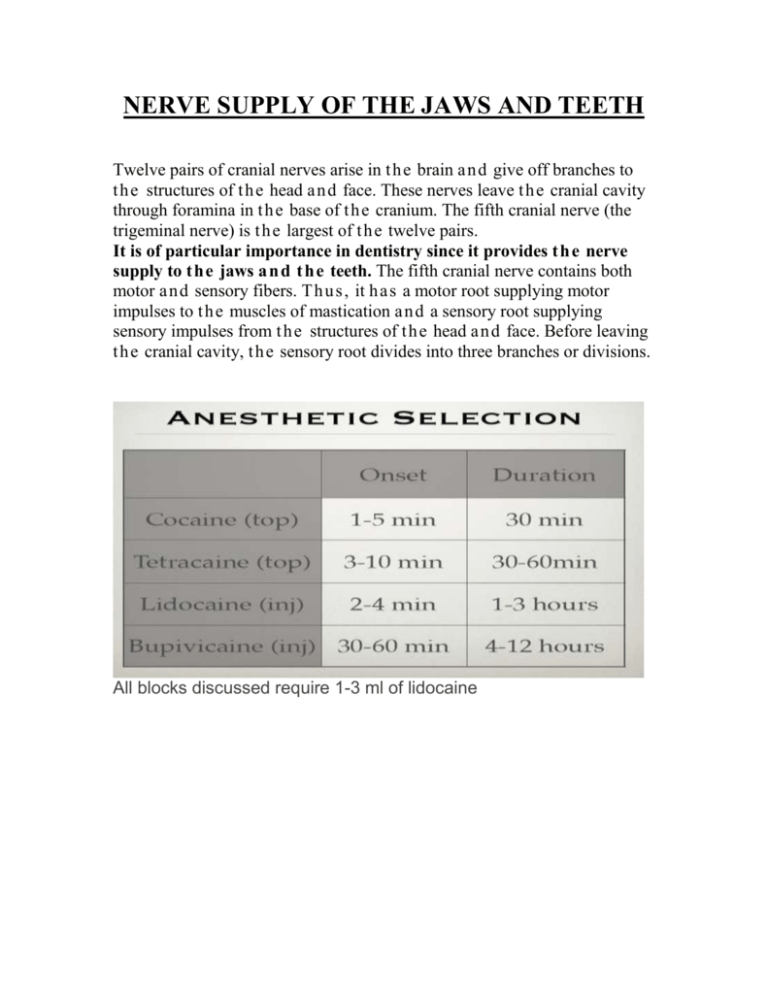
NERVE SUPPLY OF THE JAWS AND TEETH Twelve pairs of cranial nerves arise in t h e brain a n d give off branches to t h e structures of t h e head a n d face. These nerves leave t h e cranial cavity through foramina in t h e base of t h e cranium. The fifth cranial nerve (the trigeminal nerve) is t h e largest of t h e twelve pairs. It is of particular importance in dentistry since it provides t h e nerve supply to t h e jaws a n d t h e teeth. The fifth cranial nerve contains both motor a n d sensory fibers. Th u s , it h a s a motor root supplying motor impulses to t h e muscles of mastication a n d a sensory root supplying sensory impulses from t h e structures of t h e head a n d face. Before leaving t h e cranial cavity, t h e sensory root divides into three branches or divisions. All blocks discussed require 1-3 ml of lidocaine Trigeminal Nerve Blocks Opthalmic Division Blocks • Supraorbital and Supratrochlear • Nasociliary Maxillary Division Blocks • Infraorbital • Nasopalatine • Sphenopalatine Mandibular Division Blocks • Inferior Alveolar • Mental • Auriculotemporal Infraorbital Nerve Block Anatomy This is the largest terminal branch of the maxillary nerve. It enters though the inferior orbital fissure and travels in the infraorbital foramen to innervate the incisors and canines and anterior gingival mucosa along with the skin and soft tissues of the cheek. It should be noted that blocking the infraorbital nerve does not provide anesthesia to all the upper dentition. Maxillary nerve block would be used for this purpose. Technique The infraorbital foramen is located in a line between the pupil and the corner of the mouth just below the infraorbital rim. Three ccs of injection at the sight of exit of the nerve is usually sufficient for adequate anesthesia. The foramen can be approached from the skin or sublabially. Inferior Alveolar and Lingual Nerve Blocks Anatomy The inferior alveolar nerve is the largest branch of the mandibular nerve and enters the mandibular canal giving off several branches in the canal to give sensation to the teeth before exiting the mental foramen to supply sensation to the chin. The lingual nerve travels anteriorly just medial to the mandible on the floor of the mouth where it is joined by the corda tympani nerve before traveling to the tongue. It also gives off supply to the submandibular and sublingual glands. Technique The inner surface of the mandible is infiltrated with 5 ccs of local injection after advancing the needle 2 inches deep, 1 inch superior and just medial to the 3rd mandibular molar. * The provider’s left index finger is placed over the coronoid process and the pterygomandibular raphe is indicated at the tip of the needle. This injection is 1/4 of the way between the pterygomandibular raphe and the coronoid process. Mental Nerve Block Anatomy The mental nerve is one of two terminal branches of the inferior alveolar nerve. It emerges from the mental canal to innervate the lower lip and gingival surface from the corner of the mouth to the midline. It is located just below or slightly posterior to the second premolar midway between the inferior and superior borders of the mandible. Technique The foramen is approached intraorally or though the skin with a 25 gauge needle and 2 to 3 ccs of local anesthetic. It should be noted that entrance into the foramen poses risk of permanent nerve damage Maxillary Nerve block Anatomy The maxillary nerve starts from the trigeminal ganglion, travels through the cavernous sinus exiting the skull at the foramen rotundum to enter the pterygopalatine fossa where it gives off several branches to the mid face. Technique A needle is inserted just below the zygomatic arch midway between the coronoid and condyle of the mandible. It is inserted perpendicular to the skin until the pterygoid plate is felt. The needle is then withdrawn and guided anteriorly towards the eye to enter the pterygopalatine fossa. Five ccs of local are injected when paresthesia of the upper jaw is illicited. It should be noted that hemorrhage of the maxillary artery can cause hematoma in the hard and soft palate. Sphenopalatine Ganglion Block Anatomy The sphenopalatine ganglion sits in the pterygopalatine fossa. Its main innervation is from the maxillary nerve, the greater superficial petrosal nerve, and sympathetics from the deep petrosal nerve. It supplies the periostium of the orbit and lacrimal gland, and gives off the posterior superior nasal nerve and the nasal palatine nerves that supply the gums, hard palate, soft palate, uvula, and part of the tonsils. Technique The greater palatine foramen is located at the posterior portion of the hard palate just medial to the gum line opposite the third molar. A needle is advanced 2 inches through the foramen and 3 cc is injected. Mandibular Nerve Block Anatomy The mandibular nerve exits the foramen oval and divides into an anterior motor branch and a posterior branch. The anterior motor branch supplies the medial pterygoid, tensor tympani, and tensor palatine muscles. The posterior branch supplies sensation for the lower third of the face and the prearicular area. Technique The patient is asked to open his mouth and the needle is advanced just below the zygomatic arch at the midpoint of the notch of the mandible until the pterygoid plate is felt. The needle is then withdrawn, and redirected posteriorly in the direction of the ear. 4-5 ccs of local are injected here and upon withdrawal of the needle. FACIAL NERVE:

